Chinese Journal of Tissue Engineering Research ›› 2016, Vol. 20 ›› Issue (28): 4203-4209.doi: 10.3969/j.issn.2095-4344.2016.28.015
Previous Articles Next Articles
Protective effect of retinal stem cell transplantation on retinal ganglion cells in glaucoma
Gu Zhi-min, Zhou Li-xiao, Qi Ruo
- Department of Ophthalmology, Fifth Affiliated Hospital of Zhengzhou University, Zhengzhou 450052, Henan Province, China
-
Revised:2016-05-19Online:2016-07-01Published:2016-07-01 -
About author:Gu Zhi-min, Attending physician, Department of Ophthalmology, Fifth Affiliated Hospital of Zhengzhou University, Zhengzhou 450052, Henan Province, China
CLC Number:
Cite this article
Gu Zhi-min, Zhou Li-xiao, Qi Ruo . Protective effect of retinal stem cell transplantation on retinal ganglion cells in glaucoma[J]. Chinese Journal of Tissue Engineering Research, 2016, 20(28): 4203-4209.
share this article

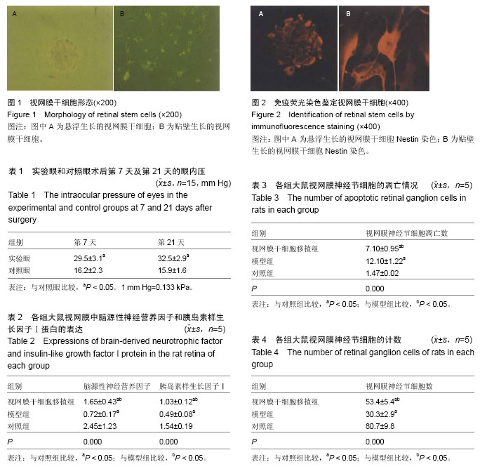
2.1 视网膜干细胞的形态 原代培养第3天出现细胞球,形状为类圆形,原代培养第5天,细胞球增多增大,折光性增强,形成立体感比较强的神经球,培养第7天以后神经球增大不明显。视网膜干细胞培养至第7天时开始传代培养,传代培养第3天出现细胞克隆,呈小圆形,第2代细胞培养24 h出现细胞团(图1)。 2.2 视网膜干细胞的鉴定 免疫荧光染色鉴定结果显示:培养细胞中96%以上细胞为Nestin阳性表达细胞(图2)。 2.3 大鼠青光眼模型的鉴定 在术后第1天有20只大鼠实验眼出现眼结膜轻度水肿,11只出现角膜轻度水肿,6只出现前房房水混浊,术后第7天时所有症状均消失,实验眼均未出现晶状体浑浊。30只对照眼均未出现异常。由表1可以看出,30只实验眼在术后第7天和第21天时的眼内压均明显高于对照眼(P < 0.05),表明青光眼大鼠模型均建造成功。 2.4 各组大鼠视网膜中脑源性神经营养因子和胰岛素样生长因子Ⅰ蛋白的表达 由表2可以看出,模型组脑源性神经营养因子和胰岛素样生长因子Ⅰ蛋白的表达均明显低于对照组(P < 0.05),视网膜干细胞移植组脑源性神经营养因子和胰岛素样生长因子Ⅰ蛋白的表达明显高于模型组(P < 0.05),但低于对照组(P < 0.05)。 2.5 视网膜神经节细胞的凋亡情况 由表3可以看出,模型组视网膜神经节细胞的凋亡数明显高于对照组(P < 0.05),视网膜干细胞移植组的视网膜神经节细胞的凋亡数明显低于模型组(P < 0.05),但高于对照组(P < 0.05)。 2.6 视网膜神经节细胞的计数 由表4可以看出,模型组视网膜神经节细胞数明显低于对照组(P < 0.05),视网膜干细胞移植组的视网膜神经节细胞数明显高于模型组(P < 0.05),但低于对照组(P < 0.05)。"
| [1] 林丁,陈琛.青光眼的视网膜神经节细胞损伤及其保护[J].中华眼科杂志,2005, 42(12):1144-1148. [2] Kang MH,Yu DY.Distribution pattern of axonal cytoskeleton proteins in the human optic nerve head. Neural Regen Res. 2015; 10 (8): 1198-1200. [3] Meyer JS, Katz ML, Maruniak JA, et al. Embryonic stem cell-derived neural progenitors incorporate into degenerating retina and enhance survival of host photoreceptors. Stem Cells. 2006;24(2):274-283. [4] Bull ND, Limb GA, Martin KR. Human Müller stem cell (MIO-M1) transplantation in a rat model of glaucoma: survival, differentiation, and integration. Invest Ophthalmol Vis Sci. 2008;49(8):3449-3456. [5] Wax MB, Tezel G, Kawase K, et al. Serum autoantibodies to heat shock proteins in glaucoma patients from Japan and the United States. Ophthalmology. 2001;108(2):296-302. [6] Javier Carreras F.Pathogenesis of glaucoma: how to prevent ganglion cell from axonal destruction? . Neural Regen Res. 2014;9 (23): 2046-2047. [7] Cheon EW, Park CH, Kang SS, et al. Betaxolol attenuates retinal ischemia/reperfusion damage in the rat. Neuroreport. 2003;14(15):1913-1917. [8] Biermann J, Lagrèze WA, Dimitriu C, et al. Preconditioning with inhalative carbon monoxide protects rat retinal ganglion cells from ischemia/ reperfusion injury. Invest Ophthalmol Vis Sci. 2010;51(7):3784-3791. [9] Vasudevan SK, Gupta V, Crowston JG. Neuroprotection in glaucoma. Indian J Ophthalmol. 2011;59 Suppl:S102-113. [10] 柳浩然,杨长虹,高俊玮,等.神经干细胞移植对大鼠视神经损伤后节细胞的保护作用[J].中国微侵袭神经外科杂志, 2006,11(5):221-224. [11] 项平,黄锦桃,李海标,等.骨髓间充质干细胞对成年大鼠视网膜节细胞再生的影响[J].解剖学杂志,2005,28(3): 252-254. [12] Weiss JN, Levy S,Malkin A.Stem Cell Ophthalmology Treatment Study (SCOTS) for retinal and optic nerve diseases: a preliminary report. Neural Regen Res. 2015; 10 (6): 982-988. [13] Yu HH,Cheng L,Cho KS.The potential of stem cell-based therapy for retinal repair. Neural Regen Res. 2014;9(11): 1100-1103. [14] Zhou X, Xia XB, Xiong SQ. Neuro-protection of retinal stem cells transplantation combined with copolymer-1 immunization in a rat model of glaucoma. Mol Cell Neurosci. 2013;54:1-8. [15] Lepski G, Maciaczyk J, Jannes CE, et al. Delayed functional maturation of human neuronal progenitor cells in vitro. Mol Cell Neurosci. 2011;47(1):36-44. [16] Böhm MR, Pfrommer S, Chiwitt C, et al. Crystallin-β-b2-overexpressing NPCs support the survival of injured retinal ganglion cells and photoreceptors in rats. Invest Ophthalmol Vis Sci. 2012;53(13):8265-8279. [17] Lawrence JM, Singhal S, Bhatia B, et al. MIO-M1 cells and similar muller glial cell lines derived from adult human retina exhibit neural stem cell characteristics. Stem Cells. 2007;25(8):2033-2043. [18] Singhal S, Bhatia B, Jayaram H, et al. Human Müller glia with stem cell characteristics differentiate into retinal ganglion cell (RGC) precursors in vitro and partially restore RGC function in vivo following transplantation. Stem Cells Transl Med. 2012;1(3): 188-199. [19] Johnson TV, Bull ND, Martin KR. Neurotrophic factor delivery as a protective treatment for glaucoma. Exp Eye Res. 2011;93(2):196-203. [20] Voulgari-Kokota A, Fairless R, Karamita M, et al. Mesenchymal stem cells protect CNS neurons against glutamate excitotoxicity by inhibiting glutamate receptor expression and function. Exp Neurol. 2012; 236(1):161-170. [21] Franquesa M, Hoogduijn MJ, Bestard O, et al. Immunomodulatory effect of mesenchymal stem cells on B cells. Front Immunol. 2012;3:212. [22] Bull ND, Johnson TV, Welsapar G, et al. Use of an adult rat retinal explant model for screening of potential retinal ganglion cell neuroprotective therapies. Invest Ophthalmol Vis Sci. 2011;52(6): 3309-3320. [23] Dalous J, Larghero J, Baud O. Transplantation of umbilical cord-derived mesenchymal stem cells as a novel strategy to protect the central nervous system: technical aspects, preclinical studies, and clinical perspectives. Pediatr Res. 2012;71(4 Pt 2):482- 490. [24] Zhao T, Li Y, Tang L, et al. Protective effects of human umbilical cord blood stem cell intravitreal transplantation against optic nerve injury in rats. Graefes Arch Clin Exp Ophthalmol. 2011;249(7): 1021-1028. [25] Chen M, Xiang Z, Cai J. The anti-apoptotic and neuro-protective effects of human umbilical cord blood mesenchymal stem cells (hUCB-MSCs) on acute optic nerve injury is transient. Brain Res. 2013;1532:63-75. [26] Jiang B, Zhang P, Zhou D, et al. Intravitreal transplantation of human umbilical cord blood stem cells protects rats from traumatic optic neuropathy. PLoS One. 2013;8(8):e69938. [27] Robinton DA, Daley GQ. The promise of induced pluripotent stem cells in research and therapy. Nature. 2012;481(7381):295-305. [28] Satarian L, Javan M, Kiani S, et al. Engrafted human induced pluripotent stem cell-derived anterior specified neural progenitors protect the rat crushed optic nerve. PLoS One. 2013;8(8):e71855. [29] Wang T, Cong R, Yang H, et al. Neutralization of BDNF attenuates the in vitro protective effects of olfactory ensheathing cell-conditioned medium on scratch-insulted retinal ganglion cells. Cell Mol Neurobiol. 2011;31(3):357-364. [30] Mead B, Logan A, Berry M, et al. Intravitreally transplanted dental pulp stem cells promote neuroprotection and axon regeneration of retinal ganglion cells after optic nerve injury. Invest Ophthalmol Vis Sci. 2013;54(12):7544-7556. [31] Eiraku M, Takata N, Ishibashi H, et al. Self-organizing optic-cup morphogenesis in three-dimensional culture. Nature. 2011;472(7341):51-56. [32] Hu Y, Ji J, Xia J, et al. An in vitro comparison study: the effects of fetal bovine serum concentration on retinal progenitor cell multipotentiality. Neurosci Lett. 2013;534:90-95. [33] Chen G, Ma J, Shatos MA, et al. Application of human persistent fetal vasculature neural progenitors for transplantation in the inner retina. Cell Transplant. 2012;21(12):2621-2634. [34] Parameswaran S, Balasubramanian S, Babai N, et al. Induced pluripotent stem cells generate both retinal ganglion cells and photoreceptors: therapeutic implications in degenerative changes in glaucoma and age-related macular degeneration. Stem Cells. 2010; 28(4):695-703. [35] Chen M, Chen Q, Sun X, et al. Generation of retinal ganglion-like cells from reprogrammed mouse fibroblasts. Invest Ophthalmol Vis Sci. 2010;51(11): 5970-5978. [36] Johnson TV, Bull ND, Martin KR. Identification of barriers to retinal engraftment of transplanted stem cells. Invest Ophthalmol Vis Sci. 2010;51(2):960-970. [37] Singhal S, Lawrence JM, Salt TE, et al. Triamcinolone attenuates macrophage/microglia accumulation associated with NMDA-induced RGC death and facilitates survival of Müller stem cell grafts. Exp Eye Res. 2010;90(2):308-315. [38] Kador KE, Goldberg JL. Scaffolds and stem cells: delivery of cell transplants for retinal degenerations. Expert Rev Ophthalmol. 2012;7(5):459-470. [39] Cho JH, Mao CA, Klein WH. Adult mice transplanted with embryonic retinal progenitor cells: new approach for repairing damaged optic nerves. Mol Vis. 2012;18: 2658-2672. [40] Reh TA, Levine EM. Multipotential stem cells and progenitors in the vertebrate retina. J Neurobiol. 1998; 36(2):206-220. [41] Reh TA, Fischer AJ. Stem cells in the vertebrate retina. Brain Behav Evol. 2001;58(5):296-305. [42] Ahmad I, Tang L, Pham H. Identification of neural progenitors in the adult mammalian eye. Biochem Biophys Res Commun. 2000;270(2):517-521. [43] Tropepe V, Coles BL, Chiasson BJ, et al. Retinal stem cells in the adult mammalian eye. Science. 2000; 287(5460):2032-2036. [44] Marquardt T, Gruss P. Generating neuronal diversity in the retina: one for nearly all. Trends Neurosci. 2002; 25(1):32-38. [45] 俞海燕,吴文涛,王薇,等.成人骨髓间充质干细胞体外向视网膜细胞的诱导分化[J].中国药理学通报, 2014,30(6): 787-791. |
| [1] | Zhang Tongtong, Wang Zhonghua, Wen Jie, Song Yuxin, Liu Lin. Application of three-dimensional printing model in surgical resection and reconstruction of cervical tumor [J]. Chinese Journal of Tissue Engineering Research, 2021, 25(9): 1335-1339. |
| [2] | Li Shanshan, Guo Xiaoxiao, You Ran, Yang Xiufen, Zhao Lu, Chen Xi, Wang Yanling. Photoreceptor cell replacement therapy for retinal degeneration diseases [J]. Chinese Journal of Tissue Engineering Research, 2021, 25(7): 1116-1121. |
| [3] | Zeng Yanhua, Hao Yanlei. In vitro culture and purification of Schwann cells: a systematic review [J]. Chinese Journal of Tissue Engineering Research, 2021, 25(7): 1135-1141. |
| [4] | Xu Dongzi, Zhang Ting, Ouyang Zhaolian. The global competitive situation of cardiac tissue engineering based on patent analysis [J]. Chinese Journal of Tissue Engineering Research, 2021, 25(5): 807-812. |
| [5] | Wu Zijian, Hu Zhaoduan, Xie Youqiong, Wang Feng, Li Jia, Li Bocun, Cai Guowei, Peng Rui. Three-dimensional printing technology and bone tissue engineering research: literature metrology and visual analysis of research hotspots [J]. Chinese Journal of Tissue Engineering Research, 2021, 25(4): 564-569. |
| [6] | Chang Wenliao, Zhao Jie, Sun Xiaoliang, Wang Kun, Wu Guofeng, Zhou Jian, Li Shuxiang, Sun Han. Material selection, theoretical design and biomimetic function of artificial periosteum [J]. Chinese Journal of Tissue Engineering Research, 2021, 25(4): 600-606. |
| [7] | Liu Fei, Cui Yutao, Liu He. Advantages and problems of local antibiotic delivery system in the treatment of osteomyelitis [J]. Chinese Journal of Tissue Engineering Research, 2021, 25(4): 614-620. |
| [8] | Li Xiaozhuang, Duan Hao, Wang Weizhou, Tang Zhihong, Wang Yanghao, He Fei. Application of bone tissue engineering materials in the treatment of bone defect diseases in vivo [J]. Chinese Journal of Tissue Engineering Research, 2021, 25(4): 626-631. |
| [9] | Zhang Zhenkun, Li Zhe, Li Ya, Wang Yingying, Wang Yaping, Zhou Xinkui, Ma Shanshan, Guan Fangxia. Application of alginate based hydrogels/dressings in wound healing: sustained, dynamic and sequential release [J]. Chinese Journal of Tissue Engineering Research, 2021, 25(4): 638-643. |
| [10] | Chen Jiana, Qiu Yanling, Nie Minhai, Liu Xuqian. Tissue engineering scaffolds in repairing oral and maxillofacial soft tissue defects [J]. Chinese Journal of Tissue Engineering Research, 2021, 25(4): 644-650. |
| [11] | Xing Hao, Zhang Yonghong, Wang Dong. Advantages and disadvantages of repairing large-segment bone defect [J]. Chinese Journal of Tissue Engineering Research, 2021, 25(3): 426-430. |
| [12] | Li Shanshan, You Ran, Guo Xiaoxiao, Zhao Lu, Wang Yanling, Chen Xi. Advances in the mechanisms of optic nerve regeneration [J]. Chinese Journal of Tissue Engineering Research, 2021, 25(23): 3740-3745. |
| [13] | Chen Siqi, Xian Debin, Xu Rongsheng, Qin Zhongjie, Zhang Lei, Xia Delin. Effects of bone marrow mesenchymal stem cells and human umbilical vein endothelial cells combined with hydroxyapatite-tricalcium phosphate scaffolds on early angiogenesis in skull defect repair in rats [J]. Chinese Journal of Tissue Engineering Research, 2021, 25(22): 3458-3465. |
| [14] | Wang Hao, Chen Mingxue, Li Junkang, Luo Xujiang, Peng Liqing, Li Huo, Huang Bo, Tian Guangzhao, Liu Shuyun, Sui Xiang, Huang Jingxiang, Guo Quanyi, Lu Xiaobo. Decellularized porcine skin matrix for tissue-engineered meniscus scaffold [J]. Chinese Journal of Tissue Engineering Research, 2021, 25(22): 3473-3478. |
| [15] | Mo Jianling, He Shaoru, Feng Bowen, Jian Minqiao, Zhang Xiaohui, Liu Caisheng, Liang Yijing, Liu Yumei, Chen Liang, Zhou Haiyu, Liu Yanhui. Forming prevascularized cell sheets and the expression of angiogenesis-related factors [J]. Chinese Journal of Tissue Engineering Research, 2021, 25(22): 3479-3486. |
| Viewed | ||||||
|
Full text |
|
|||||
|
Abstract |
|
|||||