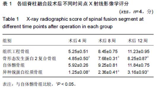
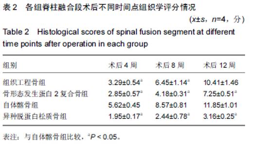
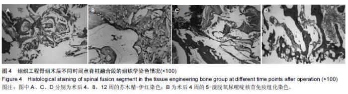
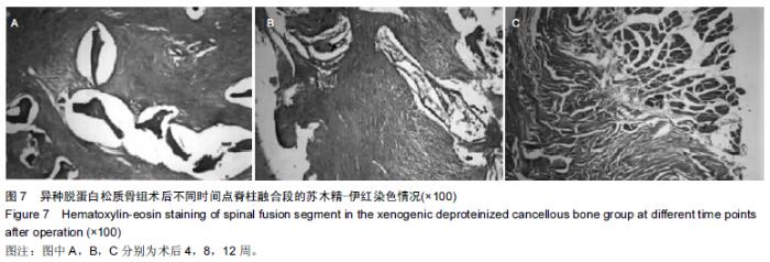
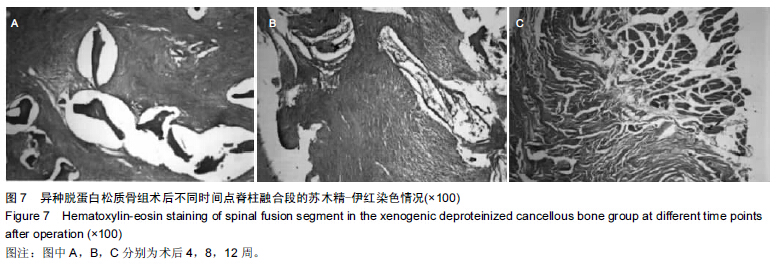
|
[1] 高春阳,李起鸿,简月奎,等.异种脱蛋白松质骨为载体构建组织工程骨用于脊柱横突间融合的实验观察[J].中国修复重建外科杂志, 2007,21(2):115-119.
[2] 高春阳,孙革,韩冬梅,等.异种脱蛋白松质骨复合自体MSCs横突间成骨融合的作用[J].中国矫形外科杂志,2009,17(11): 854-857.
[3] 高春阳.异种脱蛋白松质骨为载体构建组织工程骨髓柱横突间融合的实验研究[D].第三军医大学,2006.
[4] 尹小锋.皮质骨来源成骨细胞复合异种脱蛋白骨应用于脊柱融合的实验研究[D].南昌大学医学院,2010.
[5] 高春阳,李起鸿.异种脱蛋白松质骨作为骨组织工程载体性能研究[J].中国矫形外科杂志,2005,13(6):439-442.
[6] 高春阳,李起鸿,简月奎,等.异种脱蛋白松质骨载体的制备及性能评价[J].重庆医学,2005,34(12):1800-1802.
[7] 翟文亮,柯希煌,练克俭,等.富血小板血浆复合脱蛋白异种骨在腰椎后外侧融合术中的应用[J].脊柱外科杂志,2008,6(1):19-23.
[8] 丁真奇,练克俭,康两奇,等.脱蛋白牛松质复合骨在脊柱后外侧融合术中的应用[J].中国骨伤,2001,14(2):69-70.
[9] 杰永生,綦惠,陈磊,等.脱蛋白联合冷冻干燥法制备异种骨的实验研究[J].中国医药生物技术,2013,8(5):331-335.
[10] 朱肖奇,郭浩.酸性成纤维细胞因子复合部分脱蛋白骨修复免早期股骨头缺血性坏死的影像学评价[J].中国组织工程研究与临床康复,2009,13(42):8260-8264.
[11] 张远鹰,王金成,高忠礼,等.异种无机骨复合自体骨髓移植在颈椎前路融合术中的应用[J].中国修复重建外科杂志,2004,18(1): 25-27.
[12] 程高建.皮质骨来源成骨细胞复合异种脱蛋白骨修复兔桡骨骨缺损的实验研究[D].厦门大学,2011.
[13] 段玉金.异种脱蛋白松质骨修复兔桡骨缺损的研究[J].承德医学院学报,2012,29(2):149-152,封2.
[14] 康鹏飞.异种脱蛋白骨管复合髓内自体骨移植修复创伤性骨缺损的实验研究[D].福建医科大学,2009.
[15] 翟文亮,练克俭,丁真奇,等.钛网椎管成形植骨脊柱融合的实验研究[J].骨与关节损伤杂志,2003,18(7):464-466.
[16] 翟文亮,练克俭,宋光虎,等.脱蛋白骨复合骨形态发生蛋白对前交叉韧带重建术后腱-骨愈合的影响[J].中华创伤骨科杂志,2008, 10(1):72-75.
[17] 陈长青,程高建,练克俭,等.异种脱蛋白骨复合成骨细胞修复兔桡骨骨缺损的研究[J].中华实验外科杂志,2012,29(4):732-734.
[18] 陈长青,尹小锋,练克俭,等.成骨细胞复合异种脱蛋白骨进行腰椎后外侧融合[J].脊柱外科杂志,2011,09(1):51-55.
[19] 章庆国,赵士芳,陈文斌,等.脱蛋白骨-钛网支架成骨细胞复合物修复骨缺损的研究[J].生物医学工程学杂志,2005,22(2): 254-257.
[20] 陈长青,尹小锋,练克俭,等.成骨细胞复合异种脱蛋白骨进行腰椎后外侧融合实验研究[C].//第十一届全军骨科学术大会论文集, 2010:187-187.
[21] 蔡志刚.富集自体骨髓细胞的异种骨复合自体骨髓凝胶促进兔脊柱融合的实验研究[D].福建医科大学,2011.
[22] 刘晓阳,李广润,刘洪涛,等.硫酸钙人工骨/骨髓间充质干细胞构建组织工程化骨诱导脊柱融合[J].中国组织工程研究,2014, 18(21):3281-3286.
[23] 程维,赫兰学,郑惠,等.应用自制异种骨椎间融合器行颈椎前路椎体间融合的生物力学评价:即刻稳定效果及自身强度[J].中国组织工程研究与临床康复,2007,11(35):6991-6995.
[24] 陈永锋.复合骨髓基质干细胞的β-磷酸三钙在兔脊柱后外侧融合中的应用研究[D].第四军医大学,2012.
[25] 刘晓阳,段向东.脱钙小牛骨与兔骨髓基质干细胞移植诱导兔脊柱融合的作用研究[J].山东医药,2012,52(21):41-43.
[26] 庞清江,祝惠敏,汤涛,等.重组合异种骨在脊柱融合术中的临床应用效果[J].现代实用医学,2007,19(3):217-218.
[27] 陈永锋,雷伟,张扬,等.β-TCP/BMSCs组织工程骨细胞含量在兔脊柱后外侧融合中的作用研究[C]//第四届中华骨科杂志论坛论文集,2012:353-357.
[28] 杨民,党耕町,郭钧,等.骨形态发生蛋白在脊柱融合应用中的安全性研究[J].骨与关节损伤杂志,2004,19(2):137-139.
[29] 袁振灿.不同植骨材料椎间融合效果的实验研究[D].山东大学, 2008.
[30] 王金国,吴华,刘玉田,等.颗粒骨与条状骨在脊柱后路融合中的实验研究[J].山东医药,2008,48(6):31-32.
[31] 闵理,刘立岷,宋跃明,等.不同种类骨移植在青少年特发性脊柱侧凸后路矫形术中的中长期疗效评价[J].中国骨与关节外科,2012, 5(5):382-388.
[32] 陆宁,王岩,肖嵩华,等.异体冷冻干燥松质骨移植治疗脊柱结核的中长期随访[J].中国临床康复,2006,10(1):88-90.
[33] 任之强,丁金勇,晋大祥,等.一期后路病灶清除植骨融合内固定治疗胸腰椎结核[J].脊柱外科杂志,2014,12(6):353-356.
[34] 钱济先,高浩然,李存孝,等.后路经皮椎弓根螺钉内固定联合前路病灶清除植骨融合术治疗胸腰椎结核脊柱后凸畸形[J].中医正骨,2014,26(3):33-35.
[35] 丁江平,翁习生,王斌,等.经脊柱前路病灶清除植骨一期前路内固定术治疗脊柱结核[J].中华骨科杂志,2007,27(1):54-58.
[36] 马易群,董健.骨移植材料及植骨方式在治疗胸腰段脊柱骨折中的应用[J].中华创伤骨科杂志,2012,14(9):807-809. |