Chinese Journal of Tissue Engineering Research ›› 2015, Vol. 19 ›› Issue (18): 2849-2855.doi: 10.3969/j.issn.2095-4344.2015.18.011
Previous Articles Next Articles
Establishment of acute vertebral artery thrombosis models in dogs: micro-balloon catheter temporary isolation for embolectomy
Wei Wen-jiang1, Xiao Cheng-jiang1, Li Li-heng1, Jiang Gui-hua2
- 1Interventional Department, 2Department of Imaging, Guangdong Provincial Second People’s Hospital, Guangzhou 510317, Guangdong Province, China
-
Received:2015-02-16Online:2015-04-30Published:2015-04-30 -
Contact:Xiao Cheng-jiang, Master, Chief physician, Interventional Department, Guangdong Provincial Second People’s Hospital, Guangzhou 510317, Guangdong Province, China -
About author:Wei Wen-jiang, Master, Physician, Interventional Department, Guangdong Provincial Second People’s Hospital, Guangzhou 510317, Guangdong Province, China -
Supported by:High Technology Fund of Guangdong Province, No. 2012B031800477
CLC Number:
Cite this article
Wei Wen-jiang, Xiao Cheng-jiang, Li Li-heng, Jiang Gui-hua. Establishment of acute vertebral artery thrombosis models in dogs: micro-balloon catheter temporary isolation for embolectomy[J]. Chinese Journal of Tissue Engineering Research, 2015, 19(18): 2849-2855.
share this article
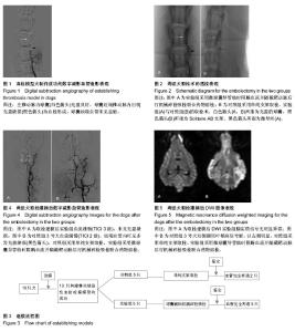
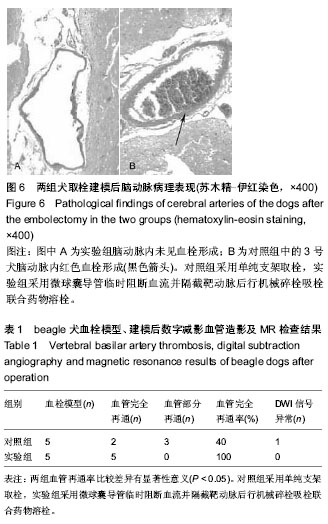
2.1 实验动物数量分析 10只犬均建模成功,均进入结果分析,无脱落。造模流程见图3。 2.2 椎动脉造影及椎动脉急性血栓栓塞模型犬鉴定结果 beagle犬优势侧椎动脉造影显示:起始部直径粗大,走行较直,约在第3颈椎水平发出2条主要分支向颅脑走行,至第1颈椎上端水平和对侧支汇合,形成向上走行的基底动脉,基底动脉与脑底动脉环相交通。往优势侧椎动脉内注入血栓后数字减影血管造影检查示:10只beagle犬优势侧椎动脉内球囊充盈良好,球囊近端椎动脉腔内均显示血栓块和闭塞,球囊远端血管未见显影(图1),证明血栓模型制作成功。 2.2 椎动脉急性血栓取栓后犬数字减影血管造影复查结果 对照组中每只犬均有血栓取出,撤出抽瘪的球囊后行数字减影血管造影检查示:其中2只犬椎-基底动脉与颅内动脉显影良好,血流通畅(TICI 3级),未见明显充盈缺损残留,也无血管痉挛与闭塞征象;另外3只犬椎-基底动脉与颅内动脉显影不佳,血流缓慢(TICI 2级),其中1只犬(3号)椎-基底动脉与颅内动脉中可见多处小点状充盈缺损及颅内动脉显影差,对比剂返流(图4)。"

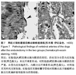
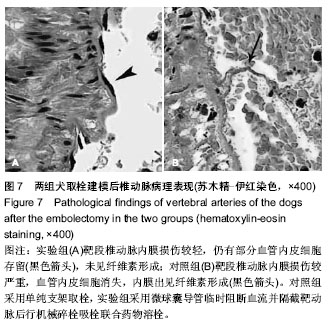
肉眼观察到实验组每只犬均有较多血栓碎块取出。撤出保护球囊后行数字减影血管造影检查示:5只犬椎-基底动脉与颅内动脉显影良好,血流通畅(TICI 3级),无明确充盈缺损残留,无血管痉挛与闭塞(图4,表1)。 2.3 椎动脉急性血栓取栓后犬苏醒情况 对照组4只犬与实验组5只犬建模后3 h均苏醒并行动自如,无明显活动障碍。但对照组中的3号犬(1/5)建模后5 h才苏醒,流涎较多,不能行走。 2.4 椎动脉急性血栓取栓后犬磁共振扫描结果 10只beagle犬制作血栓模型前的T1WI,T2WI(FLAIR)与DWI信号无异常。取栓建模后12 h:对照组4只犬的T1WI,T2WI(FLAIR)与DWI信号无明显异常,3号犬DWI示双侧颞顶叶稍高信号影,以左侧明显(图5,表1);实验组5只犬的T1WI,T2WI (FLAIR)与DWI信号无异常。 2.5 椎动脉急性血栓取栓后犬病理检测结果 苏木精-伊红染色检测显示,除对照组3号犬左侧大脑颞叶动脉腔内见纤维血栓(图6)外,其余实验犬的颅内动脉均未显示残留血栓。对照组5只犬靶段椎动脉内膜损伤较严重,血管内皮细胞消失,腔内见纤维素血栓;实验组5只犬靶段椎动脉内膜损伤较轻,大部分血管内皮完整,未见纤维素形成(图7)。"
2.6 椎动脉急性血栓取栓操作时间比较 对照组取栓时间为(11.7±1.2) min,实验组取栓时间为(13.0±0.8) min,两组取栓时间比较差异无显著性意义(P > 0.05)。"
| [1] Hayakawa M. Intravenous thrombolysis for acute ischemic stroke: past, present and future. Rinsho Shinkeigaku. 2014; 54(12):1197-1199. [2] Zhai YK, Zhu WJ, Hou HL, et al. Efficacy of telemedicine for thrombolytic therapy in acute ischemic stroke: a meta-analysis. J Telemed Telecare. 2015. [3] Hlavica M, Diepers M, Garcia-Esperon C, et al. Pharmacological recanalization therapy in acute ischemic stroke-Evolution, current state and perspectives of intravenous and intra-arterial thrombolysis. J Neuroradiol. 2015. [4] Friedricn B, Gawlitza M, Schob S, et al. Distance to thrombus in acute middle cerebral artery occlusion: a predictor of outcome after intravenous thrombolysis for acute ischemic stroke. Stroke. 2015. [5] Turc G, Isabel C, Calvet D. Intravenous thrombolysis for acute ischemic stroke. Diagn Interv Imaging. 2014;95(12): 1129-1133. [6] Lin C, Li N, Wang K, et al. Efficacy and safety of endovascular treatment versus intravenous thrombolysis for acute ischemic stroke: a meta-analysis of randomized controlled trials. PLoS One. 2013;8(10):e77849. [7] Singh B, Parsaik AK, Prokop LJ, et al. Endovascular therapy for acute ischemic stroke: a systematic review and meta-analysis. Mayo Clin Proc. 2013;88(10):1056-1065. [8] Bae GS, Kwon HJ, Kang CW, et al. Mechanical thrombectomy using a solitaire stent in acute ischemic stroke; initial experience in 40 patients. J Cerebrovasc Endovasc Neurosurg. 2012;14(3):164-169. [9] Serrone JC, Jimenez L, Ringer AJ. The role of endovascular therapy in the treatment of acute ischemic stroke. Neurosurgery. 2014;74 Suppl 1:S133-S141. [10] Turk AR, Campbell JM, Spiotta A, et al. An investigation of the cost and benefit of mechanical thrombectomy for endovascular treatment of acute ischemic stroke. J Neurointerv Surg. 2014;6(1):77-80. [11] Kang DH, Hwang YH, Kim YS, et al. Direct thrombus retrieval using the reperfusion catheter of the penumbra system: forced-suction thrombectomy in acute ischemic stroke. AJNR Am J Neuroradiol. 2011;32(2):283-287. [12] Kappelhof M, Marquering HA, Berkhemer OA, et al. Intra-arterial treatment of patients with acute ischemic stroke and internal carotid artery occlusion: a literature review. J Neurointerv Surg. 2015;7(1):8-15. [13] Castano C, Dorado L, Guerrero C, et al. Mechanical thrombectomy with the Solitaire AB device in large artery occlusions of the anterior circulation: a pilot study. Stroke. 2010;41(8):1836-1840. [14] Pi Y, Zhang L, Yang Q, et al. Neurothrombectomy for the treatment of acute ischemic stroke in 1530 patients. J Clin Neurosci. 2012;19(10):1363-1368. [15] Walcott BP, Boehm KM, Stapleton CJ, et al. Retrievable stent thrombectomy in the treatment of acute ischemic stroke: analysis of a revolutionizing treatment technique. J Clin Neurosci. 2013;20(10):1346-1349. [16] Davalos A, Pereira VM, Chapot R, et al. Retrospective multicenter study of Solitaire FR for revascularization in the treatment of acute ischemic stroke. Stroke. 2012;43(10): 2699-2705. [17] Yin NS, Benavides S, Starkman S, et al. Autopsy findings after intracranial thrombectomy for acute ischemic stroke: a clinicopathologic study of 5 patients. Stroke. 2010;41(5): 938-947. [18] Mokin M, Khalessi AA, Mocco J, et al. Endovascular treatment of acute ischemic stroke: the end or just the beginning? Neurosurg Focus. 2014;36(1):E5. [19] 罗中华,宦怡,贺洪德,等.急性缺血性脑卒中介入取栓器血栓模型的机械特性比较[J].介入放射学杂志,2013,22(4):317-321. [20] Lee SH, Kim SY, Woo DC, et al. Differential neurochemical responses of the canine striatum with pentobarbital or ketamine anesthesia: a 3T proton MRS study. J Vet Med Sci. 2010;72(5):583-587. [21] Rink C, Christoforidis G, Abduljalil A, et al. Minimally invasive neuroradiologic model of preclinical transient middle cerebral artery occlusion in canines. Proc Natl Acad Sci U S A. 2008; 105(37):14100-14105. [22] Christoforidis GA, Rink C, Kontzialis MS, et al. An endovascular canine middle cerebral artery occlusion model for the study of leptomeningeal collateral recruitment. Invest Radiol. 2011;46(1):34-40. [23] Gifford E, Drazin D, Dalfino JC, et al. The effectiveness of microballoon angioplasty in treating middle cerebral artery occlusion beyond the bifurcation. AJNR Am J Neuroradiol. 2010;31(8):1541-1548. [24] 李克,吉训明,凌锋,等.急性脑动脉闭塞的血管造影分级及溶栓治疗疗效评估系统[J].中国脑血管病杂志,2006,3(12):573-576. [25] 李贵福,马朝晖,罗望池,等.Solitaire AB型支架用于急性脑动脉闭塞取栓术31例[J].介入放射学杂志,2012,21(2):98-102. [26] Roth C, Papanagiotou P, Behnke S, et al. Stent-assisted mechanical recanalization for treatment of acute intracerebral artery occlusions. Stroke. 2010;41(11):2559-2567. [27] Papanagiotou P, Roth C, Walter S, et al. Treatment of acute cerebral artery occlusion with a fully recoverable intracranial stent: a new technique. Circulation. 2010;121(23):2605-2606. [28] Shindo A, Kawanishi M, Kawakita K, et al. Treatment of acute cerebral artery occlusion using the penumbra system: our early experience. Neurol Med Chir (Tokyo). 2014;54(6): 441-449. [29] Nayak S, Ladurner G, Killer M. Treatment of acute middle cerebral artery occlusion with a Solitaire AB stent: preliminary experience. Br J Radiol, 2010;83(996): 1017-1022. [30] Mokin M, Kass-Hout T, Levy EI. Solitaire FR-a promising new device for acute ischemic stroke treatment. World Neurosurg. 2012;78(6):557-558. [31] Humphries W, Hoit D, Doss V T, et al. Distal aspiration with retrievable stent assisted thrombectomy for the treatment of acute ischemic stroke. J Neurointerv Surg. 2015;7(2):90-94. [32] Mccabe JJ, Phillips TJ, Phatouros C, et al. Mechanical thrombectomy with the Solitaire AB device in large intracerebral artery occlusions. J Med Imaging Radiat Oncol. 2013;57(2):149-155. [33] Miteff F, Faulder KC, Goh AC, et al. Mechanical thrombectomy with a self-expanding retrievable intracranial stent (Solitaire AB): experience in 26 patients with acute cerebral artery occlusion. AJNR Am J Neuroradiol. 2011;32(6): 1078-1081 |
| [1] | Min Youjiang, Yao Haihua, Sun Jie, Zhou Xuan, Yu Hang, Sun Qianpu, Hong Ensi. Effect of “three-tong acupuncture” on brain function of patients with spinal cord injury based on magnetic resonance technology [J]. Chinese Journal of Tissue Engineering Research, 2021, 25(在线): 1-8. |
| [2] | Zhang Tongtong, Wang Zhonghua, Wen Jie, Song Yuxin, Liu Lin. Application of three-dimensional printing model in surgical resection and reconstruction of cervical tumor [J]. Chinese Journal of Tissue Engineering Research, 2021, 25(9): 1335-1339. |
| [3] | Chen Jinping, Li Kui, Chen Qian, Guo Haoran, Zhang Yingbo, Wei Peng. Meta-analysis of the efficacy and safety of tranexamic acid in open spinal surgery [J]. Chinese Journal of Tissue Engineering Research, 2021, 25(9): 1458-1464. |
| [4] | Zeng Yanhua, Hao Yanlei. In vitro culture and purification of Schwann cells: a systematic review [J]. Chinese Journal of Tissue Engineering Research, 2021, 25(7): 1135-1141. |
| [5] | Xu Dongzi, Zhang Ting, Ouyang Zhaolian. The global competitive situation of cardiac tissue engineering based on patent analysis [J]. Chinese Journal of Tissue Engineering Research, 2021, 25(5): 807-812. |
| [6] | Wu Zijian, Hu Zhaoduan, Xie Youqiong, Wang Feng, Li Jia, Li Bocun, Cai Guowei, Peng Rui. Three-dimensional printing technology and bone tissue engineering research: literature metrology and visual analysis of research hotspots [J]. Chinese Journal of Tissue Engineering Research, 2021, 25(4): 564-569. |
| [7] | Xu Jianxia, Wang Zhaoxu, Wang Chunren. Blood compatibility of disposable blood perfusion device in vitro [J]. Chinese Journal of Tissue Engineering Research, 2021, 25(4): 588-592. |
| [8] | Chang Wenliao, Zhao Jie, Sun Xiaoliang, Wang Kun, Wu Guofeng, Zhou Jian, Li Shuxiang, Sun Han. Material selection, theoretical design and biomimetic function of artificial periosteum [J]. Chinese Journal of Tissue Engineering Research, 2021, 25(4): 600-606. |
| [9] | Liu Fei, Cui Yutao, Liu He. Advantages and problems of local antibiotic delivery system in the treatment of osteomyelitis [J]. Chinese Journal of Tissue Engineering Research, 2021, 25(4): 614-620. |
| [10] | Li Xiaozhuang, Duan Hao, Wang Weizhou, Tang Zhihong, Wang Yanghao, He Fei. Application of bone tissue engineering materials in the treatment of bone defect diseases in vivo [J]. Chinese Journal of Tissue Engineering Research, 2021, 25(4): 626-631. |
| [11] | Zhang Zhenkun, Li Zhe, Li Ya, Wang Yingying, Wang Yaping, Zhou Xinkui, Ma Shanshan, Guan Fangxia. Application of alginate based hydrogels/dressings in wound healing: sustained, dynamic and sequential release [J]. Chinese Journal of Tissue Engineering Research, 2021, 25(4): 638-643. |
| [12] | Chen Jiana, Qiu Yanling, Nie Minhai, Liu Xuqian. Tissue engineering scaffolds in repairing oral and maxillofacial soft tissue defects [J]. Chinese Journal of Tissue Engineering Research, 2021, 25(4): 644-650. |
| [13] | Xing Hao, Zhang Yonghong, Wang Dong. Advantages and disadvantages of repairing large-segment bone defect [J]. Chinese Journal of Tissue Engineering Research, 2021, 25(3): 426-430. |
| [14] | Yi Meizhi, Luo Guanghua, Xiao Yawen, Hu Rong, Chen Xiaolong, Zhao Heng. MRI findings of anatomical variations of the talus [J]. Chinese Journal of Tissue Engineering Research, 2021, 25(24): 3888-3893. |
| [15] | Chen Siqi, Xian Debin, Xu Rongsheng, Qin Zhongjie, Zhang Lei, Xia Delin. Effects of bone marrow mesenchymal stem cells and human umbilical vein endothelial cells combined with hydroxyapatite-tricalcium phosphate scaffolds on early angiogenesis in skull defect repair in rats [J]. Chinese Journal of Tissue Engineering Research, 2021, 25(22): 3458-3465. |
| Viewed | ||||||
|
Full text |
|
|||||
|
Abstract |
|
|||||