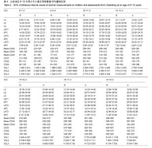
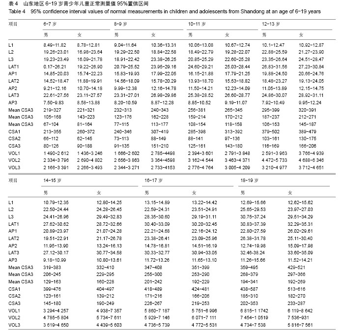
| [1]Kim MA, Kim BR, Choi JY, et al. Three-dimensional changes of the hyoid bone and airway volumes related to its relationship with horizontal anatomic planes after bimaxillary surgery in skeletal Class III patients. Angle Orthod. 2013; 83(4):623-629.
[2]Lin YC, Lin HC, Tsai HH. Changes in the pharyngeal airway and position of the hyoid bone after treatment with a modified bionator in growing patients with retrognathia. J Exp Clin Med. 2011;3(2):93-98.
[3]蒋奕,郑小华,叶果,等.正常成年人上气道CT测量值研究[J].中国耳鼻喉头颈外科,2007,14(7):435-437.
[4]Hatcher DC. Cone beam computed tomography: craniofacial and airway analysis. Dent Clin North Am. 2012;56(2):343-357.
[5]Schendel SA, Hatcher D. Automated 3-dimensional airway analysis from cone-beam computed tomography data. J Oral Maxillofac Surg. 2010;68(3):696-701.
[6]冯斌,王帅,王虎,等.57例青少年上气道的三维形态研究[J].临床口腔医学杂志,2013,29(11):657-660.
[7]郭涛,丁寅.正常骨面型儿童、成人上气道形态的X线头影测量分析[J].北京口腔医学,2006,14(4):247-250.
[8]中华人民共和国卫生部. WS/T428-2013 中华人民共和国卫生行业标准:成人体重判定[S].北京:中国标准出版社,2013.
[9]Xu Y, Zhao S, Shi J, et al. 3-dimensional computed tomographic analysis of the pharynx in adult patients with unrepaired isolated cleft palate. J Oral Maxillofac Surg. 2013;71(8):1424-1434.
[10]Sheng CM, Lin LH, Su Y, et al. Developmental changes in pharyngeal airway depth and hyoid bone position from childhood to young adulthood. Angle Orthod. 2009;79(3): 484-490.
[11]Tsai HH. Developmental changes of pharyngeal airway structures from young to adult persons. J Clin Pediatr Dent. 2007;31(3):219-221.
[12]Li H, Lu X, Shi J, et al. Measurements of normal upper airway assessed by 3-dimensional computed tomography in Chinese children and adolescents. Int J Pediatr Otorhinolaryngol. 2011; 75(10):1240-1246.
[13]Zhong Z, Tang Z, Gao X, et al. A comparison study of upper airway among different skeletal craniofacial patterns in nonsnoring Chinese children. Angle Orthod. 2010;80(2): 267-274.
[14]Samman N, Mohammadi H, Xia J. Cephalometric norms for the upper airway in a healthy Hong Kong Chinese population. Hong Kong Med J. 2003;9(1):25-30.
[15]Shen GF, Samman N, Qiu WL, et al. Cephalometric studies on the upper airway space in normal Chinese. Int J Oral Maxillofac Surg. 1994;23(4):243-247.
[16]Alves M Jr, Baratieri C, Nojima LI, et al. Three-dimensional assessment of pharyngeal airway in nasal- and mouth-breathing children. Int J Pediatr Otorhinolaryngol. 2011;75(9):1195-1199.
[17]Valiathan M, El H, Hans MG, et al. Effects of extraction versus non-extraction treatment on oropharyngeal airway volume. Angle Orthod. 2010;80(6):1068-1074.
[18]Lenza MG, Lenza MM, Dalstra M, et al. An analysis of different approaches to the assessment of upper airway morphology: a CBCT study. Orthod Craniofac Res. 2010; 13(2):96-105. |