Chinese Journal of Tissue Engineering Research ›› 2014, Vol. 18 ›› Issue (30): 4851-4856.doi: 10.3969/j.issn.2095-4344.2014.30.015
Previous Articles Next Articles
Sustained release of polyethylene glycol-modified hydrogel-packaged growth factors and endothelial progenitor cells
Han Yan-jiu, Liu Guo-hui, Ouyang Liu
- Department of Traumatic Surgery, Western District, Xiehe Hospital, Tongji Medical College, Huazhong University of Science and Technology, Wuhan 430056, Hubei Province, China
-
Revised:2014-06-13Online:2014-07-16Published:2014-08-08 -
Contact:Liu Guo-hui, Professor, Chief physician, Doctoral supervisor, Department of Traumatic Surgery, Western District, Xiehe Hospital, Tongji Medical College, Huazhong University of Science and Technology, Wuhan 430056, Hubei Province, China -
About author:Han Yan-jiu, Studying for doctorate, Attending physician, Department of Traumatic Surgery, Western District, Xiehe Hospital, Tongji Medical College, Huazhong University of Science and Technology, Wuhan 430056, Hubei Province, China -
Supported by:the National Natural Science Foundation of China, No. 81141077
CLC Number:
Cite this article
Han Yan-jiu, Liu Guo-hui, Ouyang Liu. Sustained release of polyethylene glycol-modified hydrogel-packaged growth factors and endothelial progenitor cells[J]. Chinese Journal of Tissue Engineering Research, 2014, 18(30): 4851-4856.
share this article

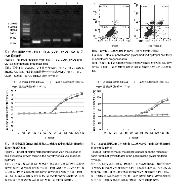
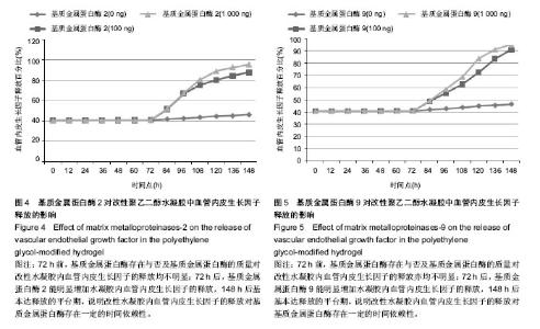
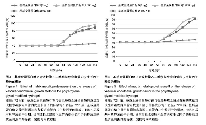
2.1 RT-PCR 法检测内皮祖细胞特异性分子标志 内皮祖细胞特异性分子标志vWF、Flk-1、Tie-2、CD34、CD133、eNOS mRNA均呈阳性表达(图1)。 2.2 基质金属蛋白酶介导的改性聚乙二醇水凝胶碱性成纤维细胞生长因子、血管内皮生长因子体外释放性能 2.2.1 不同基质金属蛋白酶对改性聚乙二醇水凝胶中碱性成纤维细胞生长因子体外释放的影响 基质金属蛋白酶的质量对改性聚乙二醇水凝胶中碱性成纤维细胞生长因子释放曲线及最大释放量的影响不明显。72 h前,基质金属蛋白酶存在与否及基质金属蛋白酶的质量对改性水凝胶中碱性成纤维细胞生长因子的释放均不明显;72 h后,基质金属蛋白酶2及基质金属蛋白酶9均能明显增加水凝胶内碱性成纤维细胞生长因子的释放,148 h后基本达释放的平台期。改性水凝胶内碱性成纤维细胞生长因子的释放对基质金属蛋白酶有一定的时间依赖性(图2,3)。 2.2.2 不同基质金属蛋白酶对改性聚乙二醇水凝胶中血管内皮生长因子体外释放的影响 通过绘制细胞因子体外缓释曲线,可以观察基质金属蛋白酶2同基质金属蛋白酶9相比使改性聚乙二醇水凝胶内血管内皮生长因子释放较为平缓,但是基质金属蛋白酶的质量对改性聚乙二醇水凝胶内血管内皮生长因子释放的影响不明显。72 h前,基质金属蛋白酶存在与否及基质金属蛋白酶的质量对改性水凝胶内血管内皮生长因子的释放亦均不明显;72 h后,基质金属蛋白酶2及基质金属蛋白酶9亦均能明显增加水凝胶内血管内皮生长因子的释放,148 h后基本达释放的平台期,说明改性水凝胶内血管内皮生长因子的释放对基质金属蛋白酶亦存在一定的时间依赖性(图4,5)。 2.3 改性聚乙二醇水凝胶封装内皮祖细胞后对细胞活性的影响 通过Annexin V-FITC/PI双染,流式细胞仪检测存活细胞数的方法。正常对照组细胞凋亡细胞比例为(5.36± 3.24)%,活细胞比例为(95.983±4.76)%;水凝胶封装后细胞凋亡比例为(9.13±4.13)%,活细胞比例为(88.17±5.17)%。经组间t 检验分析,实验组和正常对照组间凋亡细胞比例和活细胞比例差异均无显著性意义(P > 0.05),说明改性水凝胶对内皮祖细胞的活性无明显不利影响(图6)。"
"
| [1] Guan J,Sacks MS,Beckman EJ,et al.Synthesis, characterization, and cytocompatibility of elastomeric, biodegradable poly(ester-urethane)ureas based on poly(caprolactone) and putrescine.J Biomed Mater Res 2002;61(3):493-503. [2] Urbich C,Heeschen C,Aicher A,et al.Relevance of monocytic features for neovascularization capacity of circulating endothelial progenitor cells. Circulation.2003;108(20): 2511-2516. [3] Beltrami AP,Barlucchi L,Torella D,et al.Adult cardiac stem cells are multipotent and support myocardial regeneration. Cell.2003;114(6):763-776. [4] Miranville A,Heeschen C,Sengenes C,et al.Improvement of postnatal neovascularization by human adipose tissue-derived stem cells.Circulation. 2004;110(3): 349-355. [5] Ferrara N,Alitalo K.Clinical applications of angiogenic growth factors and their inhibitors. Nat Med.1999;5(12): 1359-1364. [6] Schumacher B,Pecher P,von SBU,et al.Induction of neoangiogenesis in ischemic myocardium by human growth factors: first clinical results of a new treatment of coronary heart disease. Circulation.1998;97(7):645-650. [7] Baumgartner I,Pieczek A,Manor O,et al.Constitutive expression of phVEGF165 after intramuscular gene transfer promotes collateral vessel development in patients with critical limb ischemia. Circulation.1998;97(12):1114-1123. [8] Jackson KA,Majka SM,Wang H,et al.Regeneration of ischemic cardiac muscle and vascular endothelium by adult stem cells.J Clin Invest.2001;107(11):1395-1402. [9] Mooney DJ,Vandenburgh H.Cell delivery mechanisms for tissue repair. Cell Stem Cell.2008;2(3):205-213. [10] Asahara T,Marohara T,Sullivan A,et al.Isolation of putative progenitor endothelial cells for angiogenesis. Science. 1997; 275(53):964-967. [11] Lazarous DF,Shou M,Stiber JA,et al.Pharmacodynamics of basic fibroblast growth factor: route of administration determines myocardial and systemic distribution. Cardiovasc Res.1997;36(1):78-85. [12] Kim TK,Burgess DJ.Pharmacokinetic characterization of 14C-vascular endothelial growth factor controlled release microspheres using a rat model.J Pharm Pharmacol. 2002; 54(7):897-905. [13] Kipshidze N,Chekanov V,Chawla P,et al.Angiogenesis in a patient with ischemic limb induced by intramuscular injection of vascular endothelial growth factor and fibrin platform.Tex Heart Inst J.2000;27(2):196-200. [14] Niklason LE,Gao J,Abbott WM,et al.Functional arteries grown in vitro.Science.1999;284(5413):489-493. [15] Zisch AH,Lutolf MP,Ehrbar M,et al.Cell-demanded release of VEGF from synthetic, biointeractive cell ingrowth matrices for vascularized tissue growth.FASEB J. 2003;17(15):2260-2262. [16] Ehrbar M,Metters A,Zammaretti P,et al.Endothelial cell proliferation and progenitor maturation by fibrin-bound VEGF variants with differential susceptibilities to local cellular activity.J Control Release. 2005;101(1-3):93-109. [17] Jain RK,Au P,Tam J,et al.Engineering vascularized tissue.Nat Biotechnol .2005;23(7):821-823. [18] Langer R.New methods of drug delivery. Science. 1990;249 (4976):1527-1533. [19] Ferreira LS,Gerecht S,Fuller J,et al.Bioactive hydrogel scaffolds for controllable vascular differentiation of human embryonic stem cells. Biomaterials.2007;28(17):2706-2717. [20] Lee KY,Peters MC,Anderson KW,et al.Controlled growth factor release from synthetic extracellular matrices. Nature. 2000;408(6815):998-1000. [21] Richardson TP,Peters MC,Ennett AB,et al. Polymeric system for dual growth factor delivery.Nat Biotechnol. 2001; 19(11): 1029-1034. [22] Arras M,Mollnau H,Strasser R,et al.The delivery of angiogenic factors to the heart by microsphere therapy.Nat Biotechnol. 1998;16(2):159-162. [23] Lutolf MP,Weber FE,Schmoekel HG,et al.Repair of bone defects using synthetic mimetics of collagenous extracellular matrices. Nat Biotechnol 2003;21(5):513-518. [24] Hiraoka N,Allen E,Apel IJ,et al.Matrix metalloproteinases regulate neovascularization by acting as pericellular fibrinolysins. Cell. 1998;95(3):365-377. [25] Heymans S,Luttun A,Nuyens D,et al.Inhibition of plasminogen activators or matrix metalloproteinases prevents cardiac rupture but impairs therapeutic angiogenesis and causes cardiac failure. Nat Med.1999;5(10):1135-1142. [26] Webb CS,Bonnema DD,Ahmed SH,et al.Specific temporal profile of matrix metalloproteinase release occurs in patients after myocardial infarction: relation to left ventricular remodeling.Circulation.2006;114(10):1020-1027. [27] Peterson JT,Li H,Dillon L,et al.Evolution of matrix metalloprotease and tissue inhibitor expression during heart failure progression in the infarcted rat.Cardiovasc Res.2000; 46(2):307-315. [28] Bradham WS,Gunasinghe H,Holder JR,et al.Release of matrix metalloproteinases following alcohol septal ablation in hypertrophic obstructive cardiomyopathy.J Am Coll Cardiol. 2002;40(12):2165-2173. [29] Ahmed SH,Clark LL,Pennington WR,et al.Matrix metalloproteinases/tissue inhibitors of metalloproteinases: relationship between changes in proteolytic determinants of matrix composition and structural, functional, and clinical manifestations of hypertensive heart disease.Circulation.2006; 113(17):2089-2096. [30] Vanhoutte D, Schellings M,Pinto Y,et al.Relevance of matrix metalloproteinases and their inhibitors after myocardial infarction: a temporal and spatial window. Cardiovasc Res. 2006;69(3):604-613. [31] Orn S, Manhenke C,Squire IB,et al.Plasma MMP-2, MMP-9 and N-BNP in long-term survivors following complicated myocardial infarction: relation to cardiac magnetic resonance imaging measures of left ventricular structure and function.J Card Fail. 2007;13(10):843-849. [32] Squire IB,Evans J,Ng LL,et al.Plasma MMP-9 and MMP-2 following acute myocardial infarction in man: correlation with echocardiographic and neurohumoral parameters of left ventricular dysfunction.J Card Fail.2004;10(4):328-333. [33] Kosmala W,Plaksej R,Przewlocka-Kosmala M,et al.Matrix metalloproteinases 2 and 9 and their tissue inhibitors 1 and 2 in premenopausal obese women: relationship to cardiac function.Int J Obes (Lond).2008;32(5):763-771. [34] Halapas A,Zacharoulis A,Theocharis S,et al.Serum levels of the osteoprotegerin, receptor activator of nuclear factor kappa-B ligand, metalloproteinase-1 (MMP-1) and tissue inhibitors of MMP-1 levels are increased in men 6 months after acute myocardial infarction.Clin Chem Lab Med.2008; 46(4):510-516. |
| [1] | Liu Xiaolin, Mu Xinyue, Ma Ziyu, Liu Shutai, Wang Wenlong, Han Xiaoqian, Dong Zhiheng. Effect of hydrogel-loaded simvastatin microspheres on osteoblast proliferation and differentiation [J]. Chinese Journal of Tissue Engineering Research, 2023, 27(7): 998-1003. |
| [2] | Li Cheng, Zheng Guoshuang, Kuai Xiandong, Yu Weiting. Alginate scaffold in articular cartilage repair [J]. Chinese Journal of Tissue Engineering Research, 2023, 27(7): 1080-1088. |
| [3] | Li Yue, Lyu Yan, Feng Wanying, Song Yang, Yan Yu, Guan Yongge. Preparation of hyperoside nanoparticles to repair endometrial injury [J]. Chinese Journal of Tissue Engineering Research, 2023, 27(3): 360-366. |
| [4] | Jiang Shengyuan, Deng Bowen, Liu Gang, Fan Xiao, Bai Huizhong, Tao Jingwei, Zhao Yi, Ren Jingpei, Xu Lin, Mu Xiaohong. Safety evaluation of electroactive hydrogel in the treatment of complete spinal cord transection in rats [J]. Chinese Journal of Tissue Engineering Research, 2023, 27(21): 3314-3319. |
| [5] | Xu Xin, Liu Yaowei, Mu Yunping, Wang Jianying, Li Fanghong, Zhao Zijian. Preparation and biological evaluation of decellularized dermal matrix hydrogel [J]. Chinese Journal of Tissue Engineering Research, 2023, 27(21): 3325-3331. |
| [6] | Tao Jingwei, Fan Xiao, Jiang Shengyuan, Deng Bowen, Zhang Yaqi, Liu Gang, Zuo Xinwei, Zhou Zhuoluo, Zhao Yi, Ren Jingpei, Xu Lin, Mu Xiaohong. Application of injectable hydrogel in spinal cord injury [J]. Chinese Journal of Tissue Engineering Research, 2023, 27(16): 2548-2555. |
| [7] | Peng Kun. Properties of force growth factor E peptide bionic bone matrix with polyethylene glycol derivative hydrogel as a carrier [J]. Chinese Journal of Tissue Engineering Research, 2023, 27(12): 1811-1816. |
| [8] | Zhu Biwen, Wang Dongzhi, Wu Di, Gong Tiancheng, Pan Haopeng, Lu Yuhua, Guo Yibing, Wang Zhiwei, Huang Yan. Biomimetic microenvironment constructed from gelatin methacrylamide/platelet-rich plasma hydrogel promotes the function of insulinoma cell line MIN6 in mice [J]. Chinese Journal of Tissue Engineering Research, 2023, 27(12): 1824-1831. |
| [9] | Ling Huajun, Cui Ruiwen, Wang Qiyou. 3D extracellular matrix hydrogel loaded with exosomes promotes wound repair [J]. Chinese Journal of Tissue Engineering Research, 2023, 27(12): 1900-1905. |
| [10] | Hu Jinlong, Quan Huahong, Wang Jingcheng, Zhang Pei, Zhang Jiale, Chen Pengtao, Liang Yuan. Effect of copper sulfide nanoparticles loaded thermosensitive hydrogel Pluronic F127 on infected wound healing in rats [J]. Chinese Journal of Tissue Engineering Research, 2023, 27(12): 1927-1931. |
| [11] | Wang Xinxin, Wang Jingxin. Mesenchymal stem cells-derived exosomes in treatment of secondary lymphedema [J]. Chinese Journal of Tissue Engineering Research, 2023, 27(10): 1603-1609. |
| [12] | Liu Siqi, Wu Mingrui, Qiao Lingran, Xie Liying, Chen Siyu, Han Zhibo, Zuo Lin. Effects of hydrogel loaded with human umbilical cord mesenchymal stem cells on diabetic wound repair in mice [J]. Chinese Journal of Tissue Engineering Research, 2023, 27(1): 21-27. |
| [13] | Huang Chuanjun, Zou Yu, Zhou Xiaoting, Zhu Yangqing, Qian Wei, Zhang Wei, Liu Xing. Transplantation of umbilical cord mesenchymal stem cells encapsulated in RADA16-BDNF hydrogel promotes neurological recovery in an intracerebral hemorrhage rat model [J]. Chinese Journal of Tissue Engineering Research, 2022, 26(4): 510-515. |
| [14] | Zhang Tong, Cai Jinchi, Yuan Zhifa, Zhao Haiyan, Han Xingwen, Wang Wenji. Hyaluronic acid-based composite hydrogel in cartilage injury caused by osteoarthritis: application and mechanism [J]. Chinese Journal of Tissue Engineering Research, 2022, 26(4): 617-625. |
| [15] | Lu Dongdong, Zhu Tianfeng, Zhang Yijian, Zhao Zhijian, Liu Yang, Shen Xu, Zhu Xuesong. 3D bio-printing methylacrylated gelatin hydrogel scaffolds promote the repair of subchondral bone defects [J]. Chinese Journal of Tissue Engineering Research, 2022, 26(34): 5454-5460. |
| Viewed | ||||||
|
Full text |
|
|||||
|
Abstract |
|
|||||