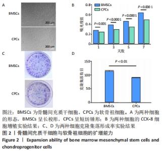
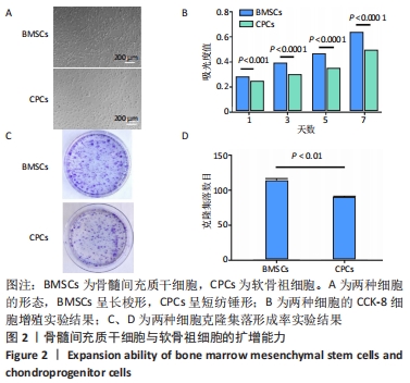
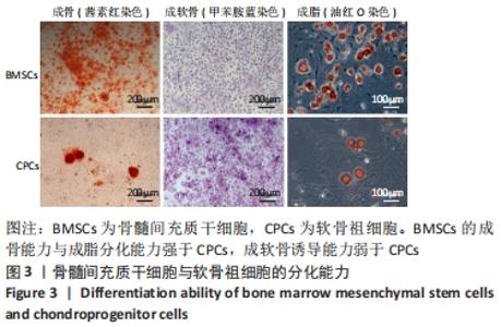
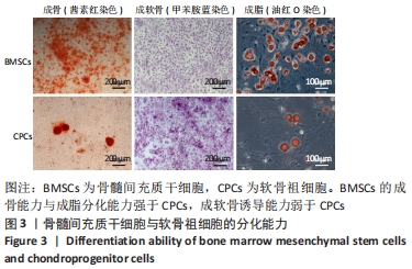
[1] BRITTBERG M, RECKER D, ILGENFRITZ J, et al. Matrix-Applied Characterized Autologous Cultured Chondrocytes Versus Microfracture: Five-Year Follow-up of a Prospective Randomized Trial. Am J Sports Med. 2018;46(6):1343-1351.
[2] IIJIMA H, AOYAMA T, ITO A, et al. Immature articular cartilage and subchondral bone covered by menisci are potentially susceptive to mechanical load. BMC Musculoskelet Disord. 2014;15(2):101.
[3] LORIES RJ, LUYTEN FP. The bone-cartilage unit in osteoarthritis. Nat Rev Rheumatol. 2011;7(1):43-49.
[4] FRAQUET N, FAIZON G, ROSSET P, et al. Long bones giant cells tumors: treatment by curretage and cavity filling cementation. Orthop Traumatol Surg Res. 2009;95(6):402-406.
[5] PARK JH, CHAE IJ, HAN SB, et al. Arthroscopic burring of exposed cement following curettage and cavity filling cementation for chondroblastoma of the proximal tibia. Knee Surg Relat Res. 2015; 27(1):61-64.
[6] HOHMANN E, TETSWORTH K. Large osteochondral lesions of the femoral condyles: Treatment with fresh frozen and irradiated allograft using the Mega OATS technique. Knee. 2016;23(3):436-441.
[7] HANGODY L, KISH G, KARPATI Z, et al. Arthroscopic autogenous osteochondral mosaicplasty for the treatment of femoral condylar articular defects. A preliminary report. Knee Surg Sports Traumatol Arthrosc. 1997;5(4):262-267.
[8] PITTENGER MF, MACKAY AM, BECK SC, et al. Multilineage potential of adult human mesenchymal stem cells. Science. 1999;284(5411):143-147.
[9] DOWTHWAITE GP, BISHOP JC, REDMAN SN, et al. The surface of articular cartilage contains a progenitor cell population. J Cell Sci. 2004; 117(Pt 6):889-897.
[10] RAMASAMY R, TONG CK, SEOW HF, et al. The immunosuppressive effects of human bone marrow-derived mesenchymal stem cells target T cell proliferation but not its effector function. Cell Immunol. 2008;251(2):131-136.
[11] TABATABAEI F, MOHARAMZADEH K, TAYEBI L. Fibroblast encapsulation in gelatin methacryloyl (GelMA) versus collagen hydrogel as substrates for oral mucosa tissue engineering. J Oral Biol Craniofac Res. 2020; 10(4):573-577.
[12] MORONI L, BURDICK JA, HIGHLEY C, et al. Biofabrication strategies for 3D in vitro models and regenerative medicine. Nat Rev Mater. 2018; 3(5):21-37.
[13] 刘伟,陈剑,宋佳,等.兔骨髓间充质干细胞的分离、培养及鉴定[J].中国实验诊断学,2013,17(8):1366-1369.
[14] CHEN P, ZHENG L, WANG Y, et al. Desktop-stereolithography 3D printing of a radially oriented extracellular matrix/mesenchymal stem cell exosome bioink for osteochondral defect regeneration. Theranostics. 2019;9(9):2439-2459.
[15] KOELLING S, KRUEGEL J, IRMER M, et al. Migratory chondrogenic progenitor cells from repair tissue during the later stages of human osteoarthritis. Cell Stem Cell. 2009;4(4):324-335.
[16] GONG H, FEI H, XU Q, et al. 3D-engineered GelMA conduit filled with ECM promotes regeneration of peripheral nerve. J Biomed Mater Res A. 2020;108(3):805-813.
[17] TABATABAEI F, MOHARAMZADEH K, TAYEBI L. Fibroblast encapsulation in gelatin methacryloyl (GelMA) versus collagen hydrogel as substrates for oral mucosa tissue engineering. J Oral Biol Craniofac Res. 2020; 10(4):573-577.
[18] SHAHABIPOUR F, OSKUEE RK, DEHGHANI H, et al. Cell-cell interaction in a coculture system consisting of CRISPR/Cas9 mediated GFP knock-in HUVECs and MG-63 cells in alginate- GelMA based nanocomposites hydrogel as a 3D scaffold. J Biomed Mater Res A. 2020;108(8):1596-1606.
[19] ZHAO X, LIU S, YILDIRIMER L, et al. Injectable Stem Cell-Laden Photocrosslinkable Microspheres Fabricated Using Microfluidics for Rapid Generation of Osteogenic Tissue Constructs. Adv Funct Mater. 2016;26(17):2809-2819.
[20] RATHEESH G, VAQUETTE C, XIAO Y. Patient-Specific Bone Particles Bioprinting for Bone Tissue Engineering. Adv Healthc Mater. 2020; 9(23):e2001323.
[21] HUANG L, ZHANG Z, GUO M, et al. Biomimetic Hydrogels Loaded with Nanofibers Mediate Sustained Release of pDNA and Promote In Situ Bone Regeneration. Macromol Biosci. 2021;21(4):e2000393.
[22] GAO J, DING X, YU X, et al. Cell-Free Bilayered Porous Scaffolds for Osteochondral Regeneration Fabricated by Continuous 3D-Printing Using Nascent Physical Hydrogel as Ink. Adv Healthc Mater. 2021;10(3): e2001404.
|
