Chinese Journal of Tissue Engineering Research ›› 2022, Vol. 26 ›› Issue (34): 5498-5503.doi: 10.12307/2022.462
Previous Articles Next Articles
Construction of chitosan/mineralized collagen porous scaffold, osteogenic differentiation in vitro and biocompatibility
Sun Xirao, Bao Jiaxin, Wang Chengyue
- Second Affiliated Hospital of Jinzhou Medical University, Jinzhou 121000, Liaoning Province, China
-
Received:2021-05-07Accepted:2021-07-10Online:2022-12-08Published:2022-04-15 -
Contact:Wang Chengyue, Chief physician, Second Affiliated Hospital of Jinzhou Medical University, Jinzhou 121000, Liaoning Province, China -
About author:Sun Xirao, Master, Attending physician, Second Affiliated Hospital of Jinzhou Medical University, Jinzhou 121000, Liaoning Province, China -
Supported by:the Natural Science Foundation of Liaoning Province, No. 2019-MS-141 (to SXR); Key Research Project of Natural Science Foundation of Liaoning Province, No. JYTZD2020004 (to WCY)
CLC Number:
Cite this article
Sun Xirao, Bao Jiaxin, Wang Chengyue. Construction of chitosan/mineralized collagen porous scaffold, osteogenic differentiation in vitro and biocompatibility[J]. Chinese Journal of Tissue Engineering Research, 2022, 26(34): 5498-5503.
share this article
Add to citation manager EndNote|Reference Manager|ProCite|BibTeX|RefWorks
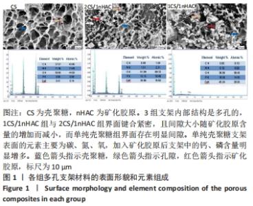
2.1 各组多孔支架材料的微观结构表征 扫描电镜下可见,3组支架内部结构是多孔的,1CS/1nHAC组与2CS/1nHAC组界面键合紧密,且间隙大小随矿化胶原含量的增加而减小,而单纯壳聚糖支架组界面存在明显间隙;同时随着壳聚糖含量的减少,矿化胶原颗粒在CS/nHAC组支架中产生的团聚现象更加明显,见图1。单纯壳聚糖支架表面的元素主要为碳、氮、氧,加入矿化胶原后支架中的钙、磷含量明显增多,表明矿化胶原与壳聚糖结合良好。"

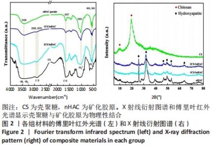
各组多孔支架材料的X射线衍射、傅里叶红外光谱结果,见图2。"
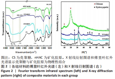
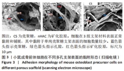
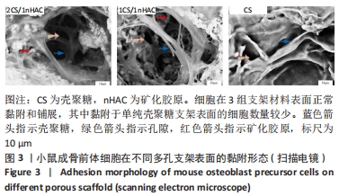
3组多孔支架中含有典型的壳聚糖的X射线衍射峰,在10°和20°左右出现宽峰,峰的强度随着壳聚糖含量的减少而降低。在傅里叶红外光谱中,所有样品在3 440 cm-1处的吸收峰归属于羟基和氨基,来自壳聚糖和Ⅰ型胶原;对于单纯的纯壳聚糖多孔支架,在3 293 cm-1处的振动被分配为羟基拉伸,该振动可能与氨基拉伸在90°时重叠;与矿化胶原粉体相比,所有CS/nHAC材料在2 930,2 870 cm-1处都出现了吸收峰,这是由于C-H极性共价键在壳聚糖中的振动吸收峰出现了伸缩;所有样品的特征峰大约为1 657 cm-1,与存在于壳聚糖和Ⅰ型胶原中的羰基(amide Ⅰ)有反应;除单纯壳聚糖多孔支架外,其他样品在1 033,604,565 cm-1处出现吸收带,与PO43-离子对应,表明存在羟基磷灰石,与X射线衍射图谱结果一致。上述结果表明,矿化胶原和壳聚糖已成功地结合[8-9]。 2.2 多孔支架材料表面的细胞黏附 骨组织工程支架必须对细胞无毒副作用,实验采用直接共培养实验分析不同材料与细胞直接接触时的黏附状态。 图3为在不同多孔支架材料上培养1 d的小鼠成骨前体细胞扫描电镜观察结果。单纯壳聚糖支架表面的细胞拉伸面积增大,细胞完全伸展;2CS/1nHAC和1CS/1nHAC多孔支架表面的细胞密度高于单纯壳聚糖支架,细胞丝足相互连接,材料表面的小鼠成骨前体细胞具有良好的黏附性,并伸出大量的伪足,细胞在材料表面的孔隙中相互连接并向内生长。此外,壳聚糖和矿化胶原作为生物相容性优良的材料,也为细胞的黏附和生长提供了良好的周边环境。"
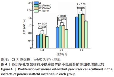
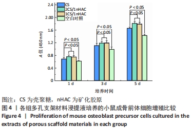
2.3 细胞增殖实验结果 各组材料浸提液培养MC3T3-E1细胞后的细胞增殖情况,见图4。3组多孔支架浸提液培养1,3,5 d后的A值均高于空白对照组(P < 0.05);2CS/1nHAC和1CS/1nHAC多孔支架浸提液组培养1,3,5 d后的A值高于单纯壳聚糖多孔支架组(P < 0.05),表明2CS/1nHAC和1CS/1nHAC多孔支架浸提液对小鼠成骨前体细胞增殖具有促进作用。"
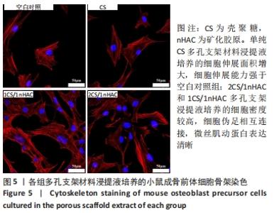
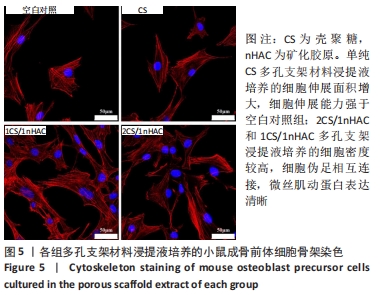
2.4 细胞骨架染色结果 正常条件下培养的小鼠成骨前体细胞形态不规则,有多个丝状伪足,细胞微丝肌动蛋白呈束状结构;单纯壳聚糖多孔支架材料浸提液培养的细胞伸展面积增大,细胞伸展能力强于空白对照组;2CS/1nHAC和1CS/1nHAC多孔支架浸提液培养的细胞密度较高,细胞伪足相互连接,微丝肌动蛋白表达清晰,见图5。"
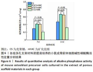
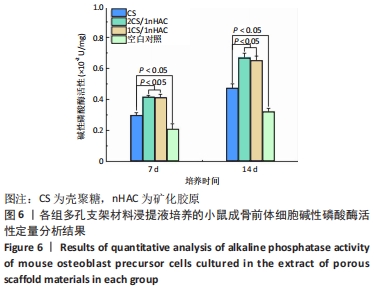
2.5 碱性磷酸酶活性检测结果 碱性磷酸酶是骨骼形成的早期标志物。在整个培养期间,3组多孔支架浸提液培养细胞的碱性磷酸酶活性均高于空白对照组(P < 0.05),2CS/1nHAC和1CS/1nHAC多孔支架浸提液培养细胞的碱性磷酸酶活性高于单纯壳聚糖多孔支架(P < 0.05),见图6,表明各组多孔支架浸提液对小鼠成骨前体细胞的碱性磷酸酶活性有长期刺激作用。"

2.6 多孔支架材料的细胞相容性 由细胞增殖与黏附实验可知,CS/nHAC多孔支架具有良好的细胞相容性。"

| [1] ARMIENTO AR, STODDART MJ, ALINI M, et al. Biomaterials for articular cartilage tissue engineering: Learning from biology. Acta Biomater. 2018;65:1-20. [2] 张雪梅,马征,吴宓勋.纳米羟基磷灰石前体与胶原自组装成类骨质复合材料的表征[J].中国组织工程研究,2020,24(10):1534-1539. [3] BRETSCHNEIDER H, QUADE M, LODE A, et al. Chemotactic and Angiogenic Potential of Mineralized Collagen Scaffolds Functionalized with Naturally Occurring Bioactive Factor Mixtures to Stimulate Bone Regeneration. Int J Mol Sci. 2021;22(11):5836. [4] DE TULLIO I, CAPUTI S, PERFETTI G, et al. A Human Clinical and Histomorphometrical Study on Different Resorbable and Non-Resorbable Bone Substitutes Used in Post-Extractive Sites. Materials (Basel). 2019;12(15):2408. [5] LI Z, DU T, RUAN C, et al. Bioinspired mineralized collagen scaffolds for bone tissue engineering. Bioactive Materials. 2021;6(5):1491-1511. [6] 陈琬雯.基于硒活性的壳聚糖复合物的制备研究[D].无锡:江南大学,2020. [7] 刘婉霞.壳聚糖基复合材料的制备及其吸附钴离子的研究[D].北京:北京建筑大学,2020. [8] DREIFKE MB, EBRAHEIM NA, JAYASURIYA AC. Investigation of potential injectable polymeric biomaterials for bone regeneration. J Biomed Mater Res A. 2013;101:2436-2447. [9] GAIHRE B, USWATTA S, JAYASURIYA AC. Nano-scale characterization of nano-hydroxyapatite incorporated chitosan particles for bone repair. Colloids Surf B Biointerfaces. 2018;165:158-164. [10] RUIXIN L, CHENG X, YINGJIE L, et al. Degradation behavior and compatibility of micro, nanoHA/chitosan scaffolds with interconnected spherical macropores. Int J Biol Macromol. 2017;103:385-394. [11] SHAKIR M, ZIA I, REHMAN A, et al. Fabrication and characterization of nanoengineered biocompatible n-HA/chitosan-tamarind seed polysaccharide: Bio-inspired nanocomposites for bone tissue engineering. Int J Biol Macromol. 2018;111:903-916. [12] CHEN GG, QI XM, GUAN Y, et al. High strength hemicellulose-based nanocomposite film for food packaging applications. ACS Sustain Chem Eng. 2016;4(4):1985-1993. [13] YANG C, GAO XT, YOUNIS MR, et al. Non-invasive monitoring of in vivo bone regeneration based on alkaline phosphatase-responsive scaffolds. Chem Eng J. 2021;408:127959. [14] 庄均珺.生物功能化胶原涂层的可控制备及其评价[D].杭州:浙江大学,2018. [15] AMINI AR, LAURENCIN CT, NUKAVARAPU SP. Bone tissue engineering: recent advances and challenges. Crit Rev Biomed Eng. 2012;40(5):363-408. [16] MAJI K, DASGUPTA S, PRAMANIK K, et al. Preparation and characterization of gelatin-chitosan-nano beta-TCP based scaffold for orthopaedic application. Mater Sci Eng C Mater Biol Appl. 2018;86:83-94. [17] 刘来俊,张宇,李超婧,等.用于骨组织再生的仿生骨膜的研究进展[J].中国生物医学工程学报,2020,39(4):493-503. [18] LING T, LIN J, TU JJ, et al. Mineralized collagen coatings formed by electrochemical deposition. J Mater Sci Mater Med. 2013;24(12):2709-2718. [19] 李博,王硕,赵勇刚,等.仿生矿化胶原骨材料用于儿童颅骨再生修复的最新研究进展[J].中国修复重建外科杂志,2021,17(2):49-54. [20] TW SUN, WL YU, YJ ZHU, et al. Porous Nanocomposite Comprising Ultralong Hydroxyapatite Nanowires Decorated with Zinc‐Containing Nanoparticles and Chitosan: Synthesis and Application in Bone Defect Repair. Chem Eur J. 2018;24:8809. [21] LIAO J, LI Y, LI H, et al. Preparation, bioactivity and mechanism of nano-hydroxyapatite/sodium alginate/chitosan bone repair material. J Appl Biomater Funct Mater. 2018;16(1):28-35. [22] KONG ZQ, YU MF, CHENG K, et al. Incorporation of chitosan nanospheres into thin mineralized collagen coatings for improving the antibacterial effect. Colloids Surf B Biointerfaces. 2013;111:536-541. [23] KONG M, CHEN XG, XING K, et al. Antimicrobial properties of chitosan and mode of action: a state of the art review. Int J Food Microbiol. 2010;144(1):51-63. [24] DAI C, LI Y, PAN W, et al. Three-Dimensional High-Porosity Chitosan/Honeycomb Porous Carbon/Hydroxyapatite Scaffold with Enhanced Osteoinductivity for Bone Regeneration. ACS Biomater Sci Eng. 2020; 6(1):575-586. [25] LI M, JIA WB, ZHANG XL, et al. Hyaluronic acid oligosaccharides modified mineralized collagen and chitosan with enhanced osteoinductive properties for bone tissue engineering. Carbohydr Polym. 2021;260:117780. [26] BERNHARDT A, LODE A, MIETRACH C, et al. In vitroosteogenic potential of human bone marrow stromal cells cultivated in porous scaffolds from mineralized collagen. J Biomed Mater Res. 2009;90(3):852-862. [27] WANG S, YANG YD, KOONS GL, et al. Tuning pore features of mineralized collagen/PCL scaffolds for cranial bone regeneration in a rat model. Mater Sci Eng C Mater Biol Appl. 2020;106:110186. [28] JIANG L, LI Y, XIONG C, et al. Preparation and properties of bamboo fiber/nano-hydroxyapatite/poly(lactic-co-glycolic) composite scaffold for bone tissue engineering. ACS Appl Mater Interfaces. 2017;9(5): 4890-4897. [29] 李锐.羟基磷灰石/壳聚糖复合二甲双胍用于大鼠骨缺损的生物学特点[J].中国组织工程研究,2021,25(28):4460-4464. [30] SADEGHINIA A, SOLTANI S, AGHAZADEH M, et al. Design and fabrication of clinoptilolite-nanohydroxyapatite/chitosan-gelatin composite scaffold and evaluation of its effects on bone tissue engineering. J Biomed Mater Res A. 2020; 108(2):221-233. [31] 李扬,刘怡卓,沈超,等.生物玻璃/壳聚糖三维多孔支架的抗感染性能和生物相容性研究[J].生物骨科材料与临床研究,2020, 17(2):49-54. [32] 陈黎,黄蕾,余梦瑶,等.用于生物人工肝反应器构建三维支架材料的选择与优化[J].中国组织工程研究,2021,25(28):4429-4434. |
| [1] | Gao Yujin, Peng Shuanglin, Ma Zhichao, Lu Shi, Cao Huayue, Wang Lang, Xiao Jingang. Osteogenic ability of adipose stem cells in diabetic osteoporosis mice [J]. Chinese Journal of Tissue Engineering Research, 2022, 26(7): 999-1004. |
| [2] | Liang Xuezhen, Yang Xi, Li Jiacheng, Luo Di, Xu Bo, Li Gang. Bushen Huoxue capsule regulates osteogenic and adipogenic differentiation of rat bone marrow mesenchymal stem cells via Hedgehog signaling pathway [J]. Chinese Journal of Tissue Engineering Research, 2022, 26(7): 1020-1026. |
| [3] | Le Guoping, Zhang Ming, Xi Licheng, Luo Hanwen. Preparation and in vitro evaluation of vancomycin hydrochloride@polylactic acid-glycolic acid copolymer-chitosan-hyaluronic acid composite sustained-release microspheres [J]. Chinese Journal of Tissue Engineering Research, 2022, 26(4): 528-534. |
| [4] | He Guanyu, Xu Baoshan, Du Lilong, Zhang Tongxing, Huo Zhenxin, Shen Li. Biomimetic orientated microchannel annulus fibrosus scaffold constructed by silk fibroin [J]. Chinese Journal of Tissue Engineering Research, 2022, 26(4): 560-566. |
| [5] | Long Zhisheng, Xiong Long, Gong Feipeng, Li Jingtang, Zeng Jianhua, Deng Ying, Lan Min, Kong Weihao, Chen Gang. Effect of artificial bone with multi-scale hydroxyapatite/chitosan microtubule structure on rabbit bone defect repair and angiogenesis [J]. Chinese Journal of Tissue Engineering Research, 2022, 26(34): 5436-5441. |
| [6] | Xu Lin, Cheng Gangyi, Li Hongwei, Wang Hongjian, Lin Minghui, Guo Hongwei, Lin Lianfeng, Xu Ling. Hemostatic effect of macroporous polysaccharides composite hemostatic materials on small-artery severed injury [J]. Chinese Journal of Tissue Engineering Research, 2022, 26(34): 5442-5447. |
| [7] | Li Yi, Yang Yanjun, Peng Songyun, Cheng Zhigang, Zhong Kai, Yin Tianping, Tang Lianghua. Effect of Miao medicine Jiuxian Luohan Jiegu Decoction on osteogenic differentiation and fracture healing in tibial fracture rats [J]. Chinese Journal of Tissue Engineering Research, 2022, 26(33): 5350-5356. |
| [8] | Huang Wei, Dong Panfeng, Huang Yourong, Xia Tian. Epimedium in regulating bone marrow mesenchymal stem cell differentiation and preventing osteoporosis related signaling pathways [J]. Chinese Journal of Tissue Engineering Research, 2022, 26(30): 4889-4895. |
| [9] | Dong Yi, Shan Shuai, Liu Jialin, Han Xiangzhen, He Huiyu. Circular RNA mmu_circ_0001775 knockdown improves the osteogenic ability of mouse bone marrow mesenchymal stem cells [J]. Chinese Journal of Tissue Engineering Research, 2022, 26(30): 4767-4772. |
| [10] | Ailimaierdan·Ainiwaer, Muhetaer·Huojia, Wang Ling. Effect of transforming growth factor-beta 3 and bone morphogenetic protein-2 on proliferation and osteogenic differentiation of dental pulp stem cells [J]. Chinese Journal of Tissue Engineering Research, 2022, 26(30): 4862-4866. |
| [11] | Li Xiheng, Li Xinyue, Mao Tianjiao, Yang Wanqi, Tang Liang, Li Jiang. Cannabidiol promotes proliferation and osteogenic differentiation of human periodontal ligament stem cells [J]. Chinese Journal of Tissue Engineering Research, 2022, 26(30): 4867-4872. |
| [12] | Luo Zhen, Huang Yuxi, Chai Shengting, Li Feilong, Chen Qunqun. Bushen Jianpi Huoxue Recipe is closely related to the target of histone demethylase JMJD2B in promoting osteogenic differentiation in osteoporosis: an in vitro cell experimental verification [J]. Chinese Journal of Tissue Engineering Research, 2022, 26(29): 4643-4650. |
| [13] | Liu Ming, Wang Kai. Chitosan/alginate composite scaffold combined with hawthorn leaf flavonoids for spinal cord injury repair [J]. Chinese Journal of Tissue Engineering Research, 2022, 26(28): 4466-4471. |
| [14] | Zhang Jiaying, Suo Hairui, Xu Mingen, Wang Ling. 3D printed collagen/chitosan scaffolds crosslinked by genipin [J]. Chinese Journal of Tissue Engineering Research, 2022, 26(28): 4477-4482. |
| [15] | He Jiachen, Liu Chang, Chen Chichi, Shi Qin. Preparation and in vitro evaluation of injectable microspheres loaded with cells [J]. Chinese Journal of Tissue Engineering Research, 2022, 26(28): 4483-4488. |
| Viewed | ||||||
|
Full text |
|
|||||
|
Abstract |
|
|||||