Chinese Journal of Tissue Engineering Research ›› 2014, Vol. 18 ›› Issue (8): 1198-1204.doi: 10.3969/j.issn.2095-4344.2014.08.009
Previous Articles Next Articles
Biocompatibility and osteoinductive activity of nano-hydroxyapatite/chitosan/ poly(lactide-co-glycolide) scaffolds in vitro
Wang Fei1, Zhou Hong1, Guo Yu-cheng1, Su Xiao-xia1, Rao Guo-zhou1, Zhao Xiao-peng2
- 1Department of Orthodontics, Hospital of Dentistry, Xi’an Jiaotong University, Xi’ an 710004, Shaanxi Province, China; 2the Hospital of the 205 Research Institute of Weapons Industry in China, Xi’an 710061, Shaanxi Province, China
-
Received:2013-12-03Online:2014-02-19Published:2014-02-19 -
Contact:Zhou Hong, Professor, Department of Orthodontics, Hospital of Dentistry, Xi’an Jiaotong University, Xi’ an 710004, Shaanxi Province, China -
About author:Wang Fei, Master, Attending physician, Department of Orthodontics, Hospital of Dentistry, Xi’an Jiaotong University, Xi’ an 710004, Shaanxi Province, China -
Supported by:the Major Specific Plan of “13115” Scientific Innovative Engineering Program of Shaanxi Province, No. 2008ZDKG-65
CLC Number:
Cite this article
Wang Fei, Zhou Hong, Guo Yu-cheng, Su Xiao-xia, Rao Guo-zhou, Zhao Xiao-peng. Biocompatibility and osteoinductive activity of nano-hydroxyapatite/chitosan/ poly(lactide-co-glycolide) scaffolds in vitro[J]. Chinese Journal of Tissue Engineering Research, 2014, 18(8): 1198-1204.
share this article

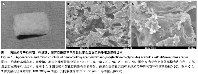
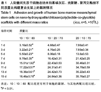
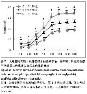
2.1 支架外观及孔隙率的测定 3种支架均为乳白色,内部及表面充满多孔状结构。应用排水法测量3种支架孔隙率均大于80%。扫描电镜可以观察到纳米羟基磷灰石/壳聚糖/聚丙交酯支架孔表面均匀分布有纳米羟基磷灰石及壳聚糖颗粒,3组支架内部孔结构没有明显差异;3种支架孔径分布均以100-300 µm为主,其间散在分布有30-50 µm不等的微孔(图1)。采用Digmizer图像测量软件测量3组复合支架孔隙的平均孔径分别为(224.14±86.47),(221.14±23.10),(226.19±47.35) µm,结果没有统计学差异,孔径大小均符合引导骨生长的支架最佳孔径。10∶10∶80、10∶20∶70组及20∶10∶70组支架抗压缩实验测试结果分别为(6.078±0.111),(4.586 ±0.159),(10.898±0.119) MPa,显示3组支架的抗压强度均与人平均松质骨抗压强度0.5-11.0 MPa相当[12]。而20∶10∶70组支架抗压强度明显大于其他两组(P < 0.01)。 2.2 人骨髓间充质干细胞与支架复合后的体外实验 细胞在支架上的黏附率及生长曲线:细胞复合 20∶10∶70支架组在第1天的黏附率及之后各个时间点测定的细胞增殖率均显著高于10∶10∶80组及10∶20∶70组(P < 0.01),见表1。3组支架的细胞增殖趋势相似,第1-3天为潜伏期,第3天进入对数增殖期,第9天后基本进入平台期(图2)。"

"
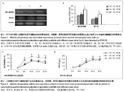
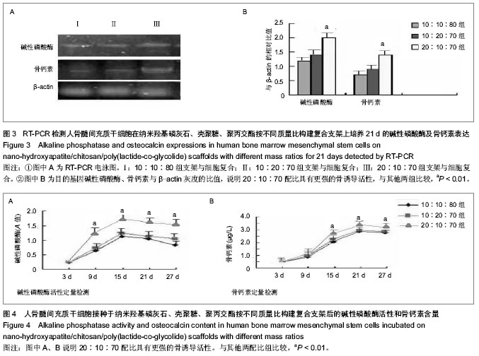
RT-PCR检测细胞碱性磷酸酶及骨钙素基因表达:3组支架细胞复合物在成骨诱导液中培养21 d后,20∶10∶70组细胞碱性磷酸酶及骨钙素的表达均强于10∶10∶80组、10∶20∶70组(P < 0.01,图3)。碱性磷酸酶活性及骨钙素含量的定量检测:细胞复合3组支架后碱性磷酸酶A值在第15天达到高峰后均开始下降,20∶10∶70组细胞碱性磷酸酶A值在第9天后显著高于其他两组(P < 0.01);细胞复合3组支架的骨钙素值在第21天达到高峰后均开始下降,20∶10∶70组细胞的骨钙素值在第15天后显著高于其他两组(P < 0.01,图4)。"
"
| [1] Baskin JZ,Eppell SJ.A selected review of the recent advances in craniomaxillofacial bone tissue engineering.Curr Opin Otolaryngol Head Neck Surg.2013;21(4):389-395.[2] Jin GZ,Kim TH,Kim JH,et al.Bone tissue engineering of induced pluripotent stem cells cultured with macrochanneled polymer scaffold.J Biomed Mater Res A. 2013;101(5): 1283- 1291.[3] Thibault RA,Mikos AG,Kasper FK.Scaffold/Extracellular matrix hybrid constructs for bone-tissue engineering.Adv Healthc Mater.2013;2(1):13-24.[4] Bao TQ,Franco RA,Lee BT.Preparation and characterization of a novel 3D scaffold from poly(?-caprolactone)/biphasic calcium phosphate hybrid composite microspheres adhesion. Biochem Eng J.2012;64:76-83.[5] Sadiasa A,Kim MS,Lee BT.Poly(lactide-co-glycolide acid)/biphasic calcium phosphate composite coating on a porous scaffold to deliver simvastatin for bone tissue engineering. J Drug Target.2013;21(8):719-729.[6] Li B,Yang J,Ma L,et al.Fabrication of poly(lactide-co-glycolide) scaffold filled with fibrin gel, mesenchymal stem cells, and poly(ethylene oxide)-b-poly(L-lysine)/TGF-beta1 plasmid DNA complexes for cartilage restoration in vivo.J Biomed Mater Res A.2013.[7] Kavya KC,Jayakumar R,Nair S,et al.Fabrication and characterization of chitosan/gelatin/nSiO2 composite scaffold for bone tissue engineering. Int J Biol Macromol.2013;59: 255-263.[8] Zhou Y,Xu L,Zhang X,et al.Radiation synthesis of gelatin/CM-chitosan/β-tricalcium phosphate composite scaffold for bone tissue engineering. Materials Science and Engineering: C.2012;32(4):994-1000.[9] Fricain JC,Schlaubitz S,Le Visage C,et al.A nano-hydroxyapatite--pullulan/dextran polysaccharide composite macroporous material for bone tissue engineering.Biomaterials.2013;34(12):2947-2959.[10] Cho JS,Rhee SH.Formation mechanism of nano-sized hydroxyapatite powders through spray pyrolysis of a calcium phosphate solution containing polyethylene glycol.J Eur Ceram Soc.2013;33(2):233-241.[11] Liu G,Li Y,Sun J,et al.In vitro and in vivo evaluation of osteogenesis of human umbilical cord blood-derived mesenchymal stem cells on partially demineralized bone matrix.Tissue Eng Part A.2010;16(3):971-982.[12] Lee RW,Volz RG,Sheridan DC.The role of fixation and bone quality on the mechanical stability of tibial knee components.Clin Orthop Relat Res.1991;(273):177-183.[13] Croisier F,Jérôme C.Chitosan-based biomaterials for tissue engineering. Eur Polymer J.2013;49(4):780-792.[14] Kim IY,Seo SJ,Moon HS,et al.Chitosan and its derivatives for tissue engineering applications.Biotechnol Adv.2008;26(1): 1-21.[15] Xu M,Li Y,Suo H,et al.Fabricating a pearl/PLGA composite scaffold by the low-temperature deposition manufacturing technique for bone tissue engineering. Biofabrication. 2010; 2(2):025002.[16] Lin ZY,Duan ZX,Guo XD,et al.Bone induction by biomimetic PLGA-(PEG-ASP)n copolymer loaded with a novel synthetic BMP-2-related peptide in vitro and in vivo.J Controlled Release.2010;144(2):190-195.[17] Minamiguchi S,Takechi M,Yuasa T,et al.Basic research on aw-AC/PLGA composite scaffolds for bone tissue engineering.J Mater Sci Mater Med.2008;19(3):1165-1172.[18] Kim SS,Sun Park M,Jeon O,et al. Poly(lactide-co-glycolide)/hydroxyapatite composite scaffolds for bone tissue engineering.Biomaterials.2006;27(8): 1399- 1409.[19] Li M,Liu W,Sun J,et al.Culturing primary human osteoblasts on electrospun poly(lactic-co-glycolic acid) and poly(lactic-co-glycolic acid)/nanohydroxyapatite scaffolds for bone tissue engineering.ACS Appl Mater Interfaces. 2013; 5(13):5921-5926.[20] Kavlock KD,Whang K,Guelcher SA,et al.Degradable segmented polyurethane elastomers for bone tissue engineering: effect of polycaprolactone content.J Biomater Sci Polym Ed.2013;24(1):77-93.[21] Zhang P,Hong Z,Yu T,et al.In vivo mineralization and osteogenesis of nanocomposite scaffold of poly(lactide-co-glycolide) and hydroxyapatite surface-grafted with poly(L-lactide).Biomaterials.2009;30(1):58-70.[22] Ngiam M,Liao S,Patil AJ,et al.The fabrication of nano-hydroxyapatite on PLGA and PLGA/collagen nanofibrous composite scaffolds and their effects in osteoblastic behavior for bone tissue engineering. Bone.2009; 45(1):4-16.[23] Jose MV,Thomas V,Johnson KT,et al.Aligned PLGA/HA nanofibrous nanocomposite scaffolds for bone tissue engineering. Acta Biomater.2009;5(1):305-315.[24] Huang YX,Ren J,Chen C,et al.Preparation and properties of poly(lactide-co-glycolide) (PLGA)/ nano-hydroxyapatite (NHA) scaffolds by thermally induced phase separation and rabbit MSCs culture on scaffolds.J Biomater Appl.2008;22(5): 409-432.[25] Wu YC,Shaw SY,Lin HR,et al.Bone tissue engineering evaluation based on rat calvaria stromal cells cultured on modified PLGA scaffolds. Biomaterials.2006;27(6):896-904.[26] Hu Q,Li B,Wang M,et al.Preparation and characterization of biodegradable chitosan/hydroxyapatite nanocomposite rods via in situ hybridization: a potential material as internal fixation of bone fracture.Biomaterials.2004;25(5):779-785.[27] Thein-Han WW,Misra RDK.Biomimetic chitosan–nanohydroxyapatite composite scaffolds for bone tissue engineering.Acta Biomater.2009;5(4):1182-1197.[28] Teng X,Ren J,Gu S.Preparation and characterization of porous PDLLA/HA composite foams by supercritical carbon dioxide technology.J Biomed Mater Res B Appl Biomater. 2007; 81(1):185-193.[29] Mooney DJ,Mazzoni CL,Breuer C,et al.Stabilized polyglycolic acid fibre-based tubes for tissue engineering.Biomaterials. 1996;17(2):115-124.[30] Whang K,Thomas CH,Healy KE,et al.A novel method to fabricate bioabsorbable scaffolds.Polymer. 1995;36(4): 837-842.[31] Harris LD,Kim BS,Mooney DJ.Open pore biodegradable matrices formed with gas foaming.J Biomed Mater Res. 1998;42(3):396-402.[32] Porter JR,Ruckh TT,Popat KC.Bone tissue engineering: a review in bone biomimetics and drug delivery strategies. Biotechnol Prog.2009;25(6):1539-1560.[33] Caverzasio J,Manen D.Essential role of Wnt3a-mediated activation of mitogen-activated protein kinase p38 for the stimulation of alkaline phosphatase activity and matrix mineralization in C3H10T1/2 mesenchymal cells. Endocrinology.2007;148(11):5323-5330.[34] Rey A,Manen D,Rizzoli R,et al.Evidences for a role of p38 MAP kinase in the stimulation of alkaline phosphatase and matrix mineralization induced by parathyroid hormone in osteoblastic cells.Bone.2007;41(1):59-67.[35] Nakamura A,Dohi Y,Akahane M,et al.Osteocalcin secretion as an early marker of in vitro osteogenic differentiation of rat mesenchymal stem cells.Tissue Eng Part C Methods.2009; 15(2):169-180.[36] Akahane M,Ueha T,Dohi Y,et al.Secretory osteocalcin as a nondestructive osteogenic marker of tissue-engineered bone.J Orthop Sci. 2011;16 (5): 622-628.[37] Pandit V,Zuidema JM,Venuto KN,et al.Evaluation of Multifunctional Polysaccharide Hydrogels with Varying Stiffness for Bone Tissue Engineering. Tissue Eng Part A.2013;19(21-22);2452-2463.[38] Vozzi G,Corallo C,Carta S,et al. Collagen-gelatin-genipin-hydroxyapatite composite scaffolds colonized by human primary osteoblasts are suitable for bone tissue engineering applications: In vitro evidences.J Biomed Mater Res A.2013. doi;10.1002/jbm.a.34823.[39] Kim J,Jeong SY,Ju YM,et al.In vitro osteogenic differentiation of human amniotic fluid-derived stem cells on a poly(lactide-co-glycolide) (PLGA)-bladder submucosa matrix (BSM) composite scaffold for bone tissue engineering.Biomed Mater.2013;8(1):014107.[40] Pavlin D,Zadro R,Gluhak-Heinrich J.Temporal pattern of stimulation of osteoblast-associated genes during mechanically-induced osteogenesis in vivo: early responses of osteocalcin and type I collagen.Connect Tissue Res.2001; 42(2):135-148.[41] Ito M.In vitro properties of a chitosan-bonded hydroxyapatite bone-filling paste.Biomaterials.1991;12(1):41-45. |
| [1] | Li Li, Ma Li. Immobilization of lactase on magnetic chitosan microspheres and its effect on enzymatic properties [J]. Chinese Journal of Tissue Engineering Research, 2021, 25(4): 576-581. |
| [2] | Li Xinping, Cui Qiuju, Zeng Shuguang, Ran Gaoying, Zhang Zhaoqiang, Liu Xianwen, Fang Wei, Xu Shuaimei. Effect of modification of β-tricalcium phosphate/chitosan hydrogel on growth and mineralization of dental pulp stem cells [J]. Chinese Journal of Tissue Engineering Research, 2021, 25(22): 3493-3499. |
| [3] | Chen Song, He Yuanli, Xie Wenjia, Zhong Linna, Wang Jian. Advantages of calcium phosphate nanoparticles for drug delivery in bone tissue engineering research and application [J]. Chinese Journal of Tissue Engineering Research, 2021, 25(22): 3565-3570. |
| [4] | Chen Siyu, Li Yannan, Xie Liying, Liu Siqi, Fan Yurong, Fang Changxing, Zhang Xin, Quan Jiayu, Zuo Lin. Thermosensitive chitosan-collagen composite hydrogel loaded with basic fibroblast growth factor retards ventricular remodeling after myocardial infarction in mice [J]. Chinese Journal of Tissue Engineering Research, 2021, 25(16): 2472-2478. |
| [5] | Liu Feng, Zhang Yu, Wang Yanli, Luo Wei, Han Chaoshan, Li Yangxin. Application of temperature-sensitive chitosan hydrogel encapsulated exosomes in ischemic diseases [J]. Chinese Journal of Tissue Engineering Research, 2021, 25(16): 2479-2487. |
| [6] | Li Jie, Xu Jianzhen, Hu Ping, Lei Qiqi, Zhang Wenning, Ao Ningjian . Preparation and performance evaluation of carboxymethyl chitosan/oxidized glucomannan/Panax notoginseng compound sponge dressing for chronic wound [J]. Chinese Journal of Tissue Engineering Research, 2021, 25(16): 2528-2534. |
| [7] | Gan Zhoujie, Pei Xibo. Enzyme-responsive nanoparticles in tumor therapy: superiority of nanoparticles in accumulation and drug release [J]. Chinese Journal of Tissue Engineering Research, 2021, 25(16): 2562-2568. |
| [8] | Feng Xiaoxia, Hou Weiwei, Jin Xiaoting, Wang Xinhua. Construction of periodontal biomimetic membrane with electrospun poly(lactic-co-glycolic acid) nanofibers and electrosprayed chitosan microspheres [J]. Chinese Journal of Tissue Engineering Research, 2020, 24(4): 511-516. |
| [9] | Li Li, Ma Li, Li He. Preparation and characterization of magnetic chitosan microspheres [J]. Chinese Journal of Tissue Engineering Research, 2020, 24(4): 577-582. |
| [10] | Li Xuan, Lu Min, Li Mingxing, Ao Meng, Tang Linmei, Zeng Zhen, Hu Jingwei, Huang Zhiqiang, Xuan Jiqing. In vitro multi-modal imaging of magnetic targeted nanoparticles and their targeting effect on hepatic stellate cells [J]. Chinese Journal of Tissue Engineering Research, 2020, 24(4): 566-571. |
| [11] | Xiang Haibin, Li Xinxia, Liang Qiuzhen, Song Xinghua. Specific bone-targeting nanoscale drug delivery system: advantages and clinical applicability [J]. Chinese Journal of Tissue Engineering Research, 2020, 24(4): 612-618. |
| [12] | Liu Haiyan, Hu Yang, Wu Xiuping, Pan Haobo, Jing Xuan. Chitosan-based polysaccharide biomaterial for prevention and treatment of oral diseases [J]. Chinese Journal of Tissue Engineering Research, 2020, 24(4): 631-636. |
| [13] | Liao Jian, Huang Xiaolin, Huo Hua, Zhou Qian, Cheng Yuting, Qi Yuhan, Wu Chao, Yang Tongjing, Liao Yunmao, Liang Xing. Effects of calcined bone/chitosan composite materials on proliferation and adhesion in osteoblasts [J]. Chinese Journal of Tissue Engineering Research, 2020, 24(34): 5447-5453. |
| [14] | Fang Yulu, Yi Bingcheng, Shen Yanbing, Tang Han, Zhang Yanzhong. Potential of corn husk fibers reinforced chitosan-based hydrogels in cartilage tissue engineering scaffold [J]. Chinese Journal of Tissue Engineering Research, 2020, 24(34): 5493-5501. |
| [15] | Liao Jian, Huang Xiaolin, Zhou Qian, Huo Hua, Qi Yuhan, Wu Chao, Shi Qianhui, Yang Tongjing, Liao Yunmao, Liang Xing. Calcined bone/chitosan composite promotes osteogenic differentiation of bone marrow mesenchymal stem cells in Sprague-Dawley rats [J]. Chinese Journal of Tissue Engineering Research, 2020, 24(31): 4941-4947. |
| Viewed | ||||||
|
Full text |
|
|||||
|
Abstract |
|
|||||