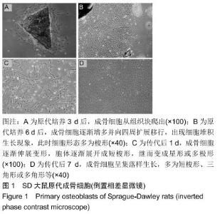
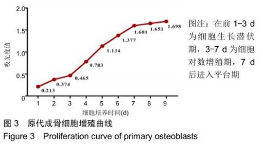
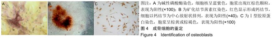
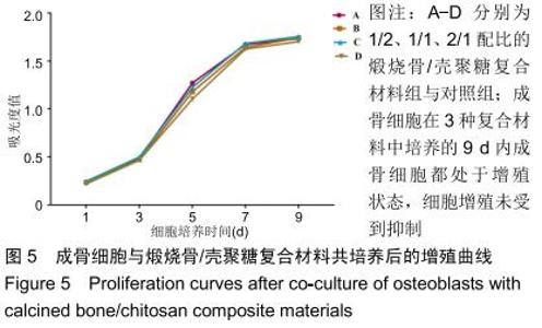
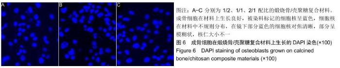
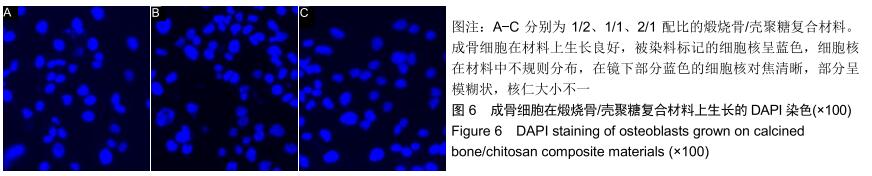
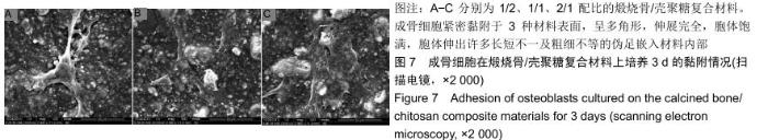
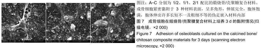
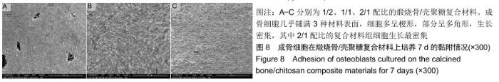
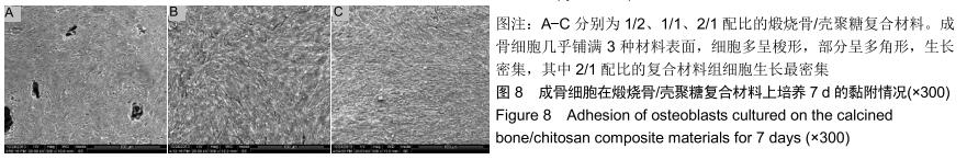
|
[1] TROELTZSCH M, TROELTZSCH M, KAUFFMANN P, et al.Clinical efficacy of grafting materials in alveolar ridge augmentation: A systematic review.J Craniomaxillofac Surg. 2016;44(10):1618-1629.
[2] SHANBHAG S, SHANBHAG V. Clinical applications of cell-based approaches in alveolar bone augmentation: a systematic review. Clin Implant Dent Relat Res.2015;17 Suppl 1:e17-e34.
[3] GRAHAM SM, LEONIDOU A, ASLAM-PERVEZ N, et al. Biological therapy of bone defects: the immunology of bone allo-transplantation.Expert Opin Biol Ther.2010;10(6):885-901.
[4] IIJIMA K, TSUJI Y, KURIKI I, et al. Control of cell adhesion and proliferation utilizing polysaccharide composite film scaffolds. Colloids Surf B Biointerfaces.2017;160:228-237.
[5] GUNARAJAH DR, SAMMAN N. Biomaterials for repair of orbital floor blowout fractures: a systematic review.J Oral Maxillofac Surg.2013;71(3):550-570.
[6] BERINGER LT, KIECHEL MA, KOMIYA Y, et al. Osteoblast biocompatibility of novel chitosan crosslinker, hexamethylene- 1,6-diaminocarboxysulfonate.J Biomed Mater Res A. 2015; 103(9):3026-3033.
[7] WU S, ZHOU Y, YU Y, et al. Evaluation of Chitosan Hydrogel for Sustained Delivery of VEGF for Odontogenic Differentiation of Dental Pulp Stem Cells.Stem Cells Int.2019;2019:1515040.
[8] CHEUNG RC, NG TB, WONG JH, et al. Chitosan: An Update on Potential Biomedical and Pharmaceutical Applications.Mar Drugs. 2015;13(8):5156-5186.
[9] PARISI L, TOFFOLI A, BIANCHI MG, et al. Functional Fibronectin Adsorption on Aptamer-Doped Chitosan Modulates Cell Morphology by Integrin-Mediated Pathway.Materials (Basel). 2019;12(5). pii: E812.
[10] VENKATESAN J, KIM SK. Chitosan composites for bone tissue engineering--an overview. Mar Drugs. 2010;8(8):2252-2266.
[11] TAN ML, SHAO P, FRIEDHUBER AM, et al. The potential role of free chitosan in bone trauma and bone cancer management. Biomaterials.2014;35(27):7828-7838.
[12] YANG CC, LIN CC, LIAO JW, et al. Vancomycin-chitosan composite deposited on post porous hydroxyapatite coated Ti6Al4V implant for drug controlled release.Mater Sci Eng C Mater Biol Appl.2013;33(4):2203-2212.
[13] ELANGO J, SARAVANAKUMAR K, RAHMAN SU, et al. Chitosan-Collagen 3D Matrix Mimics Trabecular Bone and Regulates RANKL-Mediated Paracrine Cues of Differentiated Osteoblast and Mesenchymal Stem Cells for Bone Marrow Macrophage-Derived Osteoclastogenesis.Biomolecules. 2019; 9(5).pii: E173.
[14] LU X, ZHENG B, TANG X, et al. In vitro biomechanical and biocompatible evaluation of natural hydroxyapatite/chitosan composite for bone repair.J Appl Biomater Biomech.2011;9(1):11-18.
[15] ZHAO HX, JIN H, CAI JY. Preparation and characterization of nano-hydroxyapatite/chitosan composite with enhanced compressive strength by urease-catalyzed method.Mater Lett. 2014;116(feb.1):293-295.
[16] ZHANG J, NIE J, ZHANG Q, et al. Preparation and characterization of bionic bone structure chitosan/hydroxyapatite scaffold for bone tissue engineering.J Biomater Sci Polym Ed. 2014;25(1):61-74.
[17] QU Y, AO D, WANG P, et al. Chitosan/nano-hydroxyapatite composite electret membranes enhance cell proliferation and osteoblastic expression in vitro.J Bioact Compat Polym.2014; 29(1):3-14.
[18] MARDAS N, DEREKA X, DONOS N, et al. Experimental model for bone regeneration in oral and cranio-maxillo-facial surgery.J Invest Surg.2014;27(1):32-49.
[19] El-SHERBINY IM, YAHIA S, MESSIERY MA, et al. Preparation and Physicochemical Characterization of New Nanocomposites Based on β-Type Chitosan and Nano-Hydroxyapatite as Potential Bone Substitute Materials.Int J Polym Mater.2014;63(4):213-220.
[20] VENKATESAN J, QIAN ZJ, RYU B, et al. Preparation and characterization of carbon nanotube-grafted-chitosan – Natural hydroxyapatite composite for bone tissue engineering. Carbohydrate Polym.2011;83(2):569-577.
[21] HE Y, JIN Y, WANG X, et al. An Antimicrobial Peptide-Loaded Gelatin/Chitosan Nanofibrous Membrane Fabricated by Sequential Layer-by-Layer Electrospinning and Electrospraying Techniques. Nanomaterials (Basel).2018;8(5).pii: E327.
[22] 廖健,黄晓林,周倩,等.煅烧骨/壳聚糖复合材料的制备及表征[J].中国组织工程研究,2020,24(22):2452-3459.
[23] TANG ZX, QIAN JQ, SHI LE. Preparation of chitosan nanoparticles as carrier for immobilized enzyme.Appl Biochem Biotechnol.2007; 136(1):77-96.
[24] OHGUSHI H, GOLDBERG VM, CAPLAN AI. Repair of bone defects with marrow cells and porous ceramic. Experiments in rats.Acta Orthop Scand.1989;60(3):334-339.
[25] 肖国强,胡介宇,王茜,等.特异性信号通路在骨修复和再生中的作用研究进展[J].中国综合临床,2017,33(12):1138-1142.
[26] 宁思敏,王东,孙海钰,等.大鼠成骨细胞与壳聚糖/羟基磷灰石复合支架降解产物的生物相容性[J].中国组织工程研究,2014,18(12): 1846-1851.
[27] MAO D, LI Q, BAI N, et al. Porous stable poly(lactic acid)/ethyl cellulose/hydroxyapatite composite scaffolds prepared by a combined method for bone regeneration.Carbohydr Polym.2018; 180:104-111.
[28] 赵淑珍,张婷,王彦鹏,等.复合3D支架在修复颅骨缺损中的应用进展[J].中国综合临床,2019,35(2):183-186.
[29] PAPPALARDO S, GUARNIERI R. Proliferation Assessment of Mesenchymal Stem Cells on an Acellular Dermal Matrix (AlloDerm® GBR) Used for Guided Bone Regeneration.J Biomater Tissue Eng.2013;3(5):589-596.
[30] 康明,黄杰华,张理选,等.壳聚糖/胡须/磷酸钙骨水泥复合生物材料的力学性能及对诱导多能干细胞成骨潜能的影响[J].中国修复重建外科杂志,2018,32(7):959-967.
[31] LYNCH I. Are there generic mechanisms governing interactions between nanoparticles and cells? Epitope mapping the outer layer of the protein–material interface.Physica A.2007;373:511-520.
[32] HING KA, BEST SM, TANNER KE, et al. Quantification of bone ingrowth within bone-derived porous hydroxyapatite implants of varying density.J Mater Sci Mater Med.1999;10(10/11):663-670.
[33] 朱明华,曾怡,孙涛,等.生物陶瓷与细胞外基质对成骨细胞生长及代谢影响[J].北京生物医学工程,2005,24(1):17-23.
[34] 李冀,王志强,张育敏等.人工骨传导材料的研究进展[J].中国修复重建外科杂志,2006,20(1):81-84.
[35] 张永军,杨静茹,王芳,等.羧甲基壳聚糖联合胶原膜治疗根分叉病变的疗效观察[J].河北医学,2016,22(6):999-1000.
[36] COSTA-PINTO AR, CORRELO VM, SOL PC, et al. Chitosan- poly(butylene succinate) scaffolds and human bone marrow stromal cells induce bone repair in a mouse calvaria model.J Tissue Eng Regen Med.2012;6(1):21-28.
[37] GHAEE A, NOURMOHAMMADI J, DANESH P. Novel chitosan- sulfonated chitosan-polycaprolactone-calcium phosphate nanocomposite scaffold.Carbohydr Polym.2017; 157:695-703.
[38] 刘苗,李德超,张颖.鹿瓜多肽/煅烧骨颗粒/壳聚糖复合材料修复兔下颌骨缺损的研究[J].中国体视学与图像分析,2015,20(2):175-179.
|