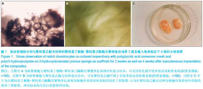
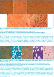
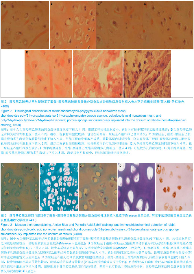
| [1] Marcacci M,Filardo G, Kon E.Treatment of cartilage lesions: What works and why? Injure.2013;44 (Suppl 1):11-15. [2] Fishman JM,DeCoppi P,Elliott MJ,et al.Airway tissue enjineering. Expert Opin Biol Ther. 2011;11(12):1623-1635.[3] 王海江,陈启富.组织工程支架材料研究进展[J].中国现代医生,2012,50(1):27-29.[4] 李德保,章庆国.软骨组织工程支架材料研究进展[J].中国美容医学,2009,18(3):408-410.[5] 孙安科,陈文弦,崔鹏程,等.以PGA 为三维支架同种异体工程化软骨的构建[J].解放军医学杂志,2001,26(10):748-749.[6] 孙安科,裴国献,胡平,等.组织工程化喉软骨的构建[J].中华耳鼻咽喉科杂志,2004,39(10):606-611.[7] 鄂征.组织培养和分子细胞学技术[M].北京:北京出版社,1999: 1-118.[8] Mikos AG,Sarakinos G,Leite SM,et al.Laminated three-dimensional biodegradable foams for ues in tissue engineering.Biomaterials.1993;14(2):323-330.[9] Zhang R,Ma PX. Poly(α-hydroxyl acids)/hydroxyapatite porous composites for bone-tissue engineering. I. Preparation and morphology.J Biomed Mater Res.1999; 44(4):446-455.[10] Kim BS,Mooney DJ.Development of biocompatible synthetic extracelluar matrices for tissue engineering.Trends Biotechnol.1998;16(5):224.[11] Shim IK,Lee SY,Park YJ,et al.Homogeneous chitosan-PLGA composite fibrous scaffolds for tissue regeneration.J Biomed Mater Res A.2008;84(1):247-255.[12] Glowacki J,Mizuno S.Collagen scaffolds for tissue engineering. Biopolymers. 2008;89(5):338-344.[13] Cohen SB,Meirisch CM,Wilson HA,et al.The use of absorbable co-polymer pads with alginate and cells for articular cartilage repair in rabbits. Biomaterials. 2003;24(15): 2653-2660.[14] Chen GQ,Wu Q.The application of polyhydroxyalkanoates as tissue engineering materials.Biomaterials. 2005;26(33): 6565-6578.[15] 刘清宇,王富友,杨柳.关节软骨组织工程支架的研究进展[J].中国修复重建外科杂志,2012,26(10):1247-1250.[16] Be k CH, Ko YJ. Characteristics of tissue-engineered cartilage on macropomus biodegradable PLGA scaffold.Laryngoscope.2006;116(10):1829-1834.[17] 吴俊,孙俊英,李海燕,等.以PHBV为支架构建组织工程化软骨[J].中国矫形外科杂志,2006,14(13):1016-1018.[18] 于娟,万涛,李世普.生物可降解聚合材料聚羟基乙酸[J].生物骨科材料与临床研究,2005,2(6):50-56.[19] Puelacher WC,Mooney DJ,langer R,et al.Design of nasoseptal cartilage replacements synthesized from biodegradable polymers and chondrocytes. Biomaterials. 1994;15(6):774-778. [20] Boyan DB,Hunnert TW,Dean DD,et al.Role of material surfaces in regulating bone and cartilage cell response. Biomaterials.1996;17(2):137-146.[21] Vacanti CA,Paige KJ,Kim W,et al.Experimental tracheal replacement using tissue engineered cartilage.J Pediart Surg.1994;27(3):201-204.[22] Vacanti CA,Upton J.Tissue-engineered morphogenesis of cartilage and bone by means of cell transplantation using synthetic biodegradable polymer Matrices. Clin Plast Surg. 1994;21(3):445-462. [23] 潘勇,黄蔚,艾玉峰,等.组织工程人工血管支架的预构[J].中华整形外科杂志,2003,19(1):44-46.[24] 李鹏程,程代薇,徐福,等.组织工程肌腱种子细胞的比较研究[J].组织工程与重建外科杂志,2006,2(3):134-136.[25] 胡晓洁,王敏,柴岗,等.组织工程技术构建兔角膜基质组织的实验研究[J].中华眼科杂志,2004,40(8):517-521.[26] 杨光辉,崔磊,刘伟,等.利用聚羟基乙酸构建组织工程皮肤的实验研究[J].中华实验外科杂志,2003,20(l1):984-985.[27] 田敏,王贻宁,陈新明,等.PGA无纺网与PDLCs的3D共培养物在裸鼠体内的生长观察[J].口腔医学研究,2005,21(2):116- 118.[28] 毛天球.组织工程的研究概括[J].实用口腔医学杂志,2000,16(2): 74-76.[29] Luo L,Wei X,Cben GQ.Physical Properties and Biocompatibility of. Poly(3-hydroxybutyrate-co-3- hydroxyhexanoate) Blended with Poly(3-hydroxybutyrate-co-4-hydroxybutyrate). J Biomater Sci Polym Ed.2009;20(11):1537-1553.[30] 田频源,米钰,尚龙安,等.聚羟基烷酸酯的研究进展[J].化工进展,2009,28(3):468-476.[31] Zhao K,Deng Y,Chen J,et al.Polyhydroxyalkanoate(PHA) scaffolds with good mechanicalproperties and biocompatibility. Biomaterials.2003;24(9):1041-1045. [32] 郭霄飞,张永红,杜美丽,等.羟基丁酸与羟基辛酸共聚物一体化骨软骨组织工程支架的制备及性能[J].中国组织工程研究,2012, 16(16):2865-2868.[33] Cool SM,Kenny B,Wu A,et al. Poly(3-hydroxybutyrate-co-3-hydroxyvalerate) composite biomaterials for bone tissue regeneration: in vitro performance assessed by osteoblast proliferation, osteoclast adhesion and resorption, and macrophage proinflammatory response.J Biomed Mater Res A.2007;82(3):599-610.[34] Veleirinho B,Coelho DS,Dias PF,et al.Nanofibrous poly (3-hydroxybutyrate-co-3-hydroxyvalerate)/chitosan scaffolds for skin regeneration.Int J Biol Macromol.2012;51(4):343-350.[35] 冷晔.聚羟基丁酸己酸酯及其在组织工程中的应用[J].生物医学工程学进展,2011,32(4):213-218.[36] 唐秀杰,胡平,吴清玉.聚羟基烷酸酯、聚丁二酸丁二酯及其共混材料的细胞相容性评价比较[J].生物医学工程学杂志,2007, 24(4): 790-793.[37] 孙安科,胡平,李万同,等.组织工程PHBHH多孔材料喉支架的制备与细胞相容性研究[J].听力学及言语疾病杂志,2011,19(6):558-561.[38] Freed LE,Marpuis JC,Nohria A,et al.Neocartilage formation in vitro and in vivo using cells cultured on synthetic biodegradable polymers.J Biomed Mater Res. 1993;27(1):11-23.[39] 周栋,胡平,汪钢,等.聚羟基丁酸及其共聚物在体内组织相容性和降解率的实验研究[J].武警医学,2003,14(4):210-212.[40] 欧阳少平,丘远征,卢晓云,等.代谢工程技术调控3一羟基丁酸与3一羟基己酸共聚酯PHBHHx的微生物合成[J].生物加工过程, 2003, 1(1):60-65. |