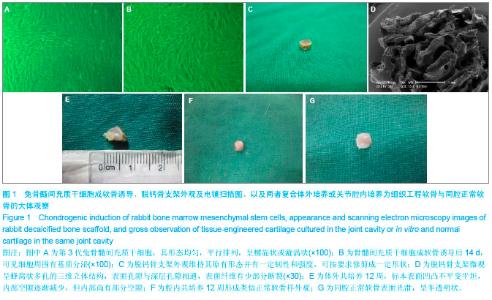
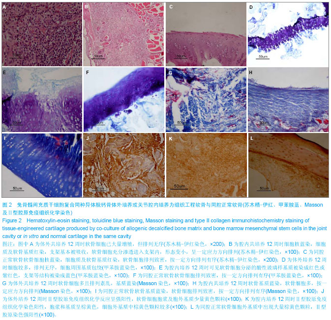
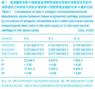
| [1] Hunter W.Of the structure and disease of articulating cartilages. Phil Trans.1743;470:514-521.[2] Paget J.Healing of injuries in various tissues.Lect Surg Path (London). 1853;1:262-268.[3] Hunziker EB.Articular cartilage repair: are the intrinsic biological constraints undermining this process insuperable? Osteoarthritis Cartilaqe.1999;7(1):15-28. [4] Buckwalter JA,Mankin HJ.Articular cartilage: degeneration and osteoarthritis,repair, regeneration, and transplantation. Instr Course Lect. 1998;47:487-504. [5] 中华人民共和国科学技术部.关于善待实验动物的指导性意见. 2006-09-30.[6] Ringe J,Kaps C,Burmester GR,et al.Stem cells for regenerative medicine: advances in the engineering of tissues and organs.Naturwissenschaften. 2002;89(8):338-351. [7] Ahmed TA,Hincke MT.Strategies for articular cartilage lesion repair and functional restoration.Tissue Eng Part B Rev.2010; 16(3):305-329.[8] Henderson I,Francisco R,Oakes B,et al.Autologous chondrocyte implantation fortreatment of focal chondal defects of the knee-a clinical, arthroscopic, MRI and histoligic evaluation at 2 years. Knee.2005;12(3):209-216.[9] Aggarwal S , Pittenger MF. Human mesenchymal stem cells modulate allogeneic immune cell responses.Blood.2005; 105(4):1815-1822.[10] Ramasamy R, Tong CK, Seow HF, et al. The immunosuppressive effects of human bone marrow-derived mesenchymal stem cells target T cell proliferation but not its effector function.Cell Immunol.2008;251(2):131-136.[11] Brown BN,Barnes CA,Kasick RT,et al.Surface characterization of extracellular matrix scaffolds.Biomaterials. 2010;31(3):428-437.[12] Mandal BB,Park SH,Gil ES,et al.Multilayered silk scaffolds for meniscus tissueengineering.Biomaterials. 2011;32(2):639- 651. [13] Martinez-Diaz S,Garcia-Giralt N,Lebourg M,et al.In vivo evaluation of 3-dimensional polycaprolactone scaffolds for cartilage repair in rabbits. Am J Sports Med.2010;38(3): 509-519. [14] Lin PB,Ning LJ,Lian QZ,et al.A study on repair of porcine articular cartilage defects with tissue-engineered cartilage constructed in vivo by composite scaffold materials.Ann Plast Surg.2010;65(4):430-436.[15] Dazzi F,Ramasamy R,Glennie S,et al.The role of mesenchymal stem cells in haemopoiesis.Blood Rev.2006; 20(3): 161-171.[16] Frenkel S,Cesare P.Degradation and repair of articular cartialge. Front Biosci.1999;(4):671-685.[17] Grgic M,Jelic M,Basic V,et al.Regeneration of articular cartilage defects in rabbits by osteogenic protein-1 (bone morphogeneticprotein-7).Acta Med Croatica.1997;51:23-27.[18] Gloria A,De Santis R,Ambrosio L.Polymer-based composite scaffolds for tissue engineering.J Appl Biomater Biomech. 2010; 8(2):57-67.[19] Nakajima H,Goto T,Horikawa O,et al.Characterization of the cells in the repair tissue of full-thickness articular cartilage defects.HistochemCell Biol.1998;109:331-338.[20] Bosnakovski D,Mizuno M,Kim G,et al.Chondrogenic differentiation of bovine bone marrow mesenchymal stem cells (MSCs) in different hydrogels: influence of collagen type II extracellular matrix on MSC chondrogenesis.Biotechnol Bioeng.2006;93(6):1152-1163.[21] Smith RL,Lin J,Trindade MC,et al.Time-dependent effects of intermittent hydrostatic pressure on articular chondrocyte type II collagen and aggrecan mRNA expression.J Rehabil Res Dev.2000;37:153-161.[22] 王刚,刘一,单玉兴,等.不同应力环境对兔骨髓间充质干细胞修复关节软骨缺损的影响[J].中国修复重建外科杂志,2004,18(2): 96-99.[23] Olmez U,Ryan LM,Kurup IV,et al.Insulin-like growth factor-1 suppresses pyrophosphate elaboration by transforming growth factor beta 1-stimulated chondrocytes and cartilage. Osteoarthritis Cartilage.1994;2:149-154. |