Chinese Journal of Tissue Engineering Research ›› 2014, Vol. 18 ›› Issue (5): 663-668.doi: 10.3969/j.issn.2095-4344.2014.05.002
Previous Articles Next Articles
Medical TH adhesive embolism for establishing a rabbit model of ischemic necrosis of lunate bone
Lu Yun-xiang, Chen Yu-xian, Zhuang Ze, Ren Jian-hua, Peng You, Shi De-hai, Wang Kun, Li Zhi-yong
- Department of Joint Surgery and Orthopedic Trauma, the Third Affiliated Hospital of Sun Yat-sen University, Guangzhou 510630, Guangdong Province, China
-
Revised:2014-01-02Online:2014-01-29Published:2014-01-29 -
Contact:Li Zhi-yong, M.D., Associate chief physician, Associate professor, Master’s supervisor, Department of Joint Surgery and Orthopedic Trauma, the Third Affiliated Hospital of Sun Yat-sen University, Guangzhou 510630, Guangdong Province, China -
About author:Lu Yun-xiang, Studying for master’s degree, Department of Joint Surgery and Orthopedic Trauma, the Third Affiliated Hospital of Sun Yat-sen University, Guangzhou 510630, Guangdong Province, China -
Supported by:the Guangdong Provincial Medical Science Foundation in China, No. A2012173
CLC Number:
Cite this article
Lu Yun-xiang, Chen Yu-xian, Zhuang Ze, Ren Jian-hua, Peng You, Shi De-hai, Wang Kun, Li Zhi-yong. Medical TH adhesive embolism for establishing a rabbit model of ischemic necrosis of lunate bone[J]. Chinese Journal of Tissue Engineering Research, 2014, 18(5): 663-668.
share this article
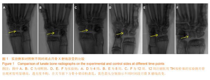
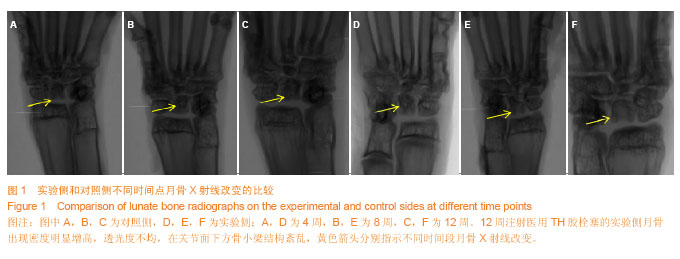
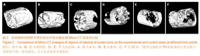
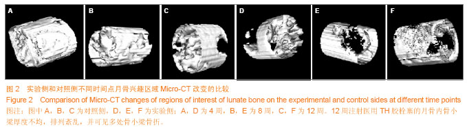
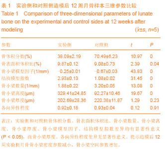
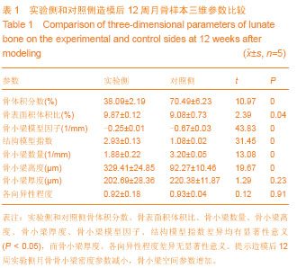
2.1 实验动物数量分析 实验共纳入30只新西兰兔,全部进入结果分析,无脱失。 2.2 X射线检查 注药后双腕关节平片显示,造模后4周实验侧与对照侧比较,未见明显差异;造模后8,12周实验侧相对于对照侧月骨密度增高;其中造模后12周实验侧月骨密度明显增高,透光度不均,在关节面下方骨小梁结构紊乱(图1)。 2.3 Micro-CT检查 Micro-CT三维重建图可见,12周实验侧月骨与对照侧相比骨小梁密度明显减低,月骨内骨小梁厚度不均,排列紊乱,并可见多处骨小梁骨折,月骨高度变低,月骨密度分布不均(图2)。造模后12周形成月骨三维兴趣区域并进行计算分析骨小梁参数,实验侧和对照侧骨体积分数、骨表面积体积比、骨小梁数量、骨小梁离度、骨小梁厚度、骨小梁模型因子、结构模型指数差异均有显著性意义,而骨小梁厚度和各向异性程度差异无显著性意义(P均 < 0.05,表1)。"
"
"
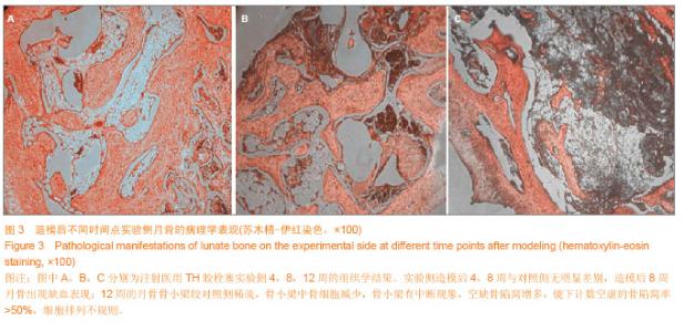
2.4 标本大体观察 造模后4周无明显差异;造模后8周月骨表面失去光泽,软骨部分失去半透明性状,颜色淡黄色;12周时实验侧软骨组织部分区域可见软骨表面黑色坏死组织,出现月骨明显的变形,并合并明显软骨退变,部分月骨较健侧明显变小。对照组各时间点月骨均呈正常形态,软骨面完整,未见破坏及缺损。 2.5 组织学检查 苏木精-伊红染色结果示,各时间点对照侧月骨显示关节软骨表面平滑,骨陷窝内骨细胞填充,骨髓内大量细胞存在;实验侧造模后12周病理改变主要集中于软骨下区,骨小梁较对照侧稀疏,骨小梁中骨细胞减少,骨小梁有中断现象,空缺骨陷窝增多,镜下计数空虚的骨陷窝率>50%,细胞排列不规则(图3)。"

| [1] Schuind F, Eslami S, Ledoux P. Kienbock’s disease. J Bone Joint Surg (Br). 2008;90(2):133-139.[2] Cross D, Matullo KS. Kienböck disease. Orthop Clin North Am. 2014;45(1):141-152.[3] Lutsky K, Beredjiklian PK. Kienböck disease. J Hand Surg Am. 2012;37(9):1942-1952.[4] Martin GR, Squire D.Long-term outcomes for Kienböck's disease. Hand (NY). 2013;8(1):23-26. [5] Ueba Y, Kakinoki R, Nakajima Y, et al. Morphology and histology of the collapsed lunate in advanced kienböck disease. Hand Surg. 2013;18(2):141-149. [6] Stahl S, Stahl AS, Meisner C, et al. Critical analysis of causality between negative ulnar variance and Kienböck disease. Plast Reconstr Surg. 2013;132(4):899-909.[7] Kirkeby L, von Varfalva Palffy L, Hansen TB. Long-term results after vascularised bone graft as treatment of Kienböck disease. J Plast Surg Hand Surg. 2014;48(1):21-23. [8] Mir X, Barrera-Ochoa S, Lluch A, et al. New surgical approach to advanced kienböck disease: lunate replacement with pedicled vascularized scaphoid graft and radioscaphoidal partial arthrodesis. Tech Hand Up Extrem Surg. 2013;17(2): 72-79. [9] Lluch A, Garcia-Elias M. Etiology of Kienböck disease. Tech Hand Up Extrem Surg.2011;15(1):33-37.[10] Dubey PP, Chauhan NK, Siddiqui MS, et al. Study of vascular supply of lunate and consideration applied to Kienböck disease. Hand Surg. 2011;16(1):9-13.[11] Lamas C, Carrera A, Proubasta I, et al. The anatomy and vascularity of the lunate: considerations applied to Kienböck's disease. Chir Main. 2007;26(1):13-20. [12] Mjoberg B. kienbock disease and negative ulnar variance. J Bone Joint Surg(Am). 2000;82:144.[13] Pichler M, Putz R. The venous drainage of the lunate bone. Surg Radiol Anat. 2003;24(6):372-376. [14] Schiltenwolf M, Martini AK, Mau HC, et al. Further investigations of the intraosseous pressure characteristics in necrotic lunates (Kienbock’s disease). J Hand Surg. 1996; 21A: 754-758.[15] Ogawa T, Ishii T, Mishima H,et al. Effectiveness of bone marrow transplantation for revitalizing a severely necrotic small bone: experimental rabbit model. J Orthop Sci. 2010; 15:381-388.[16] Shigematsu K, Hattori K, Kobata Y, et al. Treatment of Kienböck’s disease with cultured stem cell-seeded hybridtendon roll interposition arthroplasty: experimental study. J Orthop Sci. 2006;11:198-203.[17] Ikeguchi R, Kakinoki R, Aoyama T,et al. Regeneration of Osteonecrosis of Canine Scapho-lunate Using Bone MarrowStromal Cells: Possible Therapeutic Approach for Kienbock Disease. Cell Transplantation. 2006;15:411-422.[18] 黄启顺,付强,郑怀远,等.液氮冷冻并血管破坏建立犬月骨缺血性坏死动物模型的实验研究[J].中华手外科杂志,2013,29(4): 239-242.[19] 胡锦华,田相国,王广川,等.经皮经肝医用胶栓塞治疗肝硬化瘤样胃底静脉曲张临床观察[J].中华消化内镜杂志,2012,29(10): 545-548.[20] 殷晓煜,华赟鹏,梁力建,等.应用医用胶减少肝断面渗血的随机对照研究[J].中国实用外科杂志,2003,23(11):668-670. [21] 刘进,傅明,黄广鑫,等.Micro-CT软骨成像监测发育性髋关节发育不良大鼠髋关节软骨早期退变的应用价值[J].中华关节外科杂志(电子版),2013,7(2):213-219.[22] 陶云霞,王根林,袁鹏,等.两种染色法判定左前臂骨缺损修复骨组织的成熟度[J].中国组织工程研究,2012,16(37):6841-6846.[23] 闫宏伟,王坤正,时志斌,等.Legg-Perthes病动物模型设计与评价[J].中国矫形外科杂志,2002,10(8):795-796.[24] Berner A, Müller M, Fuechtmeier B.Arthroplasty of the lunate using bone marrow mesenchymalstromal cells. International Orthopaedics. 2011;35:379-387.[25] Lesley N, Lichtman D. Classification and treatment of Kienbock disease: a review of the past 100 years, and a look at the future. Handchir Mikrochir Plast Chir. 2010;42(3): 171-176.[26] Allan CH, Joshi A, Lichtman DM. Kienbock's disease: diagnosis and treatment. J Am Acad Orthop Surg. 2001;9(2): 128-136.[27] 高振国,蔡磊.5例月骨缺血性坏死的CT诊断[J].宁夏医学院学报, 2007,29(2):201-202.[28] Kim HK, Aruwajoye O, Stetler J,et al. Effects of non-weight-bearing on the immature femoral head following ischemic osteonecrosis: an experimental investigation in immature pigs. J Bone Joint Surg Am. 2012;94(24):2228- 2237.[29] Sun Y, Feng Y, Zhang C, et al. Beneficial effect of autologous transplantation of endothelial progenitor cells on steroid-induced femoral head osteonecrosis in rabbits. Cell Transplant. 2011;20(2):233-243.[30] Xie XH, Wang XL, Zhang G, et al. Impaired bone healing in rabbits with steroid-induced osteonecrosis. J Bone Joint Surg Br. 2011;93(4):558-565.[31] Ritman EL. Micro-computed tomography of thelungs and pulmonary-vascular system. Proc Am Thorac Soc. 2005;2(6): 477-480.[32] Feldkamp LA, Goldstein SA, Parfitt AM, et al.The direct examination of three-dimensional bone architecture in vitro by computed tomography.J Bone Miner Res. 1989;4(1): 3-11.[33] Han KJ, Kim JY, Chung NS, et al. Trabecular microstructureof the human lunate in Kienbock’s disease. J Hand Surg. 2012; 37E:336-341.[34] Calder JD, Pearse MF, Revell PA. The extent of osteocyte death in theproximal femur of patients with osteonecrosis of the femoral head. J Bone Joint Surg(Br). 2001;83(3):419- 422.[35] Ulrich D,van Rietbergen B,Laib A,et al. The ability of three dimensional structural indices to reflect mechanical aspects of trabecular bone. Bone. 1999;25(1):55-60.[36] Stauber M,Muller R.Age-related changes in trabecular bone microstructures:global and local morphometry. Osteoporos Int. 2006;17(4):616-626.[37] Nazarian A,Snyder BD,Zurakowski D,et al. Quantitative microcomputedtomogruphy: a non-invasive method to assess equivalentbone mineral density.Bone. 2008;43(2): 302-311.[38] 彭江,汪爱媛,孙明学,等.股骨头骨坏死样本空间结构的Micro-CT评估[J]. 中华创伤骨科杂志,2007,9(3):263-268.[39] Fan M, Jiang WX, Wang AY, et al.Effect and mechanism of zoledronate on prevention of collapse in osteonecrosis of the femoral head. Zhongguo Yi Xue Ke Xue Yuan Xue Bao. 2012; 34(4):330-336.[40] Fan M, Wang AY, Wang Y, et al. A new animal model of osteonecrosis induced by focal alternative cooling and heating insults. Zhongguo Yi Xue Ke Xue Yuan Xue Bao. 2011;33(4):375-381. |
| [1] | Liu Wei, Huang Jian. Applied research and progress of three-dimensional printing technology in joint replacement [J]. Chinese Journal of Tissue Engineering Research, 2017, 21(7): 1123-1130. |
| [2] | Yang Bao-jia, Yang Kai-shun, Yao Ru-bin. Correlation of nicotine dose and lumbar posterolateral fusion rate: imaging and biomechanical testing [J]. Chinese Journal of Tissue Engineering Research, 2016, 20(48): 7251-7260. |
| [3] | Wu Xiao-guang, Qiu Zhi-fu, Meng Jie, Zu Bing-xue, Li Meng-meng, Miao Hui. Effects of Buyanghuanwu decoction on the protein expression of PI3K, Akt, Bcl-2 and BAX in brain tissue of a rat model of cerebral hemorrhage [J]. Chinese Journal of Tissue Engineering Research, 2016, 20(40): 5933-5938. |
| [4] | Zhang Jian-jun, Shi Huan-chang. Immunosuppressed rat model of cerebral hemorrhage: construction and assessment [J]. Chinese Journal of Tissue Engineering Research, 2016, 20(40): 5939-5945. |
| [5] | Fu Feng, Zhao Ming-liang, Li Xiao-hong, Chen Chong, Wang Li-na, Sun Hong-tao, Tu Yue, Zhang Sai . Dynamic changes of brain cavity in rats after traumatic brain injury detected by MRI-based three-dimensional reconstruction [J]. Chinese Journal of Tissue Engineering Research, 2016, 20(40): 5946-5952. |
| [6] | Yu Yu-qin, Hu Nian-chun, Duan Ji-an, Li Da-peng, Liu Chang. Neuroprotective effects of sufentanil preconditioning on spinal cord injury in mouse models [J]. Chinese Journal of Tissue Engineering Research, 2016, 20(40): 5966-5972. |
| [7] | Wang Zhan, Li Hao-peng, He Xi-jing, Hao Ding-jun, Zhang Kun, Chen Ming-xia, Lei Ting. Pathological changes in the spinal cord of a model of acute cauda equina compression [J]. Chinese Journal of Tissue Engineering Research, 2016, 20(40): 5973-5978. |
| [8] | Zhao Yan-rui, Lv Wen-rui, Wang Dong, Zhou Jun-lin. Effects of carbonyl sulfide in a rat model of limb ischemia/reperfusion-induced acute lung injury [J]. Chinese Journal of Tissue Engineering Research, 2016, 20(40): 5994-6000. |
| [9] | Chen Wei-hua, Lv Guo-hua, Zhou Bin, Kang Yi-jun. Establishment of a dog model of pyogenic spinal infection [J]. Chinese Journal of Tissue Engineering Research, 2016, 20(40): 6014-6020. |
| [10] | Ge Run, Yang Lan. Protective effects of piperine on alveolar bone and collagen in a periodontitis model [J]. Chinese Journal of Tissue Engineering Research, 2016, 20(40): 6034-6040. |
| [11] | Song Qiao-qiao, Zhou Hui-liang, Zhen Hai-tao, Wang Na, Deng Jing, Wang Jin-xiang, Pan Xing-hua . Establishment and evaluation of a rhesus monkey model of experimental type 2 diabetes mellitus [J]. Chinese Journal of Tissue Engineering Research, 2016, 20(40): 6048-6053. |
| [12] | Pan Lei, Zhang Jin-biao, Cui Rong-gang, Zhao Bao-hui, Li Hua, Zhang Zhong-yong, Wang Xu-chu. Establishment of nonalcoholic fatty liver C57BL/6 mouse models [J]. Chinese Journal of Tissue Engineering Research, 2016, 20(40): 6054-6059. |
| [13] | Wang Shi-jun, Li Yu-ting, Li Chun-de. Establishment, application and progression of an animal model of cervical spondylosis [J]. Chinese Journal of Tissue Engineering Research, 2016, 20(40): 6067-6073. |
| [14] | Liu Ying-jie, Peng Jun, Liu Xiao-kang, Zhao Cheng, Yang Er-zhu, Xu Jian-guang. Experimental animal models of intervertebral fusion-induced adjacent segment degeneration [J]. Chinese Journal of Tissue Engineering Research, 2016, 20(39): 5825-5833. |
| [15] | Zhang Meng, Wei Jun-qiang, Duan Jian-wei, Yan Shi. Relationship of quadriceps tendon and the stability of external fixator in rabbit femur fracture model [J]. Chinese Journal of Tissue Engineering Research, 2016, 20(39): 5834-5839. |
| Viewed | ||||||
|
Full text |
|
|||||
|
Abstract |
|
|||||