Chinese Journal of Tissue Engineering Research ›› 2016, Vol. 20 ›› Issue (48): 7251-7260.doi: 10.3969/j.issn.2095-4344.2016.48.016
Previous Articles Next Articles
Correlation of nicotine dose and lumbar posterolateral fusion rate: imaging and biomechanical testing
Yang Bao-jia, Yang Kai-shun, Yao Ru-bin
- Department of Spinal Surgery, Affiliated Hospital of Dali University, Dali 671000, Yunnan Province, China
-
Revised:2016-09-11Online:2016-11-25Published:2016-11-25 -
Contact:Yang Kai-shun, Master, Professor, Master’s supervisor, Chief physician, Department of Spinal Surgery, Affiliated Hospital of Dali University, Dali 671000, Yunnan Province, China -
About author:Yang Bao-jia, Master, Physician, Department of Spinal Surgery, Affiliated Hospital of Dali University, Dali 671000, Yunnan Province, China -
Supported by:the General Program of Applied Research in Yunnan Province, No. 2011FZ294
CLC Number:
Cite this article
Yang Bao-jia, Yang Kai-shun, Yao Ru-bin. Correlation of nicotine dose and lumbar posterolateral fusion rate: imaging and biomechanical testing[J]. Chinese Journal of Tissue Engineering Research, 2016, 20(48): 7251-7260.
share this article
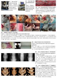
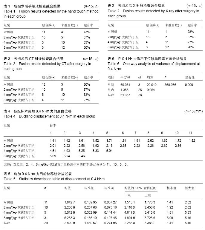
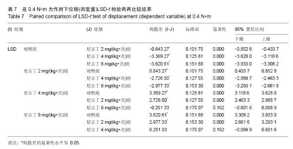
2.1 实验动物数量分析 纳入家兔60只,随机分为4组,每组15只,全部进入结果分析,无脱失。 2.2 手法触诊检查结果 术后12周空气栓塞法处死家兔,截取L4-L7节段。由2位未参与该实验过程的骨科医师手触法检查。对照组4例标本检查发现活动融合节段有轻微松动,评定为不融合;其余11例融合节段结合紧密,触诊检查无活动,评定为融合。融合成功率为73%。 实验组中,2 mg/(kg•次)尼古丁组有5例出现松动现象,认定为不融合,其余10例标本判定为融合,融合成功率为67%;4 mg/(kg•次)尼古丁组3例骨痂明显不连直接认定为不融合,有7例出现松动现象,认定为不融合,其余5例标本判定为融合,融合成功率为33%;8 mg/(kg•次)尼古丁组5例骨痂明显不连直接认定为不融合,有7例出现松动现象,认定为不融合,其余3例标本判定为融合,融合成功率为20%。 对照组与2 mg/(kg•次)尼古丁组比较,2组差异无显著性意义(P > 0.05),说明2 mg/(kg•次)的尼古丁对脊柱融合无明显影响。对照组与4 mg/(kg•次)尼古丁组比较,差异有显著性意义(P < 0.05)。对照组与8 mg/(kg•次)尼古丁组比较,差异有显著性意义(P < 0.05)。注射尼古丁的3组间采用行×列表资料χ2检验,差异有显著性意义(P < 0.05),说明随着尼古丁剂量的增加,对脊柱融合产生了影响(见表1)。 对照组融合标本骨痂体积大,硬度高,骨痂表面光滑(图5A);8 mg/(kg•次)尼古丁组融合标本痂体积小,硬度低,骨痂表面粗糙(图5B)。 2.3 X射线检查结果 所有标本经X射线检查发现:双侧横突间充满骨痂,手触法检查也为融合。单侧充满骨痂,对侧被吸收,手触测定也为不融合。对于双侧骨桥宽度不一致的标本,手触法与X射线检查结果不一致。部分手触法无明显骨痂形成的标本,X射线显示为横突间低密度影。对照组1例标本X射线检查发现单侧充满骨痂,对侧无明显骨痂,评定为不融合;其余14例双侧均充满骨痂,评定为融合,融合成功率为93%。实验组中,2 mg/(kg•次)尼古丁组有2例双侧无明显骨痂形成,认定为不融合;其余10例双侧均充满骨痂,3例双侧为骨桥相连,标本判定为融合,融合成功率为87%。4 mg/(kg•次)尼古丁组11例无明显骨痂形成,认定为不融合;其余4例标本判定为融合,融合成功率为27%。8 mg/(kg•次)尼古丁组12例无明显骨痂形成,认定为不融合;其余3例标本判定为融合,融合成功率为20%。 对照组与2 mg/(kg.次)尼古丁组比较,2组差异无显著性意义(P > 0.05),说明2 mg/(kg•次)的尼古丁对脊柱融合无明显影响。对照组与4 mg/(kg•次)尼古丁组比较,2组差异有显著性意义(P < 0.05)。对照组与 8 mg/(kg•次)尼古丁组比较,2组差异有显著性意义(P < 0.05)。注射尼古丁的3组间采用行×列表资料χ2检验,差异有显著性意义(P < 0.05),说明随着尼古丁剂量的增加,对脊柱融合产生了影响(见表2)。 X射线表现对照组新生骨痂密度较高,皮质较厚,表面光滑(图6A);尼古丁组随着剂量的增加植骨区骨痂较小,骨痂表面粗糙,不光滑(图6B-D)。 2.4 CT检查结果 对照组3例标本CT扫描及三维重建发现骨痂表面粗糙,评定为不融合;其余12例双侧均充满骨痂,评定为融合,融合成功率为80%。实验组中,2 mg/(kg•次)尼古丁组有5例双侧无明显骨痂形成,认定为不融合;其余7例双侧均充满骨痂,3例双侧为骨桥相连,标本判定为融合,融合成功率为67%。4 mg/(kg•次)尼古丁组10例无明显骨痂形成,认定为不融合;其余5例标本判定为融合,融合成功率为33%。8 mg/(kg•次)尼古丁组11例无明显骨痂形成,认定为不融合,其余4例标本判定为融合,融合成功率为27%。图7为各组家兔融合节段CT重建图像。 对照组与2 mg/(kg•次)尼古丁组比较,2组差异无显著性意义(P > 0.05),说明2 mg/(kg•次)的尼古丁对脊柱融合无明显影响。对照组与4 mg/(kg•次)尼古丁组比较,2组差异有显著性意义(P < 0.05)。对照组与8 mg/(kg•次)尼古丁组比较,2组差异有显著性意义(P < 0.05)。注射尼古丁的3组间采用行×列表资料χ2检验,差异有显著性意义(P < 0.05),说明随着尼古丁剂量的增加,对脊柱融合产生了影响(见表3)。 2.5 生物力学检查结果 本实验通过液压万能力学试验机记录在0.4 N•m力作用下脊柱模型的最大位移,见表4。 通过记录数据,利用SPSS 19.0软件包进行方差分析(表5-7),评价不同剂量尼古丁对脊柱融合的生物力学影响。 表5为各组数据的统计描述表,描述了对照组与不同剂量尼古丁实验组融合节段模型的集中趋势和离散趋势。从左到右依次为各组检测模型的例数(n)、位移数据的均数、标准差、标准误、总体均数95%可信区间、极小值和极大值。 方差齐性检验结果显示,Levene统计量为0.668, ν1=3,ν2=25,P=0.58,可认为样本所有各总体的方差齐。 表6为单因素方差分析的结果,F=368.976,P < 0.001,按α=0.05水准,拒绝H0,接收H1,差异有显著性意义。可认为不同剂量尼古丁对脊柱融合产生了影响。表7为采用LSD-t 检验进行两两比较的结果,均数差值用“*”标注者,表示所对应的2组均数差异有显著性意义(P < 0.01)。由表中可知:①所有实验组,无论低剂量或高剂量尼古丁组与对照组相比,差异均有显著性意义(P < 0.01),说明尼古丁对脊柱融合产生了影响;②2 mg/(kg•次)尼古丁组与4 mg/(kg•次)尼古丁组及 8 mg/(kg•次)尼古丁组的差异均有显著性意义(P < 0.01),说明低剂量和高剂量对脊柱融合产生的影响不一致;③4 mg/(kg•次)尼古丁组和8 mg/(kg•次)尼古丁组的差异无显著性意义(P > 0.05)。 "

"
"

| [1] Katz JN,Spratt KF,Andersson GB,et al. Epidemiology introduction. 1995 Focus Issue Meeting on Fusion. Spine.1995;20(24):76-77. [2] Henningfield JE,Fant RV,Buchhalter AR,et al. Pharmacotherapy for nicotine dependence.CA Cancer J Clin. 2005;55:281-299. [3] Sandén B, Michaëlsson K, Försth P, et al. Smokers show less improvement than non-smokers 2 years after surgery lumber spinal stenosis. Swed Spine, 2011: 1059-1064 [4] Blumenthal S,Baker J,Dossett A,et al.The role of anterior lumber fusion for internal disc disruption.Spine. 1986;13:566-569. [5] Brown CW, Orme TJ, Richardson HD. The rate of pseudarthosis(surgical nonunion) in patients who are smokers and patients who are nonsmokers:A comparison study. Spine. 1983;8: 942-943. [6] Hanley E,Levy J.Surgical treatment of isthmic lumbosacral spondylolisthesis,analysis of variables infulencing results.Spine. 1989;14:48-50. [7] Morone MA,Boden SD.Experimental posterolateral lumbar spinal fusion with a demineralized bone matrix gel.Spine. 1998;23:159-167. [8] 蔡宏歆,范顺武,赵凤东.腰椎椎间融合术后融合评价方法和标准[J]. 国际骨科学杂志,2007,28(1):22-25. [9] Baramki HG, Steffen T, Lander P, et al. The efficacy of interconnected porous hydroxyapatite in achieving posterolateral lumbar fusion in sheep. Spine. 2000; 25(9):1053-1060. [10] Rocca M,Fini M,Grggi T, et al.Biomechanicals in spinal fixation.An experimental animal study to improve the performance. Int J Artif Organs. 2000; 23(12):824-830. [11] Foster MR,Allen MJ,Schoonmaker JE, et al. Characterization of a developing lumbar arthrodesis in a sheep model with quantitive instability. Spinal J. 2002;2(4):244-250. [12] Olinger A,Vollmar B,Hildebrandt U,et al.Experimental development and validation of a technique for lumbar endoscopic and open surgery in alive porcine model. Surg Endosc. 2000;14(9):844-848. [13] 关凯,时述山,孙天胜,等.兔腰椎融合三种手术方法的比较[J]. 中国比较医学杂志,2003,13(1):33-35. [14] Boden SD,The biology of posterolateral lumbar spinal fusion. Orthop Clin North Am.1998;29:603-619. [15] Boden SD,Schimandle JH,Hutton WC.An experimental lumbar inter transverse process spinal fusion model:Radiographic, histologic,and biomechanical healing characteristics. Spine. 1995;20:412-420. [16] Schimandle JH,Boden SD. The use of animal models to study spinal fusion. Spine. 1994;19:1998-2006. [17] Cook SD, Salkeld SL, Patron LP. Low-intensity pulsed ultrasound improves spinal fusion. Spine J. 2001;1: 246-254. [18] Boden SD.Overview of the biology of lumbar spine fusion and principles for selecting a bone graft substitute.Spine. 2002;27(16Suppl 1):S26-31. [19] Aynaci O, Onder C, Piskin A. The Effect of Ultrasound on the Healing of Muscle-Pediculated Bone Graft in Spinal Fusion. Spine. 2002;27(14):1531-1535. [20] 吕以仙.有机化学[M].北京:人民卫生出版社,2004:82. [21] 张锦楠.医用化学[M].北京:人民卫生出版社, 2001: 176-177. [22] 陈雪泥.让吸烟者从我们这一代开始后继无人[J].化学教学,2003,1(2):1-2. [23] Slemenda CW. Cigarettes and Skeleton. N Engl Med. 1994;330(6):387. [24] Krall EA,Dawson HB.Smoking increase bone loss and decrease intestinal calcium absorption. J Bone Miner Res. 1999;14(2):215-220. [25] Vogel JM, Davis JW, Nomura A, et al. The effects of smoking on bone mass and the rates of bone loss among eldery Japanese-American men. J Bone Miner Res. 1997;12(9):1495-1501. [26] Krall EA. Dawson Hughes B:Smoking and bone loss among postmenopausal woman. Bone Miner Res. 1991;6:331-338. [27] 田心义,肖四旺.骨折诊断与治疗选择[M].北京:人民军医出版社,2005:6. [28] Frost HM.Wollf’s law and bone’s structural adaptation to mechanical usage. Agle Orthop. 1996;64(3):175. [29] 赵怀志,郝华,卢明书,等.X射线片骨痂的定量探讨与应用[J].中国中医骨伤科杂志,1994,2(4):8. [30] 吕玉琦,庞俊.测量桡骨远端骨折骨痂生长的微机图像分析系统[J].中华放射学杂志,1993,27(7):472. [31] 张建国,夏家骝,李华滢,等.周期性载荷对实验性骨折愈合影响的图像分析[J].中华骨科杂志,1995,15(4):220. [32] Sato T,Abe T,Nakamoto N,et al. Nicotine induces cell proliferation in association with cyclin D1 up-regulation and inhibits cell differentiation in association with p53 regulation in a murine pre-osteoblastic cell line. Biochem Biophys Res Commune. 2008;377(1): 126-130. [33] Rothem DE,Rothem L,Soudry M,et al.Nicotine modulates bone metabolism associated gene expression in osteoblast cells. Bone Miner Metab. 2009;27:555-561. [34] Rothem DE,Rothem L,Dahan A,et al.Nicotine modulation of gene expression in osteoblast cells,MG-63.Bone. 2011;48:903-909. [35] Skott M,Andreassen TT,Ulrich-Vinther M,et al.Tobacco extract but not nicotine impairs the mechanical strength of fracture healing in rats. Orthop Res. 2006; 24(7):1472-1479. [36] 白冰,朱静涛,钟丽芳.烟草影响人成骨细胞生物学性能机制的研究[J].口腔医学,2010,30(1):35-37. [37] Glowacki J,Schulten AJ,Perrott D,et al. Nicotine impairs distraction osteogeneses in the rat mandible. Int J Oral Maxillofac Surg. 2008;37(2):156-161. [38] Zheng LW, Ma L, Cheng LK. Changes in blood perfusion and bone healing induced by nicotine during distraction osteogenesis. Bone. 2008;43(2):355-361. [39] Iwaniec UT,Cullen F. Effects of Nicotine on Bone and Calciotropic Hormones in Growing Female Rats.Calif Tissue Int. 2000;67:68-74. [40] Theiss SM, Boden SD, Hair G, et al. The effect of nicotine on gene expression during spine fusion. Spine (Phila Pa 1976). 2000;25(20):2588-2594. [41] Forrest CR,Xu N,Pang CY. Evidence for nicntine-induced skin flap ischemic necrosis in the pig. Can Physiol Pharmacol. 1994;72(1):30-38. [42] Krall EA, Dawson HB. Smoking and bone loss among postmenopausal women. Bone Miner Res. 1991;6: 331-338. [43] Reitstetter R. Dependence of nicotinic acetylcholine receptor recovery from desensitization on the duration of agonist exposure. Pharm Exp Therap. 1999;289: 656-660. [44] 秦宏敏,李强,胥毅,等.尼古丁对骨髓间充质干细胞碱性磷酸酶活性的抑制作用[J].中国临床康复,2005,9(2):76-77. [45] 刘卫平,耿华鸥,周磊.尼古丁对大鼠颅骨缺损修复效能的影响[J].中国口腔种植学杂志,2006,11(3):116-118. [46] 王勇平,陈根元,末建民.吸烟大鼠骨折愈合过程中骨痂含量的变化[J].中国组织工程研究与临床康复,2008,12(50): 9889-9892. [47] 冯元桢,主编.生物力学[M].北京:科学出版社,1983. [48] Liu Yk,Ray G,Hirsch C.The resistance of the lumber spine to direct shear. Orthop Clin North Am.1975;6: 33-49. [49] Scholten R, Svensson NL, Wood RD. Three dimensional Stress analysis of the human femur. Comput Biol Med.1977;6:130. [50] Turner MS,Clough RW,Martin HC,et al.Stiffness and deflection analysis of complex structure.J Aero Sci. 1956;23:805. [51] 夏荣,赵云凤.有限元法及其进展[J].中国口腔种植学杂志,1997,2(2):96-101. |
| [1] | Shi Bin, An Jing, Chen Long-gang, Zhang Nan, Tian Ye . Influencing factors for pain after total knee arthroplasty [J]. Chinese Journal of Tissue Engineering Research, 2017, 21(7): 993-997. |
| [2] | Wang Xian-xun. Impact of local compression cryotherapy combined with continuous passive motion on the early functional recovery after total knee arthroplasty [J]. Chinese Journal of Tissue Engineering Research, 2017, 21(7): 998-1003. |
| [3] | Yuan Wei, Zhao Hui, Ding Zhe-ru, Wu Yu-li, Wu Hai-shan, Qian Qi-rong. Association between psychological resilience and acute mental disorders after total knee arthroplasty [J]. Chinese Journal of Tissue Engineering Research, 2017, 21(7): 1015-1019. |
| [4] | Chen Qun-qun, Qiao Rong-qin, Duan Rui-qi, Hu Nian-hong, Li Zhao, Shao Min. Acu-Loc®2 volar distal radius bone plate system for repairing type C fracture of distal radius [J]. Chinese Journal of Tissue Engineering Research, 2017, 21(7): 1025-1030. |
| [5] | Huang Xiang-wang, Liu Hong-zhe. A new low elastic modulus of beta titanium alloy Ti2448 spinal pedicle screw fixation affects thoracic stability: biomechanical analysis [J]. Chinese Journal of Tissue Engineering Research, 2017, 21(7): 1031-1035. |
| [6] | Xie Qiang. Three-dimensional finite element model for biomechanical analysis of stress in knee inversion and external rotation after posterior cruciate ligament rupture [J]. Chinese Journal of Tissue Engineering Research, 2017, 21(7): 1036-1040. |
| [7] | He Ze-dong, Zhao Jing, Chen Liang-yu, Li Ke, Weng Jie. Multilevel finite element analysis on the biological tribology damage of water on bone tissue [J]. Chinese Journal of Tissue Engineering Research, 2017, 21(7): 1041-1045. |
| [8] | Jiang Zi-wei, Huang Feng, Cheng Si-yuan, Zheng Xiao-hui, Sun Shi-dong, Zhao Jing-tao, Cong Hai-chen,Sun Han-qiao, Dong Hang. Design and finite element analysis of digital splint [J]. Chinese Journal of Tissue Engineering Research, 2017, 21(7): 1052-1056. |
| [9] | Wang Fei, Liu Zhi-bin, Tao Hui-ren, Zhang Jian-hua, Li Chang-hong, Cao Qiang, Zheng Jun, Liu Yan-xiong, Qu Xiao-peng. Clinical efficacy of preoperative osteotomy designs using paper-cut technology versus photoshop software for ankylosing spondylitis with kyphosis [J]. Chinese Journal of Tissue Engineering Research, 2017, 21(7): 1057-1063. |
| [10] | Li Hui, Ma Jun-yi, Ma Yuan, Zhu Xu . Establishment of a three-dimensional finite element model of ankylosing spondylitis kyphosis [J]. Chinese Journal of Tissue Engineering Research, 2017, 21(7): 1069-1073. |
| [11] | Ling Guan-han, Ou Zhi-xue, Yao Lan, Wen Li-chun, Wang Guo-xiang, Lin Heng-feng. Establishment of simulating three-dimensional model of China-Japan Friendship Hospital Classification for L type osteonecrosis of the femoral head [J]. Chinese Journal of Tissue Engineering Research, 2017, 21(7): 1074-1079. |
| [12] | Fu Wei-min, Wang Ben-jie. Assessing the degree of necrotic femoral head, and association of blood supply with pathlogical changes: study protocol for a diagnostic animal trial [J]. Chinese Journal of Tissue Engineering Research, 2017, 21(7): 1086-1091. |
| [13] | Zhang Wen-qiang, Ding Qian, Zhang Na. Associations between alpha angle and herniation pit on oblique axial magnetic resonance imaging in asymptomatic hip joints of adults [J]. Chinese Journal of Tissue Engineering Research, 2017, 21(7): 1098-1103. |
| [14] | Sun Xiao-xin1, Zhou Wei2, Zuo Shu-ping3, Liu Hao1, Song Jing-feng1, Liang Chun-yu1. Morphological characteristics for the magnetic resonance imaging assessment of discoid lateral meniscal tears in children [J]. Chinese Journal of Tissue Engineering Research, 2017, 21(7): 1104-1109. |
| [15] | Lin Han-wen, Wen Jun-mao, Huang Chao-yuan, Zhou Chi, Tang Hong-yu. Correlation between the changes in lower limb power line and pain area in the knee osteoarthritis patients: imaging evaluation [J]. Chinese Journal of Tissue Engineering Research, 2017, 21(7): 1110-1114. |
| Viewed | ||||||
|
Full text |
|
|||||
|
Abstract |
|
|||||