Chinese Journal of Tissue Engineering Research ›› 2016, Vol. 20 ›› Issue (40): 5973-5978.doi: 10.3969/j.issn.2095-4344.2016.40.007

Previous Articles     Next Articles

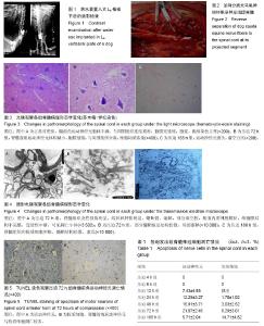
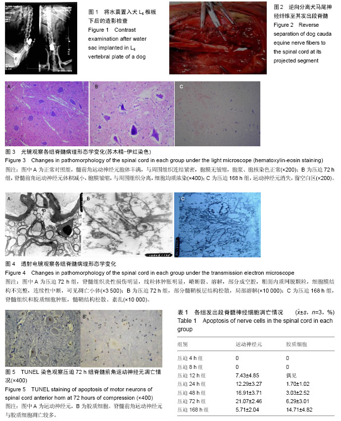
Pathological changes in the spinal cord of a model of acute cauda equina compression

Wang Zhan1, Li Hao-peng2, He Xi-jing2, Hao Ding-jun3, Zhang Kun1, Chen Ming-xia4, Lei Ting5   

  1. 1Department of Orthopedic Trauma, 3Department of Spine, Xi’an Honghui Hospital, Health Science Center, Xi’an Jiaotong University, Xi’an 710054, Shaanxi Province, China; 2Second Department of Orthopedics, the Second Clinical College of Xi’an Jiaotong University, Xi’an 710003, Shaanxi Province, China; 4Room of Electron Microscope, 5Department of Pathology, Health Science Center, Xi’an Jiaotong University, Xi’an 710061, Shaanxi Province, China
  • Revised:2016-08-07 Online:2016-09-30 Published:2016-09-30
  • About author:Wang Zhan, Master, Associate chief physician, Department of Orthopedic Trauma, Xi’an Honghui Hospital, Health Science Center, Xi’an Jiaotong University, Xi’an 710054, Shaanxi Province, China
  • Supported by:

    the National Natural Science Foundation of China, No. 30271310

Abstract:

BACKGROUND: Animal studies have shown that cauda equina compression can induce apoptosis of lumbosacral spinal cord anterior horn motor neurons.

OBJECTIVE: To explore the pathological change in lumbosacral spinal cord after acute cauda equina compression in dogs.
METHODS: A total of 27 dogs were randomly divided into nine groups, with three dogs in each group. There were one normal control group, seven experimental groups and one sham surgery group. In the experimental group, an empty water sac was implanted above epidural fat below L6 vertebral plate. Compression was given by injecting water at 4, 8, 12, 24, 48, 72 and 168 hours. In the sham surgery group, an empty water sac was implanted, but compression was not given. At the time of compression, the spinal cord sent out by cauda equina nerve and adjacent to the head end was subjected to histopathological examination. 
RESULTS AND CONCLUSION: (1) Results of light microscope: at 4-48 hours of compression, spinal cord anterior horn motor neurons did not alter. At 72 hours, motor neurons became small, cell membrane shrank and separated from surrounding tissues. Cells were homogenous and darkly stained. At 168 hours, motor neurons disappeared, but spinal cord sections of the adjacent head end did not shown abnormal motor neurons in the spinal cord anterior horn. (2) Results of electron microscope: at 12 hours, spinal cord tissue began to swell, and the swelling aggravated with prolonged time of compression. The swelling of glial cells was apparent. At 168 hours, myelin sheath structure dissolved; axons showed vacuolization; axoplasm spilled, and exhibited inflammatory injury-like changes. (3) Apoptotic results of spinal cord anterior horn motor neurons: apoptosis appeared at 12 hours of compression, became increased, and showed an increased trend at 168 hours.

中国组织工程研究杂志出版内容重点:肾移植肝移植移植;心脏移植;组织移植;皮肤移植;皮瓣移植;血管移植;器官移植组织工程

Key words:  Models, Animal, Cauda Equina, Spinal Cord, Tissue Engineering

CLC Number: