Chinese Journal of Tissue Engineering Research ›› 2013, Vol. 17 ›› Issue (42): 7375-7381.doi: 10.3969/j.issn.2095-4344.2013.42.006
Previous Articles Next Articles
Insight into nano chitosan effects on MC3T3-E1 cell growth
Wang Li-ting1, 2, Zhou Gang1, Fan Yu-bo1
- 1School of Biology and Medical Engineering, Beihang University, Beijing 100191, China
2National Research Center for Rehabilitation Technical Aids, Beijing 100176, China
-
Received:2013-04-15Revised:2013-05-30Online:2013-10-15Published:2013-10-31 -
Contact:Fan Yu-bo, Professor, School of Biology and Medical Engineering, Beihang University, Beijing 100191, China yubofan@buaa.edu.cn -
About author:Wang Li-ting☆, Studying for doctorate, Assistant researcher, School of Biology and Medical Engineering, Beihang University, Beijing 100191, China; National Research Center for Rehabilitation Technical Aids, Beijing 100176, China wlt6301@sohu.com -
Supported by:the National Natural Science Foundation of China, No. 11120101001*, 10925208*
CLC Number:
Cite this article
Wang Li-ting, Zhou Gang, Fan Yu-bo . Insight into nano chitosan effects on MC3T3-E1 cell growth[J]. Chinese Journal of Tissue Engineering Research, 2013, 17(42): 7375-7381.
share this article

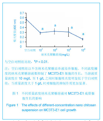
2.1 不同质量浓度材料对成骨细胞MC3T3-E1生长的影响 不同浓度梯度(10 mg/L、100 mg/L、1 g/L、10 g/L)的纳米壳聚糖溶液都抑制了MC3T3-E1细胞的生长,当溶液质量浓度在10 mg/L 至1 g/L之间时细胞吸光度明显低于空白对照组(P < 0.01);当质量浓度大于1 g/L时对细胞的抑制作用更加显著,见图1。"
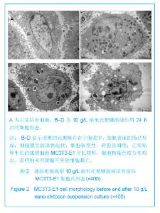
2.2 电镜观察纳米壳聚糖溶液对MC3T3-E1 成骨细胞形态 见图2。"
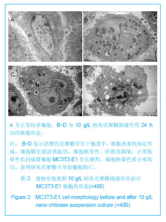
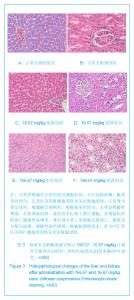
透射电镜被认为是鉴定细胞凋亡的金标准[20]。由于壳聚糖纳米颗粒存在团聚现象,图中可见团聚的壳聚糖存在于胞浆中,细胞表面的伪足形成,细胞膜呈波浪状起伏;细胞核变性、碎裂及固缩,见图2。正常贴壁生长的成骨细胞MC3T3-E1呈长梭形,细胞核染色质分布均匀,见图2A。 2.3 动物体内注射实验病理切片观察结果 各组动物肝肾组织病理变化见图3。"
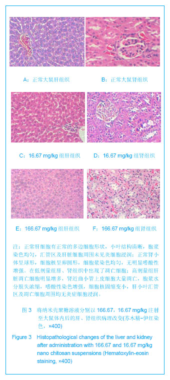
正常肝细胞有正常的多边细胞形状,小叶结构清晰,胞浆染色均匀,汇管区及肝脏细胞周围未见炎细胞浸润;正常肾小体呈球形,细胞核呈卵圆形,细胞浆染色均匀,无明显嗜酸性增强。 在两种不同剂量纳米壳聚糖溶液作用下的大鼠病理组织中观察到有凋亡细胞出现。在低剂量组肝、肾组织中出现了凋亡细胞。高剂量组肝脏凋亡细胞明显增多,肾近曲小管上皮细胞大量凋亡,胞浆水分脱失浓缩,嗜酸性染色增强,细胞核固缩变小,肝小叶汇管区及凋亡细胞周围均无炎症细胞浸润。 2.4 动物体内注射实验血清生化指标结果 各组动物血清生化指标比较见图4。"
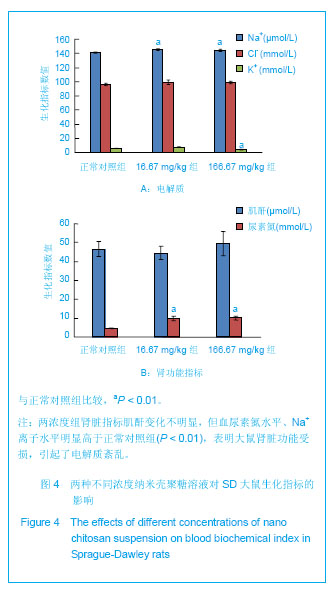

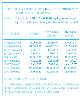
虽然结果显示高剂量组和低剂量组谷草转氨酶、谷丙转氨酶水平均低于正常对照组,但由于大鼠的正常范围是96-200 U,谷草转氨酶数据变化无临床意义。肾脏指标肌酐变化不明显,但血尿素氮水平,在高剂量组和低剂量组相比于正常对照组却有明显升高(P < 0.01)。反映身体电解质平衡的K+、Na+离子水平,在两个处理组也有明显变化,见表1。"
| [1] 蒋挺大.甲壳素[M].北京:化学工业出版社,2001: 15-19. [2] Synowiecki J,Al-Khateeb NA.Production, properties, and some new applications of chitin and its derivatives .Crit Rev Food Sci Nutr.2003; 43: 145-171. [3] Enríquez de Salamanca A,Diebold Y,Calonge M,et al. Chitosan nanoparticles as a potential drug delivery system for the ocular surface toxicity, uptake mechanism and in vivo tolerance.Invest Ophth Vis Sci.2006;47(4): 1416-1425. [4] Zheng LY,Zhu JF.Study on antimicrobial activity of chitosan with different molecular weights.Carbohyd Polym.2003;54: 527-530. [5] Yasunori M,Yoshiyuki K. Antitumor effects of various low-molecular-weight chitosans are due to increased natural killer activity of intestinal intraepithelial lymphocytes in sarcoma 180-bearing mice.J Nutr.2004; 134(4):945-950. [6] Xie Y,Zhou NJ,Gong YF,et al.Th immune response induced by H pylori vaccine with chitosan as adjuvant and its relation to immune protection.World J Gastroenterol.2007;13(10): 1547-1553. [7] Yuan XB,Yuan YB,Jiang W,et al.Preparation of rapamycin-loaded chitosan/PLA nanoparticles for immunosuppression in corneal transplantation.Int J Pharm. 2008;349(1-2): 241-248. [8] Khatri K,Goyal AK,Gupta PN,et al.Plasmid DNA loaded chitosan nanoparticles for nasal mucosal immunization against hepatitis B.Int J Pharm.2008; 354(1-2): 235-241. [9] Dailey RA,Chavez MR,Choi D.Use of a chitosan-based hemostatic dressing in dacryocystorhinostomy. Ophthal Plast Reconstr Surg.2009; 25(5): 350-353. [10] 刘昊,张永刚,郭全义,等.新型脱细胞骨基质-壳聚糖骨组织工程支架的制备及性能评价[J].军医进修学院学报,2011,32(6): 616-619. [11] 刘巍,朱新辉,张洁,等.Matrigel凝胶和壳聚糖/磷酸甘油凝胶两种支架体外构建组织工程软骨的比较[J].江苏医药,2011,37(6): 627-629. [12] Madhumathi K,Shalumon KT,Rani VV,et al. Wet chemical synthesis of chitosan hydrogel-hydroxyapatite composite membranes for tissue engineering applications.Int J Biol Macromol.2009;45(1):12-15. [13] Christenson EM,Anseth KS,van den Beucken JJ,et al. Nanobiomaterial applications in orthopedics.J Orthop Res.2007;25(1):11-22. [14] Qin F,Ye Y,Yao X,et al.Effects of nano-selenium on the capability of learning memory and the activity of Se-protein of mice. Wei Sheng Yan Jiu. 2008; 37(4):502-504. [15] Song S,Zhou F,Nordquist RE,et al.Glycated chitosan as a new non-toxic immunological stimulant.Immunopharmacol Immunotoxicol.2009;31(2):202-208. [16] Zhang C,Qu G,Sun Y,et al.Biological evaluation of N-octyl-O-sulfate chitosan as a new nano-carrier of intravenous drugs. Eur J Pharm Sci.2008;33 (4-5):415-423. [17] Nasti A,Zaki NM,de Leonardis P,et al.Chitosan/TPP and chitosan/TPP-hyaluronic acid nanoparticles: systematic optimisation of the preparative process and preliminary biological evaluation. Pharm Res. 2009; 26(8):1918-1930. [18] Kashiwazaki H,Kishiya Y,Matsuda A,et al.Fabrication of porous chitosan/hydroxyapatite nanocomposites: Their mechanical and biological properties. Biomed Mater Eng. 2009;19 (2):133-140. [19] Yoksan R,Chirachanchai S.Amphiphilic chitosan nanosphere: Studies on formation, toxicity, and guest molecule incorporation. Bioorg Med Chem. 2008; 16(5):2687-2696. [20] Elmore S.Apoptosis: A Review of Programmed Cell Death. Toxicol Pathol. 2007;35: 4495-516. [21] Mao HQ,Roy K,Troung-Le VL,et al.Chitosan-DNA nanoparticles as gene carriers:synthesis, characterization and transfection efficiency.J Controlled Release.2001; 70(3): 399-421. [22] Qi LF,Xu ZR,Jiang X,et al.Cytotoxic activities of chitosan nano particles and copper- loaded nanoparticles.Bioorg Med Chem Lett.2005;15(5):1397-1399. [23] 汪冰,丰伟悦,赵字亮,等.纳米材料生物效应及其毒理学研究进展[J].中国科学B辑,2005,35(1): 1-10. [24] Wang B,Feng WY,Zhu MT,et al.Neurotoxicity of low-dose repeatedly intranasal instillation of nano- and submicron-sized ferric oxide particles in mice.J Nanopart Res.2009;11: 41-53. [25] Inoue K,Takano H,Yanagisawa R,et al.Size effects of latex nanomaterials on lung inflammation in mice.Toxicol Appl Pharmacol.2009;234 (1):68-76. [26] Inoue K,Takano H,Koike E,et al.Effects of pulmonary exposure to carbon nanotubes on lung and systemic inflammation with coagulatory disturbance induced by lipopolysaccharide in mice.Exp Biol Med.2008;233(12): 1583-1590. [27] Wang B,Feng WY,Wang TC,et al.Acute toxicity of nano- and micro-scale zinc powder in healthy adult mice.Toxicol Lett. 2006;161(2):115-123. [28] Wei SL,Yi X,Chuan CH,et al.Toxicity of nano- and micro-sized ZnO particles in human lung epithelial cells.J Nanopart Res. 2009;11: 25-39. [29] Meirelles L,Melin L,Peltola T.Effect of hydroxyapatite and titania nanostructures on early in vivo bone response.Clin Implant Dent Relat Res. 2008; 10(4):245-54. [30] 彭黎明,王曾里.细胞凋亡的基础与临床[M].北京:人民卫生出版社,2003:363. [31] Awasthi KK,John PJ,Awasthi A,et al. Multi walled carbon nano tubes induced hepatotoxicity in Swiss albino mice. Micron. 2013;44: 359-364. [32] 陈伙德,贾振斌,邱敏,等.纳米材料在医药领域中的应用与展望[J].广东化工,2008,35(10):93-95. [33] Griffitt RJ,Hyndman K,Denslow ND,et al.Comparison of Molecular and Histological Changes in Zebrafish Gills Exposed to Metallic Nanoparticles. Toxicol Sci.2009;107(2): 404-415. [34] Watari F,Abe S,Koyama C,et al.Behavior of in vitro, in vivo and internal motion of micro/nano particles of titanium, titanium oxides and others.J Ceram Soc Jpn.2008; 116(1349): 1-5. [35] Nel A,Xia T,Madler L,et al.Toxic Potential of Materials at the Nanolevel. Science. 2006;(311):622-627. [36] Khang D,Liu-Snyder P,Pareta R,et al.Reduced responses of macrophages on nanometer surface features of altered alumina crystalline phases.Acta Biomater. 2009; 5(5): 1425-1432. [37] Waters KM,Masiello LM,Zangar RC,et al.Macrophage Responses to Silica Nanoparticles are Highly Conserved Across Particle Sizes.Toxicol Sci.2009; 107(2):553-569. [38] Jin CH,Jin YH,Wang J,et al.Comparative study of the effect on oxidative damage in rats inhaled by nano-sized and micro-sized silicon dioxide. Wei Sheng Yan Jiu.2008;37(1): 16-8,36. [39] Li R,Ning Z,Cui J,et al.Ultrafine particles from diesel engines induce vascular oxidative stress via JNK activation.Free Radic Biol Med.2009; 46(6):775-782. [40] Barmo C,Ciacci C,Canonico B,et al.In vivo effects of n-TiO(2) on digestive gland and immune function of the marine bivalve Mytilus galloprovincialis. Aquat Toxicol.2013;132-133C:9-18. [41] Min KY,Se WP.Exposing Zebrafish to Silver Nanoparticles during Caudal Fin Regeneration Disrupts Caudal Fin Growth and p53 Signaling.Mol Cell Toxicol.2008;4(4):311-317. [42] Park MR,Gurunathan S,Choi YJ,et al.Chitosan nanoparticles cause pre- and postimplantation embryo complications in mice.Biol Reprod.2013;88(4):88. [43] Yin N,Liu Q,Liu J,et al.Silver Nanoparticle Exposure Attenuates the Viability of Rat Cerebellum Granule Cells through Apoptosis Coupled to Oxidative Stress. Small. 2013; 9(9-10):1831-1841. [44] Xu P,Xu J,Liu S,et al.Nano copper induced apoptosis in podocytes via increasing oxidative stress.J Hazard Mater. 2012;30 (241):279-286. [45] 胡瑜兰,王淇,高建青,等.壳聚糖纳米粒的斑马鱼胚胎毒性及其机制研究[C].2010年中国药学大会暨第十届中国药师周论文集 [46] 黄琴琴,王永禄,李学明.纳米载体材料毒理学效应及其作用机制研究进展[J].中国药房,2011,22(21): 2004-2007. [47] 纪宗斐,张丹瑛,沈锡,等.碳纳米管的毒性研究进展[J].复旦学报:医学版,2011; 38(6): 556-559. [48] Klaus U,Catrin A,Lars-0liver K,et al.Cellular response to nanoparticles: target structures and mechanisms. Nanotoxicology. 2007;1(1): 52-71. [49] 周国强,陈春英,李玉锋,等.纳米材料生物效应研究进展[J].生物化学与生物物理进展,2008,35(9): 998-1006. [50] 韩雁,崔国权,董淑英,等.纳米氧化锌诱导血管内皮细胞凋亡及氧化应激[J].中国公共卫生,2012,28(4):94-96. |
| [1] | Wang Jing, Xiong Shan, Cao Jin, Feng Linwei, Wang Xin. Role and mechanism of interleukin-3 in bone metabolism [J]. Chinese Journal of Tissue Engineering Research, 2022, 26(8): 1260-1265. |
| [2] | Xiao Hao, Liu Jing, Zhou Jun. Research progress of pulsed electromagnetic field in the treatment of postmenopausal osteoporosis [J]. Chinese Journal of Tissue Engineering Research, 2022, 26(8): 1266-1271. |
| [3] | Le Guoping, Zhang Ming, Xi Licheng, Luo Hanwen. Preparation and in vitro evaluation of vancomycin hydrochloride@polylactic acid-glycolic acid copolymer-chitosan-hyaluronic acid composite sustained-release microspheres [J]. Chinese Journal of Tissue Engineering Research, 2022, 26(4): 528-534. |
| [4] | Cai Shengsheng, Mei Heng, Zhang Xuequan, Deng Jin, Cao Jun, He Bin. Prepared HPe6DF composite nanoparticles enhance the effect of photodynamic therapy [J]. Chinese Journal of Tissue Engineering Research, 2022, 26(10): 1566-1573. |
| [5] | Ye Xiangling, Xia Yuanjun, Wang Boqun, Kang Zhengyang, Wu Bin. Function on 3D printing poly(3-hydroxybutyrate-co-3-hydroxyvalerate)/calcium sulfate hemihydrate scaffold integrated chitosan hydrogel coating [J]. Chinese Journal of Tissue Engineering Research, 2022, 26(10): 1574-1581. |
| [6] | Yang Bingxuan, Jiang Tao, Jia Min, Li Peng, Liao Rongzhen, Li Zhao, Shao Min. Icariin promotes differentiation of mouse preosteoblasts via activating autophagy [J]. Chinese Journal of Tissue Engineering Research, 2022, 26(1): 64-69. |
| [7] | Huang Tao, Cheng Zhijian, Jia Zhiqiang, Zhao Xiaoguang, Wang Lei, Zhai Wenjing, Zhou Yongxin. Mechanism by which miR-146a regulates osteogenic differentiation of adipose derived mesenchymal stem cells [J]. Chinese Journal of Tissue Engineering Research, 2022, 26(1): 70-75. |
| [8] | Feng Zhiguo, Sun Haibiao, Han Xiaoqiang. Regulation of proliferation, differentiation and apoptosis of bone-related cells by long-stranded non-coding RNA [J]. Chinese Journal of Tissue Engineering Research, 2022, 26(1): 112-118. |
| [9] | Li Li, Ma Li. Immobilization of lactase on magnetic chitosan microspheres and its effect on enzymatic properties [J]. Chinese Journal of Tissue Engineering Research, 2021, 25(4): 576-581. |
| [10] | Wang Jing, Lang Xuemei, Wang Weiqun, Zhang Hanxiang, Zhang Yi, Wang Xin. Participation and regulatory mechanism of interleukin-1 during bone metabolism [J]. Chinese Journal of Tissue Engineering Research, 2021, 25(36): 5851-5858. |
| [11] | Zhang Zhiwen, Huang Yuliang, Zhang Lixuan, Wang Xiaofeng, Chen Ruixiong. Acellular bone matrix/chitosan scaffold combined with basic fibroblast growth factor for repairing bone defects [J]. Chinese Journal of Tissue Engineering Research, 2021, 25(34): 5439-5444. |
| [12] | Li Ye, Yang Yukun, Zhu Xiangqing, He Jie, Wang Jinxiang, Wang Yanying, Tian Chuan, Pang Rongqing, Pan Xinghua. In vivo track technique for mesenchymal stem cells: how to realize simultaneous tracing of distribution and survival [J]. Chinese Journal of Tissue Engineering Research, 2021, 25(31): 5025-5033. |
| [13] | Zhang Mingjin, Zhou Yanheng, Liu Dawei. Reopening the fused suture provides a new idea for maxillary expansion [J]. Chinese Journal of Tissue Engineering Research, 2021, 25(29): 4721-4727. |
| [14] | Li Rui. Biological characteristics of hydroxyapatite/chitosan combined with metformin for bone defect in rats [J]. Chinese Journal of Tissue Engineering Research, 2021, 25(28): 4460-4464. |
| [15] | Li Ruozhen, Tian Yaping, Wen Ning. Biological effect of magnetic fields to promote bone responses to biomaterials [J]. Chinese Journal of Tissue Engineering Research, 2021, 25(28): 4510-4515. |
| Viewed | ||||||
|
Full text |
|
|||||
|
Abstract |
|
|||||