Chinese Journal of Tissue Engineering Research ›› 2022, Vol. 26 ›› Issue (1): 70-75.doi: 10.12307/2022.012
Previous Articles Next Articles
Mechanism by which miR-146a regulates osteogenic differentiation of adipose derived mesenchymal stem cells
Huang Tao1, Cheng Zhijian2, Jia Zhiqiang3, Zhao Xiaoguang1, Wang Lei1, Zhai Wenjing2, Zhou Yongxin1
- 1First Affiliated Hospital of Xi’an Medical University, Xi’an 710077, Shaanxi Province, China; 2Second Affiliated Hospital of Xi’an Jiaotong University, Xi’an 710061, Shaanxi Province, China; 3Second Affiliated Hospital of Henan University of Science and Technology, Luoyang 471000, Henan Province, China
-
Received:2020-08-20Revised:2020-08-22Accepted:2020-10-30Online:2022-01-08Published:2021-10-25 -
Contact:Zhou Yongxin, Master, Attending physician, First Affiliated Hospital of Xi’an Medical University, Xi’an 710077, Shaanxi Province, China -
About author:Huang Tao, Master, Attending physician, First Affiliated Hospital of Xi’an Medical University, Xi’an 710077, Shaanxi Province, China -
Supported by:the National Natural Science Foundation of China, No. 81801237 (to CZJ)
CLC Number:
Cite this article
Huang Tao, Cheng Zhijian, Jia Zhiqiang, Zhao Xiaoguang, Wang Lei, Zhai Wenjing, Zhou Yongxin. Mechanism by which miR-146a regulates osteogenic differentiation of adipose derived mesenchymal stem cells[J]. Chinese Journal of Tissue Engineering Research, 2022, 26(1): 70-75.
share this article
Add to citation manager EndNote|Reference Manager|ProCite|BibTeX|RefWorks
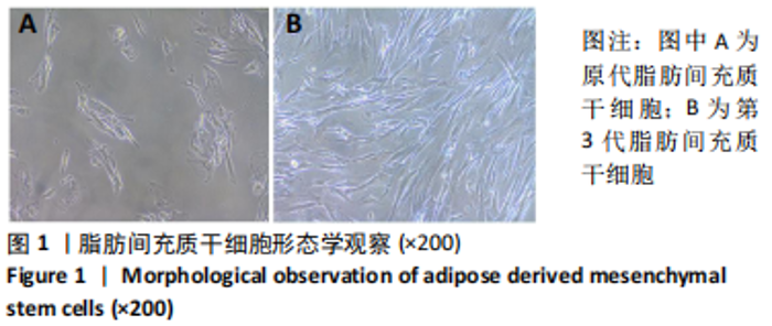
2.1 脂肪间充质干细胞的形态及鉴定结果 原代分离培养的小鼠脂肪间充质干细胞初为小圆形,细胞大小不均匀。培养24 h后可见细胞基本已经完全贴壁,贴壁细胞呈成纤维细胞形态;48 h后换液去除非贴壁细胞,此时细胞已生长出触角,呈梭形或三角形,见图1A;五六天后细胞基本达到70%-80%融合,细胞呈成纤维样生长,形态一致。传代后细胞呈典型成纤维细胞样生长,具有一定的方向性。当传至第3代时,贴壁细胞形态均一,大部分呈梭形,细胞排列呈漩涡状且增殖速度快,见图1B。"
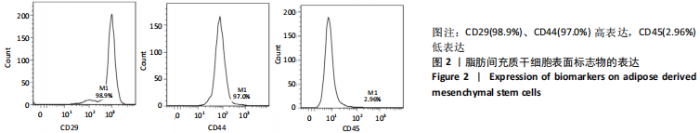
细胞传代纯化后取第3代小鼠脂肪间充质干细胞进行流式检测CD29、CD44、CD45等标志物的表达,结果显示:CD29(98.9%)、CD44(97.0%)高表达,CD45(2.96%)低表达,具有间充质干细胞特性,见图2。"
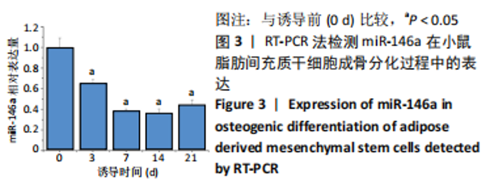
2.2 miR-146a在脂肪间充质干细胞向成骨分化过程中的表达量变化 采用RT-PCR方法检测脂肪间充质干细胞成骨诱导第0,3,7,14,21天时miR-146a的mRNA表达。与第0天相比,在第3天时miR-146a表达下降约37%,之后继续下降,并在第14天时达到最低,虽然之后miR-146a的表达有所回升,但其表达量仍低于诱导之前(P < 0.05),见图3。"


2.3 miR-146a对脂肪间充质干细胞成骨分化的影响 成骨诱导第6天,采用RT-PCR方法检测转染miR-NC、miR-146a mimic和miR-146a inhibitor之后各组细胞miR-146a mRNA表达变化,结果发现与miR-NC组相比,miR-146a mimic组细胞miR-146a的mRNA表达显著升高,miR-146a inhibitor组细胞miR-146a的mRNA表达显著减低(P < 0.05),见图4A。采用碱性磷酸酶染色比较各组细胞成骨分化程度,结果发现miR-146a mimic组显色较miR-NC组和miR-146a inhibitor组明显减弱,而miR-146a inhibitor组与miR-NC组相比则显色增强,见图4B。碱性磷酸酶活性检测结果显示,与miR-NC组相比,miR-146a mimic组碱性磷酸酶表达活性显著降低,miR-146a inhibitor组碱性磷酸酶活性显著上升(P < 0.05),见图4C。在成骨诱导后的第12天,采用茜素红染色鉴定细胞表面矿化基质的产生,与miR-NC组相比,miR-146a mimic组细胞的钙化程度显著降低,而miR-146a inhibitor组细胞的钙化程度显著升高(P < 0.05),见图4D。"


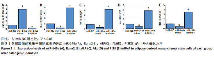
2.4 RT-PCR检测各组小鼠脂肪间充质干细胞成骨诱导后miR-146a、Runx2、ALP、AKT、PI3K的mRNA相对表达量 与miR-NC组相比,miR-146a mimic组细胞miR-146a的mRNA表达水平显著升高,miR-146a inhibitor组显著降低(P < 0.05),见图5A。与miR-NC组相比,miR-146a mimic组细胞Runx2、ALP、AKT和PI3K的mRNA表达水平显著降低,miR-146a inhibitor组显著升高(P < 0.05),见图5B-E。"

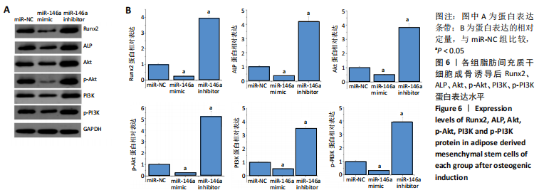
2.5 Western blot检测各组小鼠脂肪间充质干细胞成骨诱导后Runx2、ALP、Akt、p-Akt、PI3K、p-PI3K的蛋白表达 与miR-NC组相比,Runx2、ALP、Akt、p-Akt、PI3K和p-PI3K的蛋白表达水平在miR-146a mimic组显著降低,而在miR-146a inhibitor组显著升高(P < 0.05),见图6。"
| [1] KEENEY M, CHUNG MT, ZIELINS ER, et al. Scaffold-mediated BMP-2 minicircle DNA delivery accelerated bone repair in a mouse critical-size calvarial defect model. J Biomed Mater Res A. 2016;104(8):2099-2107. [2] 兰伟伟,陈维毅,黄棣.骨软骨组织工程研究进展[J].生物医学工程学杂志,2019,36(3):504-510. [3] ATASOY-ZEYBEK A, KOSE GT. Gene Therapy Strategies in Bone Tissue Engineering and Current Clinical Applications. Adv Exp Med Biol. 2018; 1119: 85-101. [4] 张爽,刘石,汪宇峰,等.间充质干细胞修复骨关节炎软骨损伤的临床应用意义[J].中国组织工程研究,2018,22(33):5379-5385. [5] DAI R, WANG Z, SAMANIPOUR R, et al. Adipose-Derived Stem Cells for Tissue Engineering and Regenerative Medicine Applications. Stem Cells Int. 2016;2016:6737345. [6] HAO W, LIU H, ZHOU L, et al. MiR-145 regulates osteogenic differentiation of human adipose-derived mesenchymal stem cells through targeting FoxO1. Exp Biol Med (Maywood). 2018;243(4):386-393. [7] ZHANG Z, MA Y, GUO S, et al. Low-intensity pulsed ultrasound stimulation facilitates in vitro osteogenic differentiation of human adipose-derived stem cells via up-regulation of heat shock protein (HSP)70, HSP90, and bone morphogenetic protein (BMP) signaling pathway. Biosci Rep. 2018; 38(3):BSR20180087. [8] UDDIN A, CHAKRABORTY S. Role of miRNAs in lung cancer. J Cell Physiol. 2018 Apr 20. doi: 10.1002/jcp.26607. [9] LOPEZ-BERTONI H, KOZIELSKI KL, RUI Y, et al. Bioreducible Polymeric Nanoparticles Containing Multiplexed Cancer Stem Cell Regulating miRNAs Inhibit Glioblastoma Growth and Prolong Survival. Nano Lett. 2018;18(7): 4086-4094. [10] HUANG S, WANG S, BIAN C, et al. Upregulation of miR-22 promotes osteogenic differentiation and inhibits adipogenic differentiation of human adipose tissue-derived mesenchymal stem cells by repressing HDAC6 protein expression. Stem Cells Dev. 2012;21(13):2531-2540. [11] QURESHI AT, MONROE WT, DASA V, et al. miR-148b-nanoparticle conjugates for light mediated osteogenesis of human adipose stromal/stem cells. Biomaterials. 2013;34(31):7799-7810. [12] WANG Z, XIE Q, YU Z, et al. A regulatory loop containing miR-26a, GSK3β and C/EBPα regulates the osteogenesis of human adipose-derived mesenchymal stem cells. Sci Rep. 2015;5:15280. [13] ZHANG WB, ZHONG WJ, WANG L. A signal-amplification circuit between miR-218 and Wnt/β-catenin signal promotes human adipose tissue-derived stem cells osteogenic differentiation. Bone. 2014;58:59-66. [14] HAO X, XIA L, QU R, et al. Association between miR-146a rs2910164 polymorphism and specific cancer susceptibility: an updated meta-analysis. Fam Cancer. 2018;17(3):459-468. [15] 匡威,谭家莉,张红梅,等.miR-146a调控骨髓间充质干细胞成骨分化的机制研究[J].生物医学工程与临床,2011,15(5):413-416. [16] MA M, WANG X, CHEN X, et al. MicroRNA-432 targeting E2F3 and P55PIK inhibits myogenesis through PI3K/AKT/mTOR signaling pathway. RNA Biol. 2017;14(3):347-360. [17] LUO G, XU B, HUANG Y. Icariside II promotes the osteogenic differentiation of canine bone marrow mesenchymal stem cells via the PI3K/AKT/mTOR/S6K1 signaling pathways. Am J Transl Res. 2017;9(5):2077-2087. [18] 李洋,陈前昭,邵英,等.骨形态蛋白9诱导干细胞骨向分化与环氧酶-2及PI3K/Akt的关系研究[J].中国药理学通报,2017,33(7): 908-915. [19] 支力强,杨一欣,许毛,等.SIRT1通过PI3K/AKT通路促进成骨细胞分化的相关机制研究[J].实用骨科杂志,2018,24(6):523-526. [20] 王雪鹏,李茂强,边振宇,等. PI3K/Akt信号通路在骨髓间充质干细胞增殖及成骨分化调控中的作用[J].中华骨质疏松和骨矿盐疾病杂志,2014,7(3):250-257. [21] LI J, HU C, HAN L, et al. MiR-154-5p regulates osteogenic differentiation of adipose-derived mesenchymal stem cells under tensile stress through the Wnt/PCP pathway by targeting Wnt11. Bone. 2015;78: 130-141. [22] LANGER R, VACANTI JP. Tissue engineering. Science. 1993;260(5110): 920-926. [23] BAJEK A, GURTOWSKA N, OLKOWSKA J, et al. Adipose-Derived Stem Cells as a Tool in Cell-Based Therapies. Arch Immunol Ther Exp (Warsz). 2016;64(6):443-454. [24] CHEN QJ, CHEN L, WU SK, et al. rhPDGF-BB combined with ADSCs in the treatment of Achilles tendinitis via miR-363/PI3 K/Akt pathway. Mol Cell Biochem. 2018;438(1-2):175-182. [25] KRZESNIAK NE, SARNOWSKA A, FIGIEL-DABROWSKA A, et al. Secondary release of the peripheral nerve with autologous fat derivates benefits for functional and sensory recovery. Neural Regen Res. 2021;16(5):856-864. [26] 刘润恒,刘于冬,陈卓凡.微小RNA在骨分化过程中的作用机制[J].国际口腔医学杂志,2017,44(1):108-113. [27] QIAO L, LIU D, LI CG, et al. MiR-203 is essential for the shift from osteogenic differentiation to adipogenic differentiation of mesenchymal stem cells in postmenopausal osteoporosis. Eur Rev Med Pharmacol Sci. 2018;22(18):5804-5814. [28] KIM YJ, BAE SW, YU SS, et al. miR-196a regulates proliferation and osteogenic differentiation in mesenchymal stem cells derived from human adipose tissue. J Bone Miner Res. 2009;24(5):816-825. [29] LACCI KM, DARDIK A. Platelet-rich plasma: support for its use in wound healing. Yale J Biol Med. 2010;83(1):1-9. [30] FKIH M’HAMED I, PRIVAT M, TRIMECHE M, et al. miR-10b, miR-26a, miR-146a And miR-153 Expression in Triple Negative Vs Non Triple Negative Breast Cancer: Potential Biomarkers. Pathol Oncol Res. 2017; 23(4):815-827. [31] DONG Z, YU C, REZHIYA K, et al. Downregulation of miR-146a promotes tumorigenesis of cervical cancer stem cells via VEGF/CDC42/PAK1 signaling pathway. Artif Cells Nanomed Biotechnol. 2019;47(1): 3711-3719. [32] HAN Z, CAO J, WANG Y, et al. Quercetin Suppresses Proliferation and Motility Through Modulating Hippo Pathway Via Upregulating Mir-146a-5p in Gastric Cancer. J Biomaterials Tissue Engineering. 2019;9:82-88. [33] KIM DY, KIM EJ, JANG WG. Piperine induces osteoblast differentiation through AMPK-dependent Runx2 expression. Biochem Biophys Res Commun. 2018;495(1):1497-1502. [34] NARAYANAN A, SRINAATH N, ROHINI M, et al. Regulation of Runx2 by MicroRNAs in osteoblast differentiation. Life Sci. 2019;232:116676. [35] KHALID AB, SLAYDEN AV, KUMPATI J, et al. GATA4 Directly Regulates Runx2 Expression and Osteoblast Differentiation. JBMR Plus. 2018; 2(2):81-91. [36] COSTA RLB, HAN HS, GRADISHAR WJ. Targeting the PI3K/AKT/mTOR pathway in triple-negative breast cancer: a review. Breast Cancer Res Treat. 2018;169(3):397-406. [37] PORTA C, PAGLINO C, MOSCA A. Targeting PI3K/Akt/mTOR Signaling in Cancer. Front Oncol. 2014;4:64. [38] ZHANG Y, YANG JH. Activation of the PI3K/Akt pathway by oxidative stress mediates high glucose-induced increase of adipogenic differentiation in primary rat osteoblasts. J Cell Biochem. 2013;114(11): 2595-2602. |
| [1] | Wang Baojuan, Zheng Shuguang, Zhang Qi, Li Tianyang. Miao medicine fumigation can delay extracellular matrix destruction in a rabbit model of knee osteoarthritis [J]. Chinese Journal of Tissue Engineering Research, 2022, 26(8): 1236-1242. |
| [2] | Wang Jing, Xiong Shan, Cao Jin, Feng Linwei, Wang Xin. Role and mechanism of interleukin-3 in bone metabolism [J]. Chinese Journal of Tissue Engineering Research, 2022, 26(8): 1316-1322. |
| [3] | Xiao Hao, Liu Jing, Zhou Jun. Research progress of pulsed electromagnetic field in the treatment of postmenopausal osteoporosis [J]. Chinese Journal of Tissue Engineering Research, 2022, 26(8): 1323-1329. |
| [4] | An Weizheng, He Xiao, Ren Shuai, Liu Jianyu. Potential of muscle-derived stem cells in peripheral nerve regeneration [J]. Chinese Journal of Tissue Engineering Research, 2022, 26(7): 1183-1190. |
| [5] | Fan Yiming, Liu Fangyu, Zhang Hongyu, Li Shuai, Wang Yansong. Serial questions about endogenous neural stem cell response in the ependymal zone after spinal cord injury [J]. Chinese Journal of Tissue Engineering Research, 2022, 26(7): 1191-1197. |
| [6] | Gao Yujin, Peng Shuanglin, Ma Zhichao, Lu Shi, Cao Huayue, Wang Lang, Xiao Jingang. Osteogenic ability of adipose stem cells in diabetic osteoporosis mice [J]. Chinese Journal of Tissue Engineering Research, 2022, 26(7): 1047-1052. |
| [7] | Hou Jingying, Guo Tianzhu, Yu Menglei, Long Huibao, Wu Hao. Hypoxia preconditioning targets and downregulates miR-195 and promotes bone marrow mesenchymal stem cell survival and pro-angiogenic potential by activating MALAT1 [J]. Chinese Journal of Tissue Engineering Research, 2022, 26(7): 1053-1059. |
| [8] | Zhou Ying, Zhang Huan, Liao Song, Hu Fanqi, Yi Jing, Liu Yubin, Jin Jide. Immunomodulatory effects of deferoxamine and interferon gamma on human dental pulp stem cells [J]. Chinese Journal of Tissue Engineering Research, 2022, 26(7): 1060-1067. |
| [9] | Tian Chuan, Zhu Xiangqing, Yang Zailing, Yan Donghai, Li Ye, Wang Yanying, Yang Yukun, He Jie, Lü Guanke, Cai Xuemin, Shu Liping, He Zhixu, Pan Xinghua. Bone marrow mesenchymal stem cells regulate ovarian aging in macaques [J]. Chinese Journal of Tissue Engineering Research, 2022, 26(7): 1033-1039. |
| [10] | Liang Xuezhen, Yang Xi, Li Jiacheng, Luo Di, Xu Bo, Li Gang. Bushen Huoxue capsule regulates osteogenic and adipogenic differentiation of rat bone marrow mesenchymal stem cells via Hedgehog signaling pathway [J]. Chinese Journal of Tissue Engineering Research, 2022, 26(7): 1068-1074. |
| [11] | Wang Jifang, Bao Zhen, Qiao Yahong. miR-206 regulates EVI1 gene expression and cell biological behavior in stem cells of small cell lung cancer [J]. Chinese Journal of Tissue Engineering Research, 2022, 26(7): 1075-1079. |
| [12] | Liu Feng, Peng Yuhuan, Luo Liangping, Wu Benqing. Plant-derived basic fibroblast growth factor maintains the growth and differentiation of human embryonic stem cells [J]. Chinese Journal of Tissue Engineering Research, 2022, 26(7): 1080-1085. |
| [13] | Wen Dandan, Li Qiang, Shen Caiqi, Ji Zhe, Jin Peisheng. Nocardia rubra cell wall skeleton for extemal use improves the viability of adipogenic mesenchymal stem cells and promotes diabetes wound repair [J]. Chinese Journal of Tissue Engineering Research, 2022, 26(7): 1086-1092. |
| [14] | Zhu Bingbing, Deng Jianghua, Chen Jingjing, Mu Xiaoling. Interleukin-8 receptor enhances the migration and adhesion of umbilical cord mesenchymal stem cells to injured endothelium [J]. Chinese Journal of Tissue Engineering Research, 2022, 26(7): 1093-1098. |
| [15] | Luo Xiaoling, Zhang Li, Yang Maohua, Xu Jie, Xu Xiaomei. Effect of naringenin on osteogenic differentiation of human periodontal ligament stem cells [J]. Chinese Journal of Tissue Engineering Research, 2022, 26(7): 1099-1104. |
| Viewed | ||||||
|
Full text |
|
|||||
|
Abstract |
|
|||||