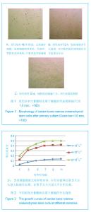
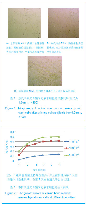
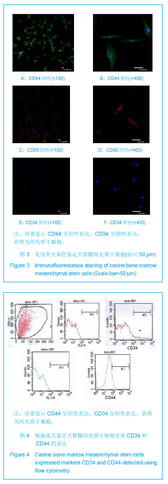
| [1] Lee HS,Huang GT,Chiang H,et al.Multipotential mesenchymal stem cells from femoral bone marrow near the site of osteonecrosis.Stem Cells.2003;21(2):190-199.
[2] Deng W,Han Q,Liao L,et al.Engrafted bone marrow-derived flk-(1+) mesenchymal stem cells regenerate skin tissue. Tissue Eng.2005;11(1-2):110-119.
[3] Oshima Y,Watanabe N,Matsuda K,et al.Behavior of transplanted bone marrow-derived GFP mesenchymal cells in osteochondral defect as a simulation of autologous transplantation.J Histochem Cytochem. 2005;53(2):207-216.
[4] Silva GV,Litovsky S,Assad JA,et al.Mesenchymal stem cells differentiate into an endothelial phenotype, enhance vascular density, and improve heart function in a canine chronic ischemia model.Circulation. 2005;111(2):150-156.
[5] 姚旺祥,裴国献,姚海燕.成体干细胞在骨组织工程中的应用[J].国际骨科学杂志,2006,27(1): 7-9.
[6] 中华医学会骨科分会显微修复学组及中国修复重建外科专业委员会骨缺损及骨坏死学组.成人股骨头坏死诊疗标准专家共识(2012年版)[J].中国骨与关节科,2012,5(2):185-192.
[7] Trotha R,Hanck T,Konig W,et al.Rapid ribesequencing- an effective diagnostic tool for detecting microbial infection.Infect. 2001;29:12-16.
[8] Pountos I,Corsadden D,Emery P,et al.Mesenchymal stem cell tissue engineering: techniques for isolation, expansion andapplication.Injury. 2007;38 Suppl 4: S23-S33.
[9] 曲彦隆,杨卫良,孟祥文,等.梯度降解软骨支架材料应力刺激下与骨髓基质干细胞复合三维培养的实验研究[J].中国矫形外科杂志,2006,14(10):772-774.
[10] 刘长安,王江泳,张卫平,等.骨髓基质干细胞移植治疗兔股骨头缺血性坏死的实验研究[J].中国矫形外科杂志,2007,15(1):58-60.
[11] Pan X,Xiao D,Zhang X,et al.Study of rotating permanent magnetic field to treat steroid-induced osteonecrosis of femoral head.Int Orthop. 2009;33(3):617-623.
[12] Rodríguez JP,González M,Ríos S,et al.Cytoskeletal organization of human mesenchymal stem cells (MSC) changes during their osteogenic differentiation.J Cell Biochem. 2004;93: 721-731.
[13] Oswald J,Boxberger S,Jorgensen B,et al.Mesenchymal stem cells can be differentiated into endothelial cells in vitro.Stem Cells.2004;22: 377-384.
[14] Asada T,Kushida T,Umeda M.Prevention of corticosteroid-induced osteonecrosis in rabbits by intra-bone marrow injection of autologous bone marrow cells. Rheumatology. 2008;47(5):591.
[15] Hernigou P,Beaujean F,Lambotte JC.Decrease in the mesenchymal stem cell pool in the proximal femur in corticosteroid-induced osteonecrosis. J Bone Joint Surg(Br). 1999;81:349-355.
[16] Hernigou P,Beaujean F.Treatment of osteonecrosis with autologous bone marrow grafting.Clin Orthop Relat Res. 2002; 405:14-23.
[17] Hernigou P,Poignard A,Manicom O,et al.The use of percutaneous autologous bone marrow transplantation in nonunion and avascular necrosis of bone.J Bone Joint Surg (Br).2005;87(7):896-902.
[18] Gangji V,Hauzeur JP.Treatment of osteonecrosis of the femoral head with implantation of autologous bone-marrow cells. Surgical technique.J Bone Joint Surg Am.2005;87 Suppl 1(Pt 1):106-112.
[19] Calder P,Revell P,Pearse MF.The extent of osteocyte death in osteonecrosis of the femoral head.J Bone Joint Surg Br. 2001; 83(3):419-422.
[20] Johnstone B,Yoo JU.Autologous mesenchym alprogenitor cells in articular cartilage repair.Clin Orthop.1999;(367 Suppl): S 156-162.
[21] Bobyn JD,Stackpool GJ,Hacking SA,et al.Characteristics of bone ingrowth and interface mechanics of a new porous tantalum biomaterial. J Bone Joint Surg Br.1999;81(5): 907-914.
[22] Hacking SA,Bobyn JD,Toh K,et al.Fibrous tissue ingrowth and attachment to porous tantalum.J Biomed Mater Res.2000; 52(4): 631-638. |