Chinese Journal of Tissue Engineering Research ›› 2013, Vol. 17 ›› Issue (40): 7084-7089.doi: 10.3969/j.issn.2095-4344.2013.40.010
Previous Articles Next Articles
Isoproterenol influence on stem/progenitor cells of submandibular glands: Proliferative number or capability?
Tang Yue-peng1, Huang Gui-lin2, Yao Li2, Zhang Ni-ni2, Yi Jie2
- 1 Medical School of Yongzhou Vocational Technical College, Yongzhou 425000, Hunan Province, China; 2 Department of Oral and Maxillofacial Surgery, Stomatological Hospital, Zunyi Medical College, Zunyi 563003, Guizhou Province, China
-
Online:2013-10-01Published:2013-10-31 -
Contact:Huang Gui-lin, M.D., Professor, Chief physician, Department of Oral and Maxillofacial Surgery, Stomatological Hospital, Zunyi Medical College, Zunyi 563003, Guizhou Province, China chaojiehuanghgl@163.com -
About author:Tang Yue-peng★, Master, Attending physician, Lecturer, Medical School of Yongzhou Vocational Technical College, Yongzhou 425000, Hunan Province, China yuepengt@sina.com -
Supported by:the Science and Technology Development Program of Guizhou Science and Technology Department, No. SY[2011]3016*; the Science and Technology Foundation of Guizhou Province, No. LKZ[2011]23*
CLC Number:
Cite this article
Tang Yue-peng, Huang Gui-lin, Yao Li, Zhang Ni-ni, Yi Jie. Isoproterenol influence on stem/progenitor cells of submandibular glands: Proliferative number or capability?[J]. Chinese Journal of Tissue Engineering Research, 2013, 17(40): 7084-7089.
share this article
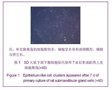
2.1 实验动物数量分析 16只SD大鼠随机分成异丙肾上腺素注射组和对照组,顺利建立实验模型,无大鼠死亡,每只大鼠下颌下腺经机械+酶消化后按400个/cm2密度接种6瓶进行原代细胞培养。 2.2 下颌下腺细胞的单克隆增殖 两组SD大鼠下颌下腺细胞原代培养48 h后,细胞开始分裂。培养第5天,可形成50多个细胞的单克隆集落,7 d后,单克隆集落的细胞数超过100个,甚至超过500个。细胞呈多角形或卵圆形,铺路石样生长,见图1。"

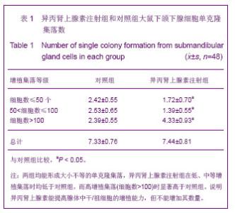
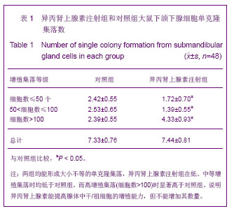
2.3 细胞集落分析 原代培养7 d后,异丙肾上腺素注射组与对照组均能形成大小不等的单克隆集落,按照集落内细胞数的多少进行分类计数,其结果见表1。"
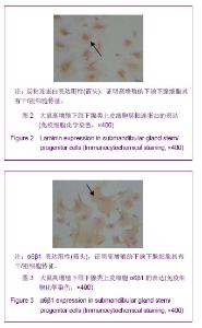
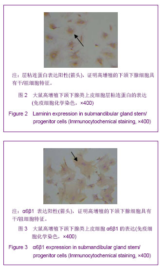
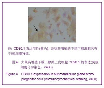
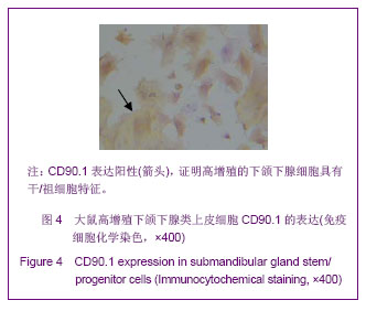
2.4 下颌下腺细胞爬片免疫细胞化学检测 将两组原代培养中细胞数超过500的集落分别选择性消化后接种到细胞爬片,待贴壁后,对爬片上的细胞进行免疫细胞化学检测。两组下颌下腺类上皮集落均表达层粘连蛋白、α6β1、CD90.1,见图2-4,两组表达程度一致。"
"
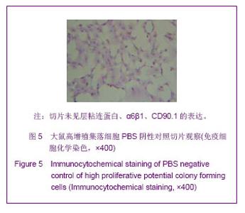
PBS对照阴性结果,见图5。"

| [1]Riva A, Puxeddu R, Loy F, et al. Serous and mucous cells of human submandibular salivary gland stimulated in vitro by isoproterenol, carbachol and clozapine: an LM, TEM, and HRSEM Study. Eur J Morphol. 2003;41(2):83-87. [2]Barka T. Induced cell proliferation:The effect of isoproterenol. Exp Cell Res. 1965;37:662-679. [3]Schneyer CA, Finley HW, Finley SC. Increased chromosome number of rat parotid cells after isoproterenol. Proc Soc Exp Biol Med.1967;125(3):722-728. [4]Radley JM. Ultrastructural changes in the rat submaxillary gland following isoprenaline. Z Zellforsch Mikosk Anat. 1969; 97(2):196-211. [5]Denny PC, Denny PA. DNA synthesis and cell division. In:Dobrosielski-Vergona K (ed) Biology of the salivary glands. CRC Press, Boca Raton. 1993:263-281. [6]Hand AR, Ho B.Mitosis and hypertrophy of intercalated duct cells and endothelial cells in the isoproterenol-treated rat parotid gland. J Dent Res. 1985;64(8):1031-1038. [7]Van den Brenk HA, Sparrow N, Moore V. Effect of X-ray irradiation on salivary gland growth in the rat. II. Quantitative studies on sialoadenotrophism induced with isopropy Lamininoradrenaline and its modification by single doses of X-rays. Int J Radiat Biol Relat Stud Phys Chem Med.1970; 17(2):135-161. [8]Katsumata O, Sato Y, Sakai Y, et al.Intercalated duct cells in the rat parotid gland may behave as tissue stem cells. Anat Sci Int. 2009;84(3):148-154. [9]Vugman I, Hand AR. Quantitative immunocytochemical study of secretory protein expression in parotid glands of rats chronically treated with isoproterenol. Microsc Res Tech. 1995; 31(2):106-117. [10]Nezu A, Morita T, Tanimura A, et al. Comparison of agonist-induced Ca2+ responses in rat submandibular acini and ducts. Arch Oral Biol.2005;50(6):585-592. [11]Lamey PJ, Ferguson MM, Marshall W. The growth and ductal epithelial cell response of mouse submandibular salivary glands to selected neurotransmitters in vitro. Arch Oral Biol. 1982;27(9):747-51. [12]黄桂林,李龙江,李学英,等.异丙肾上腺素对SD大鼠原代颌下腺细胞增殖的影响[J].实用口腔医学杂志,2005,21(5):618- 620. [13]Denny PC, Ball WD, Redman RS. Salivary glands: a paradigm for diversity of gland development. Crit Rev Oral Biol Med. 1997;8(1):51-75. [14]Hisatomi Y, Okumura K, Nakamura K, et al. Flow cytometric isolation of endodermal progenitors from mouse salivary gland differentiate into hepatic and pancreatic lineages. Hepatology. 2004;39(3):667-675. [15]Ihrler S, Zietz C, Sendelhofert A, et al. A morphogenetic concept of salivary duct regeneration and metaplasia. Virchows Arch. 2002;440(5):519-526. [16]Ihrler S, Blasenbreu-Vogt S, Sendelhofert A, et al. Regeneration in chronic sialadenitis: an analysis of proliferation and apoptosis based on double immunohistochemical labeling. Virchows Arch. 2004;444(4):356-361. [17]Tatsuishi Y, Hirota M, Kishi T, et al. Human salivary gland stem/progenitor cells remain dormant even after irradiation. Int J Mol Med. 2009;24(3):361-366. [18]姜群,黄桂林.鼠导管结扎诱导的下颌下腺干/祖细胞分离培养及生物学行为研究[D].2007. [19]王军胜,黄桂林,张霓霓,等.三维培养条件下胚胎鼠唾液腺上皮细胞的自组装[J].中国组织工程研究与临床康复,2011,15(40): 7549-7553. [20]吕汉孝,姜金玲,杨兆安,等.下颌下腺细胞裂解液诱导骨髓间充质干细胞向涎腺细胞转分化的实验研究[J].口腔医学研究,2011, 27(5):376-379. [21]黄晓明,罗小宁,郑亿庆,等.共培养系统下骨髓间充质干细胞分化为颌下腺腺泡细胞的实验研究[J].解放军医学杂志,2009, 49(5): 562-565. [22]Sato A, Okumura K, Matsumoto S, et al. Isolation, tissue locaization, and cellular characterization of progenitors derived from adult human salivary glands. Cloning Stem Cells.2007;9(2):191-205. [23]蒋练,黄桂林,姜群,等.腺体组织损伤后体外分离下颌下腺干/祖细胞后的克隆化培养[J].中国组织工程研究与临床康复,2009, 13(49):9765-9768. [24]黄桂林,姜群,王天果,等.损伤下颌下腺模型中唾液腺干/祖细胞分离及特征的初步研究[J].实用口腔医学杂志,2009,25(2): 174-177. [25]Matsumoto S, Okumura K, Ogata A, et al. Isolation of Tissue Progenitor Cells from Duct-Ligated Salivary Glands of Swine. Cloning Stem Cells. 2007;9(2):176-190. [26]姚礼,黄桂林. 不同方法诱导SD大鼠下颌下腺干/祖细胞增殖研究[D]. 遵义医学院,2011. [27]王春宇,黄桂林,张霓霓,等.人下颌下腺干/祖细胞的分离及培养[J].中国组织工程研究与临床康复,2010,14(45):8464-8468. [28]Petersen BE, Goff JP, Greenberger JS, et al. Hepatic oval cells express the hematopoietic stem cell marker Thy-1 in the rat. Hepatology. 1998;27(2):433-445. [29] Masson NM, Currie IS, Terrace JD, et al. Hepatic progenitor cells in human fetal liver express the oval cell marker Thy-1. Am J Physiol.Gastrointest Liver Physiol. 2006;291(1): G45-54. [30]Okumura K, Nakamura K, Hisatomi Y, et al. Salivary gland progenitor cells induced by duct ligation differentiate into hepatic and pancreatic lineages. Hepatology. 2003;38(1): 104-113. [31]David R, Shai E, Aframian DJ, et al. Isolation and Cultivation of Integrin a6β1–Expressing Salivary Gland Graft Cells: A Model for Use with an Artificial Salivary Gland. Tissue Eng Part A. 2008;14(2):331-337. [32]Burford-Mason AP, Cummins MM, Brown DH, et al. Immunohistochemical analysis of the proliferative capacity of duct and acinar cells during ligation-induced atrophy and subsequent regeneration of rat parotid gland. J Oral Pathol Med. 1993;22(10):440-446. [33]Kishi T, Takao T, Fujita K, et al. Clonal proliferation of multipotent stem/progenitor cells in the neonatal and adult salivary glands. Biochem Biophys Res Commun. 2006;340 (2):544-552. [34]范凯,王超,宋浩鑫,等.氯化胆碱对异丙肾上腺素诱导大鼠心肌成纤维细胞增殖及胶原合成的影响[J].哈尔滨医科大学学报,2013, 60(1): 46-48. [35]Lombaert IM, Knox SM, Hoffman MP. Salivary gland progenitor cell biology provides a rationale for therapeutic salivary gland regeneration. Oral Dis. 2011;17(5):445-449. [36]Lombaert IM, Hoffman MP. Epithelial stem/progenitor cells in the embryonic mouse submandibular gland. Front Oral Biol. 2010;14:90-106. [37]Tatsuishi Y, Hirota M, Kishi T, et al. Human salivary gland stem/progenitor cells remain dormant even after irradiation. Int J Mol Med. 2009;24(3):361-366. |
| [1] | Wang Jing, Xiong Shan, Cao Jin, Feng Linwei, Wang Xin. Role and mechanism of interleukin-3 in bone metabolism [J]. Chinese Journal of Tissue Engineering Research, 2022, 26(8): 1260-1265. |
| [2] | Xiao Hao, Liu Jing, Zhou Jun. Research progress of pulsed electromagnetic field in the treatment of postmenopausal osteoporosis [J]. Chinese Journal of Tissue Engineering Research, 2022, 26(8): 1266-1271. |
| [3] | Tian Chuan, Zhu Xiangqing, Yang Zailing, Yan Donghai, Li Ye, Wang Yanying, Yang Yukun, He Jie, Lü Guanke, Cai Xuemin, Shu Liping, He Zhixu, Pan Xinghua. Bone marrow mesenchymal stem cells regulate ovarian aging in macaques [J]. Chinese Journal of Tissue Engineering Research, 2022, 26(7): 985-991. |
| [4] | Hou Jingying, Guo Tianzhu, Yu Menglei, Long Huibao, Wu Hao. Hypoxia preconditioning targets and downregulates miR-195 and promotes bone marrow mesenchymal stem cell survival and pro-angiogenic potential by activating MALAT1 [J]. Chinese Journal of Tissue Engineering Research, 2022, 26(7): 1005-1011. |
| [5] | Zhou Ying, Zhang Huan, Liao Song, Hu Fanqi, Yi Jing, Liu Yubin, Jin Jide. Immunomodulatory effects of deferoxamine and interferon gamma on human dental pulp stem cells [J]. Chinese Journal of Tissue Engineering Research, 2022, 26(7): 1012-1019. |
| [6] | Liang Xuezhen, Yang Xi, Li Jiacheng, Luo Di, Xu Bo, Li Gang. Bushen Huoxue capsule regulates osteogenic and adipogenic differentiation of rat bone marrow mesenchymal stem cells via Hedgehog signaling pathway [J]. Chinese Journal of Tissue Engineering Research, 2022, 26(7): 1020-1026. |
| [7] | Wang Jifang, Bao Zhen, Qiao Yahong. miR-206 regulates EVI1 gene expression and cell biological behavior in stem cells of small cell lung cancer [J]. Chinese Journal of Tissue Engineering Research, 2022, 26(7): 1027-1031. |
| [8] | Liu Feng, Peng Yuhuan, Luo Liangping, Wu Benqing. Plant-derived basic fibroblast growth factor maintains the growth and differentiation of human embryonic stem cells [J]. Chinese Journal of Tissue Engineering Research, 2022, 26(7): 1032-1037. |
| [9] | Wen Dandan, Li Qiang, Shen Caiqi, Ji Zhe, Jin Peisheng. Nocardia rubra cell wall skeleton for extemal use improves the viability of adipogenic mesenchymal stem cells and promotes diabetes wound repair [J]. Chinese Journal of Tissue Engineering Research, 2022, 26(7): 1038-1044. |
| [10] | Zhu Bingbing, Deng Jianghua, Chen Jingjing, Mu Xiaoling. Interleukin-8 receptor enhances the migration and adhesion of umbilical cord mesenchymal stem cells to injured endothelium [J]. Chinese Journal of Tissue Engineering Research, 2022, 26(7): 1045-1050. |
| [11] | Luo Xiaoling, Zhang Li, Yang Maohua, Xu Jie, Xu Xiaomei. Effect of naringenin on osteogenic differentiation of human periodontal ligament stem cells [J]. Chinese Journal of Tissue Engineering Research, 2022, 26(7): 1051-1056. |
| [12] | Xiong Tinglin, Ying Menghui, Zhang Lisha, Zhang Xiaogang, Yang Yan. Electrophysiological characteristics of cardiomyocytes differentiated from induced pluripotent stem cells [J]. Chinese Journal of Tissue Engineering Research, 2022, 26(7): 1063-1067. |
| [13] | Wang Xinmin, Liu Fei, Xu Jie, Bai Yuxi, Lü Jian. Core decompression combined with dental pulp stem cells in the treatment of steroid-associated femoral head necrosis in rabbits [J]. Chinese Journal of Tissue Engineering Research, 2022, 26(7): 1074-1079. |
| [14] | Fang Xiaolei, Leng Jun, Zhang Chen, Liu Huimin, Guo Wen. Systematic evaluation of different therapeutic effects of mesenchymal stem cell transplantation in the treatment of ischemic stroke [J]. Chinese Journal of Tissue Engineering Research, 2022, 26(7): 1085-1092. |
| [15] | Guo Jia, Ding Qionghua, Liu Ze, Lü Siyi, Zhou Quancheng, Gao Yuhua, Bai Chunyu. Biological characteristics and immunoregulation of exosomes derived from mesenchymal stem cells [J]. Chinese Journal of Tissue Engineering Research, 2022, 26(7): 1093-1101. |
| Viewed | ||||||
|
Full text |
|
|||||
|
Abstract |
|
|||||