Chinese Journal of Tissue Engineering Research ›› 2013, Vol. 17 ›› Issue (24): 4502-4508.doi: 10.3969/j.issn.2095-4344.2013.24.019
Previous Articles Next Articles
Reconstruction of quadriceps femoris by anatomizing the ventral roots of spinal nerves in rats with spinal cord injury
Li Wei1, Zhong Gui-bin2
- 1 Department of Spine and Joint, Jinan Third People’s Hospital, Jinan 250101, Shandong Province, China
2 Department of Orthopedics, Renji Hospital of Shanghai Jiao Tong University, Shanghai 200001, China
-
Received:2012-10-30Revised:2012-12-17Online:2013-06-11Published:2013-06-11 -
Contact:Zhong Gui-bin, M.D., Associate professor, Department of Orthopedics, Renji Hospital of Shanghai Jiao Tong University, Shanghai 200001, China zhonggb2004@yahoo.com.cn -
About author:Li Wei★, Master, Attending physician, Department of Spine and Joint, Jinan Third People’s Hospital, Jinan 250101, Shandong Province, China liwei19721229@163.com -
Supported by:the National Natural Science Foundation of China, No. 30801157*
CLC Number:
Cite this article
Li Wei, Zhong Gui-bin. Reconstruction of quadriceps femoris by anatomizing the ventral roots of spinal nerves in rats with spinal cord injury[J]. Chinese Journal of Tissue Engineering Research, 2013, 17(24): 4502-4508.
share this article
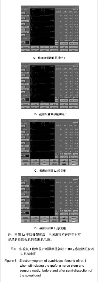
在同侧L2半切脊髓前后,电刺激移植神经干时可记录到股四头肌的收缩肌电图,在同侧L2半切脊髓前后,电刺激L1感觉根时可同样在股四头肌记录到肌电图,见图6,7。"

"
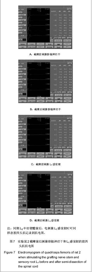

记录到股四头肌的收缩肌电图即可以记录到股四头肌的收缩动作。该脊髓旁路可以作为大鼠股四头肌的支配神经,在经过训练后有希望成为自主神经对股四头肌进行自主支配,有意识的使股四头肌产生收缩动作,为截瘫的下肢提供伸膝迈步动作。"

| [1] Zhang SC, Qu CY, Zhang XS, et al. Zhonghua Miniao Waike Zazhi. 2001;22(4):220-222.张少成,瞿创予,张雪松,等. 神经移植术治疗截瘫神经性膀胱的尿动力学观察[J]. 中华泌尿外科杂志, 2001,22(4): 220-222.http://www.cnki.com.cn/Article/CJFDTotal-ZHMN200104013.htm[2] Xiao CG, Du MX, Li B, et al. An artificial somatic-autonomic reflex pathway procedure for bladder control in children with spina bifida. J Urol. 2005;173(6):1850-1851.http://www.ncbi.nlm.nih.gov/pubmed/15879861[3] Gu YD. Shanghai: Shanghai Medical Universty Press. 1992.顾玉东.臂丛神经损伤与疾病的诊治[M].上海:上海医科大学出版社,1992.http://www.fudanpress.com/root/showdetail.asp?bookid=2069[4] Zhang SC, Xu SG, Ma YH, et al. Shanghai Dier Junyi Daxue Xuebao. 2004;7(4):803-804.张少成,许硕贵,马玉海,等. 硬脊膜内松解自体周围神经植入治疗脊髓陈旧性不完全性断裂伤[J].上海第二军医大学学报, 2004,7(4):803-804.http://www.39kf.com/cooperate/qk/ORTHOPEDIC-JOURNAL-OF-CHINA/0920/2010-01-13-629768.shtml[5] Dam-Hieu P, Liu S, Choudhri T, et al. Regeneration of primary sensory axons into the adult rat spinal cord via a peripheral nerve graft bridging the lumbar dorsal roots to the dorsal column. J Neurosci Res. 2002;68(3):293-304.http://www.ncbi.nlm.nih.gov/pubmed/12111859[6] Golden KL, Pearse DD, Blits B,et al. Transduced Schwann cells promote axon growth and myelination after spinal cord injury. Exp Neurol. 2007;207(2):203-217.http://www.ncbi.nlm.nih.gov/pubmed/17719577[7] Park IH, Zhao R, West JA, et al. Reprogramming of human somatic cells to pluripotency with defined factors. Nature. 2008;451(7175):141-146.http://www.ncbi.nlm.nih.gov/pubmed/23260147[8] Eftekharpour E, Karimi-Abdolrezaee S, Wang J, et al. Myelination of congenitally dysmyelinated spinal cord axons by adult neural precursor cells results in formation of nodes of Ranvier and improved axonal conduction. J Neurosci. 2007; 27(13):3416-3428.http://www.ncbi.nlm.nih.gov/pubmed/17392458[9] Sun Q, Zheng J, Zhao C. Nerve transplantation and accompanying peripheral vessels for repair of long nerve defect. Zhongguo Xiufu Chongjian Waike Zazhi. 2012;26(7): 832-836.http://www.ncbi.nlm.nih.gov/pubmed/22905621[10] Xia L, Hao SY, Li DZ, et al. Zhongguo Zuzhi Gongcheng Yanjiu. 2012;16(47):821-8825.夏雷,郝淑煜,李德志,等. 种植神经干细胞-许旺细胞的共聚物支架移植修复大鼠损伤脊髓[J]. 中国组织工程研究, 2012,16(47): 821-8825.http://www.cnki.com.cn/Article/CJFDTotal-XDKF201247025.htm[11] Kan R, Sheng WB. Zhongguo Zuzhi Gongcheng Yanjiu. 2013; 17(3):501-508.阚瑞,盛伟斌. 人工合成高分子支架材料治疗脊髓损伤[J]. 中国组织工程研究,2013,17(3):501-508.http://www.cqvip.com/Read/Read.aspx?id=45155203[12] Chen X, Yu ZJ. Jujie Shoushuxue Zazhi. 2013;21(2):199-201.陈祥,余资江. 电针与骨髓问充质干细胞移植在脊髓损伤修复及再生中的作用[J]. 局解手术学杂志,2013,21(2):199-201.http://www.cqvip.com/QK/97466X/201302/45280624.html[13] Yin H, Re XJ, Jiang T., et al. Zhonghua Chuangshang Zazhi. 2013;9(3):278-283.阴洪,任先军,蒋涛,等.超声辅助脱细胞脊髓支架的生物学特性研究[J]. 中华创伤杂志,2013,29(3):278-283.http://www.cnki.com.cn/Article/CJFDTotal-SWGK200804003.htm[14] Wei XK, Wen YM, Zhang T, et al. Zhonguo Xiufu Chongjian Waike Zazhi. 2012;26(11):1362-1368.魏祥科,文益民,张涛,等. BMSCs联合去细胞肌肉生物支架修复大鼠脊髓半切损伤的实验研究[J]. 中国修复重建外科杂志, 2012, 26(11):1362-1368.http://www.cnki.com.cn/Article/CJFDTotal-ZXCW201211030.htm[15] Wang D,Fan YH,Zhang JJ,et al. Transplantation of Nogo-66 receptor gene-silencecl cells in a poly(D,L-lactic-co-glycolic acid) scaffoldfor the treatment of spinal cord injur. Neural Regeneration Research. 2013,8(8):677-685.http://d.wanfangdata.com.cn/Periodical_zgsjzsyj-e201308001.aspx[16] Li N,Liu B, Rong LM, et al. Zhonghua Chuangshang Guke Zazhi. 2012;14(3):241-245.李宁,刘斌,戎利民,等. 静电纺丝聚乳酸聚乙醇酸/聚乙二醇纳米纤维作为组织工程支架的体外细胞相容性研究[J]. 中华创伤骨科杂志,2012,14(3):241-245.http://d.wanfangdata.com.cn/Periodical_zhcsgkzz201203014.aspx[17] Guérout N, Paviot A, Bon-Mardion N, et a1.Co-transplantation of olfactory ensheathing cells from mucosa and bulb origin enhances functional recovery after peripheral nerve lesion. PLoS One. 2011;6(8):e22816. http://www.docin.com/p-342712149.html[18] Gu SH, Xu WD, Xu L, et a1.Regenerated host axons form synapses with neurons derived from neural stem cells transplanted into peripheral nerves. J Int Med Res. 2010; 38(5): 1721-1729.http://www.ncbi.nlm.nih.gov/pubmed/21309486[19] Yu P, Huang L, Zou J, et al. Immunization with recombinant Nogo-66 receptor (NgR) promotes axonal regeneration and recovery of function after spinal cord injury in rats. Neurobiol Dis. 2008;32(3):535-542.http://www.ncbi.nlm.nih.gov/pubmed/18930141[20] Nouhaud FX, Caremel R, Leroi AM, et al. Bladder reinnervation with creation of a "somato-autonomic" reflex pathway in spinal cord injured or spina bifida, a new way for treatment?. Prog Urol. 2011;21(8):501-507.http://www.ncbi.nlm.nih.gov/pubmed/21872150[21] Liu S, Blanchard S, Bigou S, et a1.Neurotrophin 3 improves delayed reconstruction of sensory pathways after cervical dorsal root injury. Neurosurgery. 2011;68(2):450-461.http://www.ncbi.nlm.nih.gov/pubmed/21135740[22] Lin H, Hou C, Chen A, et al. Reinnervation of atonic bladder after conus medullaris injury using a modified nerve crossover technique in canines. World Neurosurg. 2010;73(5):582-586.http://www.ncbi.nlm.nih.gov/pubmed/20920947[23] Liu S, Bohl D, Blanchard S, et al. Combination of microsurgery and gene therapy for spinal dorsal root injury repair. Mol Ther. 2009n;17(6):992-1002.http://www.ncbi.nlm.nih.gov/pubmed/19240691[24] Dam-Hieu P, Liu S, Tadié M. Experimental bypass surgery between the spinal cord and caudal nerve roots for spinal cord injuries. Neurochirurgie. 2004;50(5):500-514.http://www.ncbi.nlm.nih.gov/pubmed/15654303[25] Ando T, Sato S, Toyooka T, et al. Photomechanical Wave-Driven Delivery of siRNAs Targeting Intermediate Filament Proteins Promotes Functional Recovery after Spinal Cord Injury in Rats. PLoS One. 2012;7(12):e51744.http://www.ncbi.nlm.nih.gov/pubmed/23272155[26] Fawatt J.Repair of spinal cord injuries:where we are,where are we going.Spinal cord. 2002;40(2):615-623.http://www.ncbi.nlm.nih.gov/pubmed/12483491[27] Bregman BS, Kunkel-Bagden E, Schnell L, et al. Recovery from spinal cord injury mediated by antibodies to neurite growth inhibitors. Nature. 1995;378(6556):498-501.http://www.ncbi.nlm.nih.gov/pubmed/7477407[28] Ferguson AR, Huie JR, Crown ED, et al. Central nociceptive sensitization vs. spinal cord training: opposing forms of plasticity that dictate function after complete spinal cord injury. Front Physiol. 2012;3:396.http://www.ncbi.nlm.nih.gov/pubmed/23060820[29] Yang YL. Effect of adenovirus-mediated basic fibroblast growth factor gene transfer in vivo on oligodendrocyte cell numbers throughout ventrolateral white matter following spinal cord injury in rats. Zhongguo Yi Xue Ke Xue Yuan Xue Bao. 2012;34(4):348-352.http://www.ncbi.nlm.nih.gov/pubmed/22954116 |
| [1] | Wu Guang-wen, Ye Jin-xia, Zheng Chun-song, Chen Wen-lie, Liu Xian-xiang, Ye Hong-zhi . Effect of Tougu Xiaotong capsule on articular cartilage changes in rat models of osteoarthritis [J]. Chinese Journal of Tissue Engineering Research, 2014, 18(49): 7924-7929. |
| [2] | Wang Pei-pei, Yang Peng . Lower limb walking state under different road conditions verified with virtual reality technology [J]. Chinese Journal of Tissue Engineering Research, 2013, 17(35): 6293-6299. |
| [3] | Yin Xiao-peng, Xu Hui-fen, He Hui-yu. Comparison of the biocompatibility of three kinds of antigen-extracted xenogeneic cancellous bone matrices [J]. Chinese Journal of Tissue Engineering Research, 2013, 17(29): 5275-5281. |
| [4] | Xie Hui, Yang Fei, Zhao De-wei, Wang Ben-jie, Cui Da-ping, Wang Wei, Huang Shi-bo. Chondrocyte differentiation of dog bone marrow mesenchymal stem cells co-cultured with Bio-gide collagen membrane in vitro [J]. Chinese Journal of Tissue Engineering Research, 2013, 17(29): 5282-5289. |
| [5] | Lin Wei, Li Xu-dong, Tang Kuang-yun, Hu Jing. Zinc ions improve the stability of titanium implants under the condition of osteoporosis [J]. Chinese Journal of Tissue Engineering Research, 2013, 17(29): 5296-5302. |
| [6] | Li Wei-dong, Cui Zhi-ming, Xu Guan-hua, Fan Jian-bo, Bao Guo-feng, Sun Yu-yu, Wang Ling-ling. Biocompatibility of olfactory ensheathing cells and poly-L-lactic acid reinforced by chitosan in vitro [J]. Chinese Journal of Tissue Engineering Research, 2013, 17(29): 5316-5322. |
| [7] | Wei Wei, Shi Geng-hu, Li Yu-tang, Zhang Bing, Gao Chuang. Research and application progress of visual fixation component separation [J]. Chinese Journal of Tissue Engineering Research, 2013, 17(24): 4553-4560. |
| [8] | . Culture of bone marrow mesenchymal stem cells plus allogenic decalcified bone matrix in the knee cavity of rabbits for tissue engineered cartilage [J]. Chinese Journal of Tissue Engineering Research, 2013, 17(24): 4371-4379. |
| [9] | Xi Yue. Molecular mechanism of spinal cord injury detected using microarray technology [J]. Chinese Journal of Tissue Engineering Research, 2013, 17(24): 4529-4538. |
| [10] | Wen Cai-xiong. Resistance training and protein supplement: Improvement of elderly muscle protein metabolism [J]. Chinese Journal of Tissue Engineering Research, 2013, 17(24): 4545-4552. |
| [11] | Lu Shou-ming, Lu Shou-liang, Sun Tian-wei, Zhang Hang, Wang Qi-ming. Estrogen effects on serum interleukin-8 and interleukin-10 expression in ovariectomized rats with osteoporosis [J]. Chinese Journal of Tissue Engineering Research, 2013, 17(24): 4394-4400. |
| [12] | Adila•Azhati, Zhao Long, Wang Qian, Ma Yi-tong. Cardiac function and electrophysiological characteristics in myocardial infarction rats after tissue engineered cardiac patch transplantation [J]. Chinese Journal of Tissue Engineering Research, 2013, 17(24): 4401-4408. |
| [13] | Shi Yu-lu, Li Xiao-yuan, Cao Mei-na, Yu Shu-yuan, Wang Ping. Simultaneous isolation of myocardial cells and cardiac fibroblasts from neonatal rats [J]. Chinese Journal of Tissue Engineering Research, 2013, 17(24): 4414-4420. |
| [14] | Wang Qi-feng, Wang Zhi-gang, Huang Jing. Left ventricular papillary muscle ablation in canines by ultrasound ablation catheter [J]. Chinese Journal of Tissue Engineering Research, 2013, 17(24): 4409-4413. |
| [15] | Chen Li-ying, Fan Kun-wu, Wang Jin-shui, Yu Xu-ming, Xu Cheng. Gene expression of the skin and sweat glands in different development stages of embryo [J]. Chinese Journal of Tissue Engineering Research, 2013, 17(24): 4421-4428. |
| Viewed | ||||||
|
Full text |
|
|||||
|
Abstract |
|
|||||