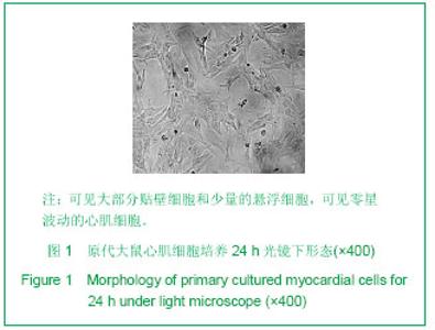
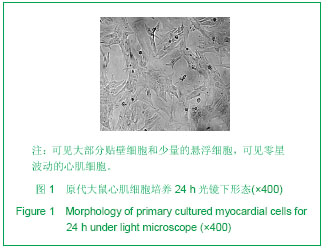
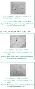
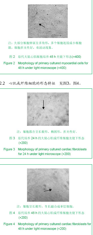
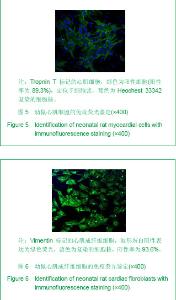
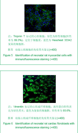
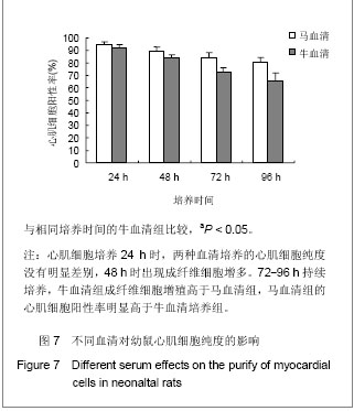
| [1] Harary L,Farly B.In vitro studies of single isolated beating heart cells.Science.1960;131(3414):1674-1675.http://www.ncbi.nlm.nih.gov/pubmed/14399673[2] Liu JP, Cao HG, An ZX, et al. Xibei Nonglin Keji Daxue Xuebao (Ziran Kexueban). 2005;33(1):9-12.刘俊平,曹鸿国,安志兴,等. 大鼠心肌细胞体外培养特性研究[J]. 西北农林科技大学学报(自然科学版),2005,33(1):9-12.http://www.cnki.com.cn/Article/CJFDTOTAL-XBNY200501003.htm[3] Zhang J, Zhu JZ, Zhang RY, et al. Zhongguo Yiliao Qianyan. 2012;7(10):8-10.张晶,朱劲舟,张瑞岩,等.乳小鼠心肌细胞体外培养方法的优化[J].中国医疗前沿,2012,7(10):8-10.http://www.cnki.com.cn/Article/CJFDTOTAL-YLQY201210006.htm[4] Cerbai E, Sartiani L, De Paoli P, et al. Isolated cardiac cells for electropharmacological studies. Pharmacol Res. 2000;42(1):1-8.http://www.ncbi.nlm.nih.gov/pubmed/10860628[5] Ren J, Wold LE. Measurement of Cardiac Mechanical Function in Isolated Ventricular Myocytes from Rats and Mice by Computerized Video-Based Imaging. Biol Proced Online. 2001;3:43-53.http://www.ncbi.nlm.nih.gov/pubmed/12734580[6] Louch WE, Hake J, Jølle GF, et al. Control of Ca2+ release by action potential configuration in normal and failing murine cardiomyocytes. Biophys J. 2010;99(5):1377-1386.http://www.ncbi.nlm.nih.gov/pubmed/20816049[7] Wang Q, Wang DJ, Li QG, et al. Zhongguo Zuzhi Gongcheng Yanjiu yu Linchuang Kangfu. 2007;11(6):1168-1169.王强,王东进,李庆国,等. 改良的原代心肌细胞培养方法[J]. 中国组织工程研究与临床康复,2007,11(6):1168-1169.http://www.cnki.com.cn/Article/CJFDTOTAL-XDKF200706049.htm[8] Yang HY, Dong W, Ding AS, et al. Jushi Yixue Kexueyuan Yuankan. 2010;34(1): 30-33.阳海鹰,丁巍,丁爱石,等.新生小鼠心肌细胞培养模型的建立及其在毒性评价研究中的应用[J]. 军事医学科学院院刊,2010,34(1): 30-33.http://www.cnki.com.cn/Article/CJFDTOTAL-JSYX201001008.htm[9] Wang CH. Zhongguo Xinzang Qibo yu XIndian Shengli Zazhi. 2003;17(3):203-231.汪长华.心肌细胞的培养及注意事项[J].中国心脏起搏与心电生理杂志,2003,17(3):203-231.http://www.cnki.com.cn/Article/CJFDTOTAL-ZGXZ200303022.htm[10] Yamauchi Y, Harada A, Kawahara K.Changes in the fluctuation of interbeat intervals in spontaneously beating cultured cardiac myocytes: experimental and modeling studies. Biol Cybern. 2002;86(2):147-154. http://www.ncbi.nlm.nih.gov/pubmed/11908840[11] Chen KY, Cai SC, Chen ZW. Zhongguo Xumu Shouyi. 2012; 39(5):115-118.陈克研,蔡书成,陈振文.新生Wistar大鼠心肌细胞的分离培养与鉴定[J].中国畜牧兽医,2012,39(5):115-118.http://www.cnki.com.cn/Article/CJFDTOTAL-GWXK201205029.htm[12] Zhao BJ, Cai ZJ, Yu SQ, et al. Zhongguo Linchuang Kangfu. 2005;9(19):100-101.赵璧君,蔡振杰,俞世强,等. 差速贴壁技术与常规培养方法对窦房结细胞培养中提高梭形细胞纯化率的影响[J].中国临床康复, 2005,9(19):100-101.http://www.cnki.com.cn/Article/CJFDTOTAL-XDKF200519046.htm[13] Li B, Wu HY, Piao HL, et al. Zhongguo Shiyan Zhenduanxue. 2012;16(2):200-203.李博,吴慧颖,朴虎林,等. 新生大鼠心肌细胞原代培养方法的改良[J].中国实验诊断学,2012,16(2):200-203.http://www.cnki.com.cn/Article/CJFDTOTAL-ZSZD201202005.htm[14] Louch WE, Sheehan KA, Wolska BM. Methods in cardiomyocyte isolation, culture, and gene transfer. J Mol Cell Cardiol. 2011;51(3):288-298.http://www.ncbi.nlm.nih.gov/pubmed/21723873[15] Yan QJ, Li LJ. Disi Junyi Daxue Xuebao. 2002;23(10):923-923.严泉剑,李六金. 生长曲线拟合及细胞群体倍增时间的简化计算[J]. 第四军医大学学报,2002,23(10):923-923.http://www.cnki.com.cn/Article/CJFDTOTAL-DSJY200210026.htm[16] Golden HB, Gollapudi D, Gerilechaogetu F, et al. Isolation of cardiac myocytes and fibroblasts from neonatal rat pups. Methods Mol Biol. 2012;843:205-214.http://www.ncbi.nlm.nih.gov/pubmed/22222535[17] Wang M, Li YJ, Liu SY, et al. ZHonguo Yishi Zazhi. 2013;2: 182-185.王梅,李拥军,刘素云,等. 高糖、胰岛素诱导胰岛素抵抗大鼠心肌细胞模型的建立[J]. 中国医师杂志,2013,2:182-185.http://www.doc88.com/p-986393443875.html[18] Li ZG, Wang GX, Liu SS, et al. Zhongguo Gonggong Weisheng. 2013;29(1):69-71.李兆钢,王国贤,刘珊珊,等. α-硫辛酸对高糖诱导下乳鼠心肌细胞作用[J]. 中国公共卫生,2013,29(1):69-71.http://www.cnki.com.cn/Article/CJFDTotal-SYYZ201009024.htm[19] Hua Y, Xu B. Xinxueguan Bingxue Jinzhan. 2012;33(6): 748-750.花颖,徐标. 心肌细胞再生和干细胞移植的研究现状[J]. 心血管病学进展,2012,33(6):748-750. http://www.cqvip.com/QK/93023X/201206/44148358.html [20] Lu Y, Kong LC, Zhang ZH, et al. Zhongguo Zuzhi Gongcheng Yanjiu. 2012;16(50):9425-9430.卢艳,孔令彩,张兆华,等. 黄芩素通过抗氧化及调节细胞内钙离子浓度抑制大鼠缺氧/复氧诱导的心肌细胞凋亡[J]. 中国组织工程研究, 2012,16(50):9425-9430.http://www.cnki.com.cn/Article/CJFDTotal-XXGB201206020.htm[21] Xue YY, Gong H, Yan Y, et al. Zhongguo Fenzi Xinzangxue Zazhi. 2012;12(6):350-355. 薛媛媛,龚惠,闫媛,等. 机械牵张对诱导多能干细胞向心肌细胞分化的研究[J]. 中国分子心脏病学杂志,2012,12(6):350-355.http://www.cnki.com.cn/Article/CJFDTotal-ZGFB201206009.htm[22] Hao YR, Li GS. Lingnan Xinxueguanbing Zazhi. 2001;7(2): 137-139.郝亚荣,李庚山.乳鼠心肌细胞培养[J].岭南心血管病杂志,2001, 7(2):137-139.http://www.bioon.com/experiment/cellular12/291120.shtml[23] Ma FF, Shen XL, Lin LF, et al. Xinxueguan kangfu Yixue Zazhi. 2009;18(4):125-129.马芳芳,沈晓丽,林立芳,等.新生大鼠心肌细胞的原代培养[J].心血管康复医学杂志 2009,18(4):125-129.http://journal.9med.net/html/qikan/nkx/xxgkfyxzz/20094182/kflz/20090512092514358_475129.html[24] Wang JY, Fan HM, Liu ZM, et al. Shanghai Yixue. 2008;31(5): 314-317.汪进益,范慧敏,刘中民,等.大鼠心肌成纤维细胞体外培养的生物学特性及转基因研究[J].上海医学,2008,31(5):314-317.http://www.cnki.com.cn/Article/CJFDTOTAL-SHYX200805007.htm[25] Xia JL, Zhu ZA, Zhang XT, et al. Shengming Kexue Yanjiu. 2009;13(3):236-239.夏机良,朱泽安,张湘涛,等.新生小鼠心肌细胞分离培养的改良及其鉴定[J].生命科学研究 2009,13(3):236-239.http://www.cnki.com.cn/Article/CJFDTOTAL-SMKY200903011.htm[26] Yonemochi H, Yasunaga S, Teshima Y, et al. Mechanism of adre- nergic receptor up-regulation induced by ACE inhibition in cultured neonatal rat cardiacmyocytes.Circ.1998; 97(22): 2268-2273. http://www.ncbi.nlm.nih.gov/pubmed/9631877[27] Clark WA,Decker ML,Janes DM,et al. Cell contact as an independent factor moduletating cardiac myocytes hypertrophy and survival in long-term primary culture.J Mol Cell Cardiol.1998;30(1):139-155. http://www.ncbi.nlm.nih.gov/pubmed/9500872[28] Lawson MA, Purslow PP. Differentiation of myoblasts in serum-free media: effects of modified media are cell line-specific. Cells Tissues Organs. 2000;167(2-3):130-137.http://www.ncbi.nlm.nih.gov/pubmed/10971037[29] Mørk HK, Sjaastad I, Sande JB, et al. Increased cardiomyocyte function and Ca2+ transients in mice during early congestive heart failure. J Mol Cell Cardiol. 2007;43(2): 177-186. http://www.ncbi.nlm.nih.gov/pubmed/17574269[30] ter Keurs HE, Shinozaki T, Zhang YM, et al. Sarcomere mechanics in uniform and non-uniform cardiac muscle: a link between pump function and arrhythmias. Prog Biophys Mol Biol. 2008;97(2-3):312-331.http://www.ncbi.nlm.nih.gov/pubmed/18394686[31] Brand NJ, Lara-Pezzi E, Rosenthal N, et al. Analysis of cardiac myocyte biology in transgenic mice: a protocol for preparation of neonatal mouse cardiac myocyte cultures. Methods Mol Biol. 2010;633:113-124.http://www.ncbi.nlm.nih.gov/pubmed/20204624[32] Fujita H, Endo A, Shimizu K, et al. Evaluation of serum-free differentiation conditions for C2C12 myoblast cells assessed as to active tension generation capability. Biotechnol Bioeng. 2010;107(5):894-901. http://www.ncbi.nlm.nih.gov/pubmed/20635352[33] Fan D, Takawale A, Lee J, et al. Cardiac fibroblasts, fibrosis and extracellular matrix remodeling in heart disease. Fibrogenesis Tissue Repair. 2012;5(1):15.http://www.ncbi.nlm.nih.gov/pubmed/22943504[34] Lijnen PJ, Piccart Y, Coenen T, et al. Angiotensin II-induced mitochondrial reactive oxygen species and peroxiredoxin-3 expression in cardiac fibroblasts. J Hypertens. 2012;30(10): 1986-1991. http://www.ncbi.nlm.nih.gov/pubmed/22828084[35] vatis K, van Linthout S, Miteva K, et al. Mesenchymal stromal cells but not cardiac fibroblasts exert beneficial systemic immunomodulatory effects in experimental myocarditis. PLoS One. 2012;7(7):e41047.http://www.ncbi.nlm.nih.gov/pubmed/22815907 |