Chinese Journal of Tissue Engineering Research ›› 2013, Vol. 17 ›› Issue (18): 3333-3340.doi: 10.3969/j.issn.2095-4344.2013.18.015
Previous Articles Next Articles
Autologous tendon graft with preserved distal insertions for the treatment of old patellar tendon rupture
Zhu Hua-qiang1, Liu Yun2, Chen Fa1, Liu De-huai3
- 1 Department of Orthopedics, Bobai County People’s Hospital, Yulin 537600, Guangxi Zhuang Autonomous Region, China
2 Department of Spinal Surgery, the First Affiliated Hospital of Guangxi Medical University, Nanning 530021, Guangxi Zhuang Autonomous Region, China
3 Department of Orthopedics, Minzu Hospital of Guangxi Zhuang Autonomous Region, Nanning 530000, Guangxi Zhuang Autonomous Region, China
-
Received:2013-02-09Revised:2013-03-13Online:2013-04-30Published:2013-04-30 -
Contact:Liu De-huai, Master, Attending physician, Department of Orthopedics, Minzu Hospital of Guangxi Zhuang Autonomous Region, Xining 530000, Guangxi Zhuang Autonomous Region, China Liudehuaiguke@163.net -
About author:Zhu Hua-qiang, Attending physician, Department of Orthopedics, Bobai County People’s Hospital, Yulin 537600, Guangxi Zhuang Autonomous Region, China liuyun200450250@sina.com
CLC Number:
Cite this article
Zhu Hua-qiang, Liu Yun, Chen Fa, Liu De-huai. Autologous tendon graft with preserved distal insertions for the treatment of old patellar tendon rupture[J]. Chinese Journal of Tissue Engineering Research, 2013, 17(18): 3333-3340.
share this article
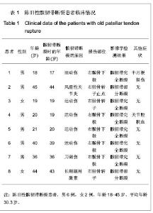
2.1 实验对象数量分析 实验共纳入8例患者,所有陈旧性髌韧带断裂患者髌韧带重建后均得到随访,无一例患者失访,随访时间为24个月。 2.2 陈旧性髌韧带断裂患者临床情况分析 8例陈旧性髌韧带断裂患者中共有运动伤5例、刀刺伤1例、长期服用激素自发性断裂1例及类风湿性关节炎相关性断裂1例。将所有8例患者损伤部位分类:髌骨下极断裂5例、胫骨转子止点断裂3例;完全断裂6例,部分断裂2例。陈旧性髌韧带断裂患者临床情况见表1。"
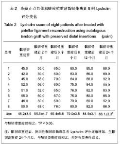
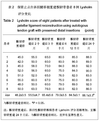
2.3 陈旧性髌韧带断裂患者保留止点的半腱肌及股薄肌重建髌韧带后运动功能的恢复 见表2。"
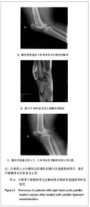
2.4 陈旧性髌韧带断裂患者保留止点的半腱肌及股薄肌重建髌韧带后髌骨恢复情况 见图2。"

| [1] Muratli HH, Celebi L, Hapa O, et al. Bilateral patellar tendon rupture in a child: a case report. Knee Surg Sports Traumatol Arthrosc. 2005;13(8):677-682. [2] Ramsey RH, Muller GE. Quadriceps tendon rupture: a diagnostic trap. Clin Orthop Relat Res. 1970;70:161-164.[3] Siwek CW, Rao JP. Ruptures of the extensor mechanism of the knee joint. J Bone Joint Surg Am. 1981;63(6):932-937.[4] Chen B, Li R, Zhang S. Reconstruction and restoration of neglected ruptured patellar tendon using semitendinosus and gracilis tendons with preserved distal insertions: two case reports. Knee. 2012;19(4):508-512.[5] McNally PD, Marcelli EA. Achilles allograft reconstruction of a chronic patellar tendon rupture. Arthroscopy. 1998;14(3): 340-344.[6] Wuller TG, Jansson KA, Bruner BW. Augmentation of the patellar ligament with a bone-patellar tendon-bone inlay allograft. Arthroscopy. 1998;14(1):89-93.[7] Rasul AT Jr, Fischer DA. Primary repair of quadriceps tendon ruptures. Results of treatment. Clin Orthop Relat Res. 1993; (289):205-207.[8] Mittal R, Singh DP, Kapoor A. Neglected patellar tendon rupture: preserve the fat pad. Orthopedics. 2011;34(2):134.[9] Chen B, Wang G, Zhang S, et al. Shiyong Guke Zazhi. 2010; 16(4):258-260.陈滨,王钢,张晟,等.保留止点半腱肌、股薄肌肌腱重建修复陈旧性髌韧带断裂2例报告[J].实用骨科杂志,2010,16(4): 258-260.[10] Bali K, Mootha AK, Krishnan V, et al. A rare case of bicondylar Hoffa fracture associated with ipsilateral tibial spine avulsion and extensor mechanism disruption. Chin J Traumatol. 2011;14(4):253-256.[11] Kang S, Chung PH, Kim YS, et al. Bifocal disruption of the knee extensor mechanism: a case report and literature review. Arch Orthop Trauma Surg. 2013;133(4):517-521.[12] O'Sullivan R, Walsh M, Kiernan D, et al. The knee kinematic pattern associated with disruption of the knee extensor mechanism in ambulant patients with diplegic cerebral palsy. Clin Anat. 2010;23(5):586-592. [13] Roidis N, Varitimidis S, Poultsides L, et al. A "biologic technique" for the treatment of a disruption of the extensor mechanism after revision total knee arthroplasty: a case report. Knee Surg Sports Traumatol Arthrosc. 2008; 16(7): 661-665.[14] Barrack RL, Butler RA, Valenzuela R. Extensor mechanism disruption after total knee arthroplasty: when the unthinkable happens. Orthopedics. 2002;25(9):981-982.[15] Schoderbek RJ Jr, Brown TE, Mulhall KJ, et al. Extensor mechanism disruption after total knee arthroplasty. Clin Orthop Relat Res. 2006;446:176-185.[16] Nanninga AJ, Josaputra HA. Tibial tuberosity fracture in adolescents--report of a case and review of the literature. Neth J Surg. 1987;39(5):144-146.[17] Brooks P. Extensor mechanism ruptures. Orthopedics. 2009; 32(9). [18] Duthon VB, Fritschy D. Knee extensor mechanism ruptures. Rev Med Suisse. 2011;7(304):1544-1548.[19] Larsen E, Lund PM. Ruptures of the extensor mechanism of the knee joint. Clinical results and patellofemoral articulation. Clin Orthop Relat Res. 1986;(213):150-153.[20] McKinney B, Cherney S, Penna J. Intra-articular knee injuries in patients with knee extensor mechanism ruptures. Knee Surg Sports Traumatol Arthrosc. 2008;16(7):633-638.[21] McGrory JE. Disruption of the extensor mechanism of the knee. J Emerg Med. 2003;24(2):163-168.[22] Bek D, Demiralp B, Kömürcü M, et al. Neglected patellar tendon rupture: a case of reconstruction without quadriceps lengthening. J Orthop Traumatol. 2008;9(1):39-42.[23] Chagar B, Boussouga M, Lazrak KH, et al. Neglected spontaneous bilateral rupture of the patellar tendon: a case report. Rev Chir Orthop Reparatrice Appar Mot. 2003;89(8): 733-737.[24] Chen CF, Chen WM, Lee KS, et al. Advanced osteoarthritic knee with neglected patellar tendon rupture treated with total patellectomy and total knee arthroplasty. J Arthroplasty. 2004; 19(6):793-796.[25] Horas U, Ernst S, Meyer C, et al. Simultaneous rupture of a patellar tendon and contralateral quadriceps tendon. Unfallchirurg. 2006;109(9):801-804.[26] Kumar S, Rachakatla N, Kerin C, et al. Simultaneous traumatic rupture of the patellar tendon and the contralateral quadriceps tendon in a healthy individual. BMJ Case Rep. 2010; 2010. [27] Muratli HH, Celebi L, Hapa O, et al. Simultaneous rupture of the quadriceps tendon and contralateral patellar tendon in a patient with chronic renal failure. J Orthop Sci. 2005;10(2): 227-232.[28] Rogers A, Rix S, Kulkarni R. Simultaneous rupture of a patellar tendon and contralateral quadriceps tendon in a healthy individual. Orthopedics. 2003;26(8):817-818.[29] Shepard GJ, Christodoulou L, Hegab AI. Neglected rupture of the patellar tendon. Arch Orthop Trauma Surg. 1999;119(3-4): 41-242.[30] Kaneko K, DeMouy EH, Brunet ME, et al. Radiographic diagnosis of quadriceps tendon rupture: analysis of diagnostic failure. J Emerg Med. 1994;12(2):225-229.[31] Lu TX, Yang H, Zeng RD. Zhonghua Guke Zazhi. 2006,26 10):698.卢天祥,杨华,曾荣东.类风湿性关节炎并发双侧髌韧带断裂一例报告[J].中华骨科杂志,2006;26(10):698.[32] Clark SC, Jones MW, Choudhury RR, et al. Bilateral patellar tendon rupture secondary to repeated local steroid injections. J Accid Emerg Med. 1995;12(4):300-301.[33] Karadimas EJ, Kotzamitelos D, Kakagia DD, et al. Spontaneous rupture of the patellar tendon and the contralateral quadriceps tendon, associated with lupus erythematosus: analysis of the literature. Case Rep Orthop. 2011;2011:569363.[34] Chiou HM, Chang MC, Lo WH. One-stage reconstruction of skin defect and patellar tendon rupture after total knee arthroplasty. A new technique. J Arthroplasty. 1997;12(5): 575-579.[35] Crossett LS, Sinha RK, Sechriest VF, et al. Reconstruction of a ruptured patellar tendon with achilles tendon allograft following total knee arthroplasty. J Bone Joint Surg Am. 2002; 84-A(8):1354-1361.[36] ElGuindy A, Lustig S, Servien E, Fary C, et al. Treatment of chronic disruption of the patellar tendon in Osteogenesis Imperfecta with allograft reconstruction. Knee. 2011;18(2): 121-124.[37] Grecomoro G, Camarda L, Martorana U. Simultaneous chronic rupture of quadriceps tendon and contra-lateral patellar tendon in a patient affected by tertiary hyperparatiroidism. J Orthop Traumatol. 2008;9(3):159-162. [38] Grim C, Lorbach O, Engelhardt M. Quadriceps and patellar tendon ruptures. Orthopade. 2010;39(12):1127-1134.[39] Jab?oński JJ, Jarmuziewicz P, Dru?bicki M. Reconstruction of chronic patellar tendon rupture with semitendinosus tendon: case report. Ortop Traumatol Rehabil. 2011;13(6):607-615.[40] Labib SA, Wilczynski MC, Sweitzer BA. Two-layer repair of a chronic patellar tendon rupture: a novel technique and literature review. Am J Orthop (Belle Mead NJ). 2010;39 (6): 277-282.[41] Nguene-Nyemb AG, Huten D, Ropars M. Chronic patellar tendon rupture reconstruction with a semitendinosus autograft. Orthop Traumatol Surg Res. 2011;97(4):447-450. [42] Goertzen M, Schulitz KP. Comparison of combined extra- and intra-articular stabilization versus isolated arthroscopic semitendinosus repair after rupture of the anterior cruciate ligament. Sportverletz Sportschaden. 1993;7(1):7-12.[43] Gomez T, Ratzlaff C, McConkey JP, et al. Semitendinosus repair augmentation of acute anterior cruciate ligament rupture. Can J Sport Sci. 1990;15(2):137-142.[44] Kühne JH, Krüger-Franke M, Refior HJ. Reconstruction of acute anterior cruciate ligament rupture by suture and semitendinosus tendon augmentation. Oper Orthop Traumatol. 1997;9(1):37-47.[45] Weber K, Schmidgen A, Wentzensen A. Patellar tendon rupture after anterior cruciate ligament reconstruction with autologous patellar tendon-bone transplant. A case report. Unfallchirurg. 2000;103(12):1124-1127.[46] Mihalko WM, Vance M, Fineberg MJ. Patellar tendon repair with hamstring autograft: a cadaveric analysis. Clin Biomech (Bristol, Avon). 2010;25(4):348-351. [47] Spindler KP, Kuhn JE, Freedman KB, et al. Anterior cruciate ligament reconstruction autograft choice: bone-tendon-bone versus hamstring: does it really matter? A systematic review. Am J Sports Med. 2004;32(8):1986-1995.[48] Haasters F, Ockert B, Mutschler W, et al. Late patellar tendon rupture 10 years after anterior cruciate ligament reconstruction using a bone-patellar tendon-bone graft. Unfallchirurg. 2009;112(8):728-733.[49] Isiklar ZU, Varner KE, Lindsey RW, et al. Late reconstruction of patellar ligament ruptures using Ilizarov external fixation. Clin Orthop Relat Res. 1996;(322):174-178.[50] Mandelbaum BR, Bartolozzi A, Carney B. A systematic approach to reconstruction of neglected tears of the patellar tendon. A case report. Clin Orthop Relat Res. 1988;(235): 268-271.[51] Pan KL, Masbah O, Razak M. Late reconstruction of the patellar tendon. Med J Malaysia. 2001;56 Suppl C:73-75. |
| [1] | Yang Xin, Jin Zhe, Feng Xu, Lu Bing. The current situation of knowledge and attitudes towards organ, eye tissue, body donation of residents in Shenyang [J]. Chinese Journal of Tissue Engineering Research, 2021, 25(5): 779-784. |
| [2] | Xu Hui, Kang Bingxin, Zhong Sheng, Gao Chenxin, Zhao Chi, Qiu Guowei, Sun Songtao, Xie Jun, Xiao Lianbo, Shi Qi. Pressing local acupoints plus adjustion of the knee joint in a sitting position for treating knee osteoarthritis: a randomized controlled trial [J]. Chinese Journal of Tissue Engineering Research, 2021, 25(2): 216-221. |
| [3] | Ma Long, Tan Xiaoxin, Sun Guoshao. A 5-year follow-up on sagittal alignment and radiological outcomes of consecutive three-level anterior cervical discectomy and fusion and hybrid surgery [J]. Chinese Journal of Tissue Engineering Research, 2021, 25(12): 1879-1885. |
| [4] | Zhang Junhui, Xu Hualiang, Chang Hong, Lin Zhujian, Song Yancheng . Zero-profile anterior cervical ROI-C cage versus traditional fusion cage combined with titanium plate in treatment of two-level cervical spondylotic myelopathy [J]. Chinese Journal of Tissue Engineering Research, 2020, 24(21): 3329-3335. |
| [5] |
Zhang Chunlin, Liu Yang, Shang Lijie, Yan Xu, Ning Yongming, Li Dongzhe, Dong Chao, Cao Zhengming.
Observation of herniated
cervical intervertebral disc volume based on quantitative volume measurement “monitoring” based on PACS software |
| [6] | Li You, Zheng Bengbeng, Wang Jiaming, Ma Yongsheng. Research and application of artificial cervical disc [J]. Chinese Journal of Tissue Engineering Research, 2020, 24(12): 1941-1948. |
| [7] | Yin Yating, Zhang Aijun, Wang Hao, Wang Pingping, Li Jianhua, Wen Minmin, Jin Peisheng. Clinical application of autogenous adipose-derived stromal vascular fraction-assisted fat transplantation in the improvement of facial skin quality [J]. Chinese Journal of Tissue Engineering Research, 2019, 23(9): 1428-1433. |
| [8] | Zhong Qiusheng1, Xia Weichao1, Guo Meizhen1, Zhu Haiqing1, Zhong Cuiqiong1, Shao Jieqi1, He Xiaohong2, Chen Xiumin2. Sandwiched Moxibustion plus Bushen Quhan recipe for treating knee osteoarthritis: a randomized controlled trial [J]. Chinese Journal of Tissue Engineering Research, 2019, 23(35): 5670-5675. |
| [9] | Cheng Wendan, Wu Han, Zhang Jisen, Zhang Xin, Zhang Shuo, Li Ziyu, Wu Yibo, Bai Wenyi, Jing Juehua. Total hip arthroplasty by direct anterior approach in the lateral position in the treatment of ankylosed hips: early therapeutic effects [J]. Chinese Journal of Tissue Engineering Research, 2019, 23(28): 4429-4434. |
| [10] | Fang Genqiang, Zhao Zhengli, Jin Xianhui, Zhang Qingsheng, Cui Shengjie, Wei Wei, Yan Guanghui, Wu Jiaqi, Zhao Lei. Clinical efficacy of high viscosity bone cement vertebroplasty for treating osteoporotic vertebral compression fractures [J]. Chinese Journal of Tissue Engineering Research, 2019, 23(22): 3475-3480. |
| [11] | Cai Qiucheng, Fan Hongkai, Xiong Rihui, Jiang Yi. Intravenous administration of bone marrow mesenchymal stem cells protects liver function following fatty liver transplantation from donors after cardiac death [J]. Chinese Journal of Tissue Engineering Research, 2019, 23(17): 2625-2629. |
| [12] | Jiang Shanshan, Wang Feng, Yu Limei. Immunomodulatory properties of mesenchymal stem cells and their application in organ transplantation [J]. Chinese Journal of Tissue Engineering Research, 2019, 23(1): 103-109. |
| [13] | Liu Teng-fei, Zhou Jian-kang, Huang Tuan-jie, Xing Qu, Cheng Kang, Li Peng, Li Dong-peng, Yang Bo, Ma Shan-shan, Guan Fang-xia . MG53 protein protects against multiorgan ischemia/reperfusion injury: present and future [J]. Chinese Journal of Tissue Engineering Research, 2017, 21(20): 3248-3254. |
| [14] | Li Ling, Li Ning, Ai Zi-ye, Yao Ya-jun, Wei Wan-hui, He Wei-yang, Wang Yan-feng, Ye Qi-fa. Intra-patient variability of tacrolimus concentration in transplant recipients: a prognostic predictor post transplantation [J]. Chinese Journal of Tissue Engineering Research, 2016, 20(49): 7437-7422. |
| [15] | Lin Jing-xia, Su Fan, Luo Hong-shan. Transfusion of blood components in liver transplantation and abdominal multiple organ transplantation [J]. Chinese Journal of Tissue Engineering Research, 2016, 20(33): 4957-4962. |
| Viewed | ||||||
|
Full text |
|
|||||
|
Abstract |
|
|||||