Chinese Journal of Tissue Engineering Research ›› 2013, Vol. 17 ›› Issue (18): 3318-3324.doi: 10.3969/j.issn.2095-4344.2013.18.013
Previous Articles Next Articles
Insulin promotes the microvessel formation in fat grafts
Deng Ying1, Zeng Ling-huan2, Li Wei1, Wu Yi1
- 1 Department of Plastic Surgery, Chongqing Emergency Medical Center, Chongqing 400014, China
2 Department of Plastic Surgery, Sichuan Hospital of Integrated Chinese and Western Medicine, Chengdu 610041, Sichuan Province, China
-
Received:2012-08-11Revised:2012-09-26Online:2013-04-30Published:2013-04-30 -
Contact:Zeng Ling-huan, Associate chief physician, Department of Plastic Surgery, Sichuan Hospital of Integrated Chinese and Western Medicine, Chengdu 610041, Sichuan Province, China -
About author:Deng Ying, Attending physician, Department of Plastic Surgery, Chongqing Emergency Medical Center, Chongqing 400014, China dengying1206@hotmail.com -
Supported by:Project of Chongqing Health Bureau, No. 04-2-022
CLC Number:
Cite this article
Deng Ying, Zeng Ling-huan, Li Wei, Wu Yi. Insulin promotes the microvessel formation in fat grafts [J]. Chinese Journal of Tissue Engineering Research, 2013, 17(18): 3318-3324.
share this article
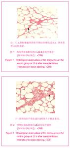
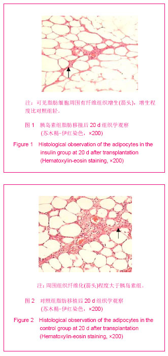
2.1 实验动物数量分析 纳入SD大鼠共48只,均分为胰岛素组和对照组,各组大鼠在实验过程中均无脱落,全部进入结果分析。 2.2 移植体观察 在大鼠肉膜层移植体周围有网状血管形成包裹移植体,移植体的脂肪色泽较移植前的脂肪色泽暗,质地比移植前的脂肪组织致密,脂肪颗粒变小。 2.3 苏木精-伊红染色观察结果 在苏木精-伊红染色下可见移植体内有成熟脂肪细胞,脂肪细胞体积变小,部分脂肪细胞有萎缩和破裂现象,脂肪细胞周围可见纤维组织增生,移植脂肪内有新生毛细血管。对照组脂肪细胞周围组织纤维化程度大于胰岛素组,见图1,2。"
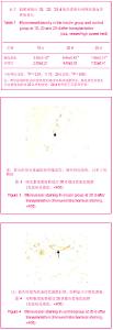
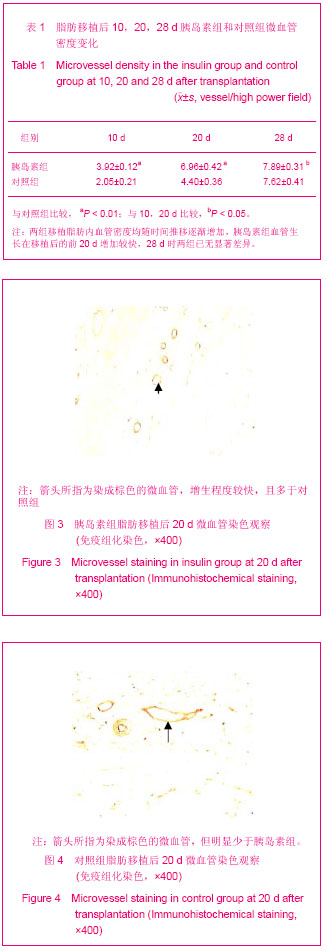
2.4 微血管免疫组化观察 免疫组化染色血管显影呈棕色,微血管显影清晰,高倍(×400)视野计数显示移植后10,20 d两组比较,差异有显著性意义,微血管密度随时间推移逐渐增多,微血管增生在移植后的前20 d增加较快,20-28 d期间新生血管增加速度变缓,见表1,图3,4。"
| [1] Metzinger S,Parrish J,Guerra A,et al.Autologous fat grafting to the lower one-third of the face.Facial Plast Surg.2012;28(1): 21-33.[2] Alencar JC,Andrade SH,Pessoa SG,et al.Autologous fat transplantation for the treatment of progressive hemifacial atrophy(Parry-Romberg syndrome:case report and review of medical literatute).An Bras Dermatol. 2011;86(4 Suppl 1): s85-88.[3] Wu Y,Li W,Deng Y.Zhongguo Meirong Yixue.2008;17(2): 180-181.吴一,李伟,邓颖.自体颗粒脂肪移植充填矫治面部凹陷[J].中国美容医学,2008,17(2): 180-181.[4] Lim AA, Fan K, Allam KA,et al.Autologous fat transplantation in the craniofacial patient: the UCLA experience.J Craniofac Surg. 2012;23(4):1061-1066.[5] Li WH,Sun ZC,Wang W.Zhongguo Zuzhi Gongcheng Yanjiu yu Linchuang Kangfu.2010;14(41):7753-7756.李卫华,孙志成,王文.碱性成纤维细胞生长因子在自体脂肪颗粒填充面部凹陷中的作用[J].中国组织工程研究与临床康复, 2010,14(41):7753-7756. [6] Lu F,Gao JH,Ogawa R,et al. Improvement of the survival of human autologous fat transplantation by using VEGF-transfected adipose-derived stem sells.Plast Reconstr Surg.2009;124(5):1437-1446.[7] Zheng ZM,Gui L,Zhang ZY,et al.Zhongguo Lichuang Kangfu. 2005; 9(42):46-47.郑宗梅,归来,张智勇,等.多隧道、多层次自体脂肪颗粒移植在矫治额颞部凹陷中的作用[J].中国临床康复,2005,9(42):46-47.[8] Gonzalez AM, Lobocki C, Kelly CP,et al.An alternative method for harvest and processing fat grafts: an in vitro study of cell viability and survival.Plast Reconstr Surg.2007;12(1): 285-294.[9] Zhang YN,Wang HT,Jiang WL,et al.Haerbin Yike Daxue Xuebao. 2011;45(1):23-26.张英男,王海涛,姜维良,等. 胰岛素对人血管内皮细胞保护作用的实验研究[J].哈尔滨医科大学学报,2011,45(1):23-26. [10] Zhou SJ,Chen S,Yu P,et al.Zhonghua Neifenmi Daixie Zazhi. 2010(10): 891-893.周赛君,陈松,于珮,等.胰岛素经VEGF-A/VEGFR2途径促进恒河猴视网膜血管内皮细胞的血管新生[J].中华内分泌代谢杂志, 2010,10:891-893. [11] Li SG,Zeng QT.Zhongguo Bingli Shengli Zazhi.2004;20(12): 2232-2235. 李书国,曾秋棠.胰岛素对牛胸主动脉内皮细胞血管内皮生长因子及其受体表达的影响[J].中国病理生理杂志,2004,20(12): 2232-2235.[12] Lu JX,Yang JT,Zang RX,et al. Zhongguo Shouyi Kexue. 2006; 36(11):915-919.卢建雄,杨具田,臧荣鑫,等.胰岛素对原代培养大鼠前体脂肪细胞增殖分化的影响[J].中国兽医科学,2006,36(11):915-919.[13] The Ministry of science and technology of the People’s Republic of China.Guidance suggestions for the care and use of laboratory animals. 2006-09-30.[14] Du XL,Luo SJ,Hao XG,et al.Zhonghua Zhengxing Waike Zazhi. 2005;21(2):128-131.杜学亮,罗少军,郝新光.等.碱性成纤维细胞生长因子在颗粒脂肪移植后血运重建过程的作用[J].中华整形外科杂志,2005,21(2): 128-131.[15] Zhu L,Li D.Zuzhi Gongcheng yu Chongjian Waike Zazhi.2012; 8(3):159-160.祝联,李丹.自体脂肪移植治疗川字纹[J].组织工程与重建外科杂志, 2012,8(3):159-160.[16] Sun ZY,Zhang W,Yu H,et al. Zhongguo Meirong Yixue.2011; 20(11): 1671-1673.孙泽钰,张伟,于海,等.自体颗粒脂肪移植填充老态鼻唇沟[J].中国美容医学,2011, 20(11):1671-1673. [17] Yang WS,Wang JQ,Guo X,et al.Zhongguo Meirong Yixue. 2012; 21(4):554-556.杨文爽,王佳琦,郭鑫,等.重睑术联合自体颗粒脂肪组织注射充填术在治疗上睑凹陷的临床应用[J].中国美容医学,2012,21(4): 554-556.[18] Xie Y,Li QF,Zheng DN.Zhongguo Xiufu Chongjian Waike Zazhi. 2007;21(12):1308-1311.谢芸,李青峰,郑丹宁.半面萎缩的自体脂肪颗粒移植治疗[J].中国修复重建外科杂志,2007,21(12):1308-1311.[19] Xie HJ.Zhongguo Meirong Zhengxing Waike Zazhi. 2007; 18(6): 411-413.谢红炬.自体脂肪颗粒移植隆乳的回顾性研究及并发症处理[J].中国美容整形外科杂志,2007,18(6):411-413.[20] Li QF.Zhongguo Meirong Yixue.2005;14(1):17-18.李青峰.自体脂肪颗粒移植临床应用回顾与分析[J].中国美容医学,2005,14(1):17-18.[21] Herold C, Ueberreiter K, Cromme F,et al.The use of mamma MRI volumetry to evaluate the rate of fat survival after autologous lipotransfer.Handchir Mikrochir Plast Chir.2010; 42(2):129-34.[22] Liu P,Liu Y,Liu L,et al.Zhongguo Meirong Yixue. 2011;20(11): 1702-1704.刘萍,刘毅,刘莉,等.颗粒脂肪移植术前注射量的评估[J].中国美容医学,2011,20(11):1702-1704.[23] Lei H,Li QF.Zhonghua Zhengxing Waike Zazhi.2005;21(5): 375-378.雷华,李青峰.葡萄糖转移方法检测脂肪颗粒活性的研究[J].中华整形外科杂志,2005,21(5):375-378.[24] Li HM,Liu DL,Wu T,et al.Zhongguo Zuzhi Gongcheng Yanjiu yu Linchuang Kangfu. 2011;15(31):5751-5755.黎洪棉,柳大烈,吴涛,等.早期应用富血小板血浆凝胶对自体脂肪组织移植存活率的影响[J].中国组织工程研究与临床康复, 2011,15(31):5751-5755. [25] Liu JF,Zhong WH,Zhang YM,et al.Zhongguo Meirong Yixue. 2008;17(4):540-543. 刘嘉锋,钟文惠,张一呜,等.重组人VEGF基因在游离组织移植中作用的实验研究[J].中国美容医学,2008,17(4):540-543. [26] Oh DS, Cheon YW, Jeon YR,et al. Activated platelet-rich plasma improves fat graft survival in nude mice: a pilot study. Dermatol Surg. 2011;37(5):619-625.[27] Tseng YH,Kriauciunas KM,Kokkotou E,et al.Differential roles of insulin receptor substrates in brown adipocyte differentiation. Mol Cell Biol. 2004;24(5):1918-1929.[28] Entingh-Pearsall A, Kahn CR. Differential roles of the insulin and insulin-like growth factor-I (IGF-I) receptors in response to insulin and IGF-I.J Biol Chem. 2004;279(36): 38016-38024. [29] Wu Y, Zeng LH, Li W, et al. Chongqiang Yike Daxue Xuebao. 2012;37(10):889-891.吴一,曾令寰,李伟,等. 特定电磁波谱对大鼠脂肪移植体内微血管形成影响的定量研究[J]. 重庆医科大学学报,2012,37(10): 889-891.[30] Zhang CC, Chen C, Jin ZL, et al. Zhongguo Zhongliu Linchuang. 2012;29(14):952-956.张翠翠,陈程,金子良,等. 重组人血管内皮抑制素对小鼠移植瘤及正常组织中血管相关因子影响的比较[J]. 中国肿瘤临床, 2012, 29(14):952-956.[31] Wu SM, Zhao YN, Yang L, et al. Zhongguo Meirong Yixue. 2008;17(5):685-688.伍尚敏,赵亚南,杨力,等.重组人血管内皮抑制素对血管内皮细胞的作用[J].中国美容医学,2008,17(5):685-688. [32] Hu XL, Lou N, Dong ZH, et al. Guoji Neifenmi Daixie Zazhi. 2012;32(2):107-110.胡晓琳,娄宁,董振华,等. 胰岛素对骨骼肌微血管的调节作用[J]. 国际内分泌代谢杂志,2012,32(2):107-110.[33] Peng Z,Jia ZH,Liu XT.Zhongguo Zuzhi Gongcheng Yanjiu yu Linchuang Kangfu. 2009;13(31):6063-6066.彭智,贾振华,刘晓韬.微渗透泵持续恒量灌注血管内皮细胞生长因子对大鼠脂肪移植体成活率的影响[J].中国组织工程研究与临床康复,2009,13(31):6063-6066.[34] Yue YG,Quan Y,Shao JS,et al.Huaxia Yixue.2011;24(6): 643-648. 岳毅刚,全媛,邵家松,等.不同浓度血管内皮细胞生长因子皮下局部注射对大鼠脂肪移植体存活率的影响[J].华夏医学,2011, 24(6): 643-648.[35] Nakano J, Kataoka H, Sakamoto J, et al. Low-level laser irradiation promotes the recovery of atrophied gastrocnemius skeletal muscle in rats. Exp Physiol. 2009;94(9):1005-1015.[36] Lei M, Liu SQ, Peng H, et al. Effect of rhVEGF gene transfection on survival of grafts after autologous free granular fat transplantation in rats. Chin J Traumatol. 2008; 11(1):49-53.[37] Shuai L, Li X, He Q, et al. Angiogenic effect of endothelial progenitor cells transfected with telomerase reverse transcriptase on peritubular microvessel in five out of six subtotal nephrectomy rats. Ren Fail. 2012;34(10):1270-1280.[38] Hiemann NE, Meyer R, Wellnhofer E, et al. Non-HLA antibodies targeting vascular receptors enhance alloimmune response and microvasculopathy after heart transplantation. Transplantation. 2012;94(9):919-924. [39] Wang Y, Lian F, Li J, et al. Adipose derived mesenchymal stem cells transplantation via portal vein improves microcirculation and ameliorates liver fibrosis induced by CCl4 in rats. J Transl Med. 2012;10:133.[40] Badeanlou L, Furlan-Freguia C, Yang G, et al. Tissue factor-protease-activated receptor 2 signaling promotes diet-induced obesity and adipose inflammation. Nat Med. 2011;17(11):1490-1497. |
| [1] | Liang Xueqi, Guo Lijiao, Chen Hejie, Wu Jie, Sun Yaqi, Xing Zhikun, Zou Hailiang, Chen Xueling, Wu Xiangwei. Alveolar echinococcosis protoscolices inhibits the differentiation of bone marrow mesenchymal stem cells into fibroblasts [J]. Chinese Journal of Tissue Engineering Research, 2021, 25(7): 996-1001. |
| [2] | Duan Liyun, Cao Xiaocang. Human placenta mesenchymal stem cells-derived extracellular vesicles regulate collagen deposition in intestinal mucosa of mice with colitis [J]. Chinese Journal of Tissue Engineering Research, 2021, 25(7): 1026-1031. |
| [3] | Yang Xin, Jin Zhe, Feng Xu, Lu Bing. The current situation of knowledge and attitudes towards organ, eye tissue, body donation of residents in Shenyang [J]. Chinese Journal of Tissue Engineering Research, 2021, 25(5): 779-784. |
| [4] | Jiang Tao, Ma Lei, Li Zhiqiang, Shou Xi, Duan Mingjun, Wu Shuo, Ma Chuang, Wei Qin. Platelet-derived growth factor BB induces bone marrow mesenchymal stem cells to differentiate into vascular endothelial cells [J]. Chinese Journal of Tissue Engineering Research, 2021, 25(25): 3937-3942. |
| [5] | Chen Ziyang, Pu Rui, Deng Shuang, Yuan Lingyan. Regulatory effect of exosomes on exercise-mediated insulin resistance diseases [J]. Chinese Journal of Tissue Engineering Research, 2021, 25(25): 4089-4094. |
| [6] | Liu Yulin, Li Guotai. Combined effects of hyperbaric oxygen, vibration training and astaxanthin on bone mineral density, glucose metabolism and oxidative stress in diabetic osteoporosis rats [J]. Chinese Journal of Tissue Engineering Research, 2021, 25(20): 3117-3124. |
| [7] | Zhang Lei, Yan Yu, Liu Yin, Xu Long, Yang Xinglei, Liu Yujia. An 8-week aerobic exercise improves obesity-induced myocardial fibrosis: role of nuclear factor-erythroid 2 p45-related factor 2 pathway [J]. Chinese Journal of Tissue Engineering Research, 2021, 25(17): 2650-2656. |
| [8] | Zhang Chuanhui, Li Jianjun, Yang Jun. Effects of dynamic pressure on the proliferation and mechanical properties of rabbit adipose mesenchymal stem cells transfected with insulin-like growth factor-1 [J]. Chinese Journal of Tissue Engineering Research, 2021, 25(13): 1982-1987. |
| [9] | Cao Linlin, Ding Kaiyang, Song Hao, Wu Guolin, Hu Maogui, Fan Dandan, Zhou Chenyang, Wang Cuicui, Feng Yuanyuan. Efficacy and influencing factors of autologous hematopoietic stem cell transplantation in the treatment of malignant lymphoma [J]. Chinese Journal of Tissue Engineering Research, 2021, 25(13): 1993-1998. |
| [10] | Liu Tao, Zhang Nini, Huang Guilin . Relationship between extracellular vesicles and radiation-induced tissue injury [J]. Chinese Journal of Tissue Engineering Research, 2021, 25(13): 2121-2126. |
| [11] | Hu Sheng, Yuan Haiyan, Hu Meng, Jin Shanhu. Effects of moderate treadmill exercise on alpha-smooth muscle actin and type IV collagen in the liver of type 2 diabetic rats [J]. Chinese Journal of Tissue Engineering Research, 2021, 25(11): 1723-1727. |
| [12] | Cao Guolong, Tian Faming, Liu Jiayin. Lovastatin combined with insulin effects on fracture healing in rat models of bilateral ovariectomized type 2 diabetic mellitus [J]. Chinese Journal of Tissue Engineering Research, 2020, 24(5): 673-681. |
| [13] | Li Xuan, Lu Min, Li Mingxing, Ao Meng, Tang Linmei, Zeng Zhen, Hu Jingwei, Huang Zhiqiang, Xuan Jiqing. In vitro multi-modal imaging of magnetic targeted nanoparticles and their targeting effect on hepatic stellate cells [J]. Chinese Journal of Tissue Engineering Research, 2020, 24(4): 566-571. |
| [14] | Zhu Shoulei, Yang Jiandong, Cai Jun, Zhang Yujie, Tian Yuan. Mechanism by which allicin inhibits proliferation and migration but promotes apoptosis of human fibroblasts [J]. Chinese Journal of Tissue Engineering Research, 2020, 24(35): 5662-5667. |
| [15] | Zhang Jian, Chen Miao, Li Weixin, Ye Yichao, Xu Huiyou, Ma Ke, Chen Xuyi, Sun Hongtao, Zhang Sai. Collagen/heparin sulfate scaffolds loaded with brain-derived neurotrophic factor promote neurological and locomotor function recovery in rats after traumatic brain injury [J]. Chinese Journal of Tissue Engineering Research, 2020, 24(34): 5538-5544. |
| Viewed | ||||||
|
Full text |
|
|||||
|
Abstract |
|
|||||