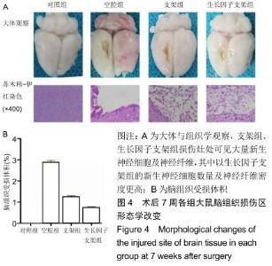
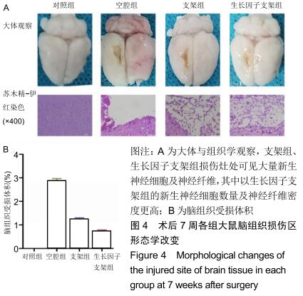
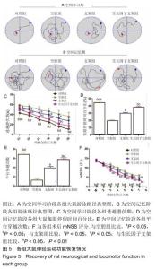
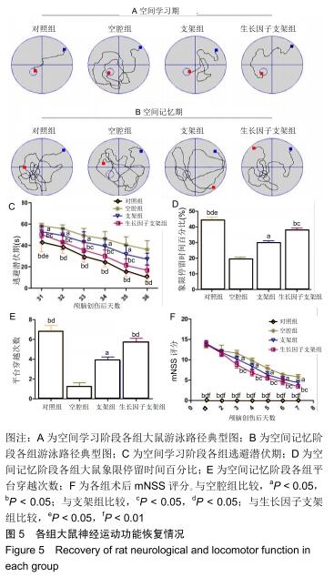
|
[1] JIANG QS, LIANG ZL, WU MJ, et al. Reduced brain-derived neurotrophic factor expression in cortex and hippocampus involved in the learning and memory deficit in molarless SAMP8 mice.Chin Med J (Engl).2011;124(10):1540-1544.
[2] MATSUMOTO T, RAUSKOLB S, POLACK M, et al. Biosynthesis and processing of endogenous BDNF: CNS neurons store and secrete BDNF, not pro-BDNF.Nat Neurosci. 2008;11(2):131-133.
[3] MITROSHINA EV, MISHCHENKO TA, USENKO AV, et al. AAV-Syn-BDNF-EGFP Virus Construct Exerts Neuroprotective Action on the Hippocampal Neural Network during Hypoxia In Vitro.Int J Mol Sci. 2018;19(8). pii:E2295.
[4] NILLESEN ST, GEUTJES PJ, WISMANS R, et al. Increased angiogenesis in acellular scaffolds by combined release of FGF2 and VEGF.J Control Release.2006;116(2):88-90.
[5] SUN B, CHEN B, ZHAO Y, et al. Crosslinking heparin to collagen scaffolds for the delivery of human platelet-derived growth factor.J Biomed Mater Res B Appl Biomater. 2009; 91(1):366-372.
[6] MOCHIZUKI M, GÜÇ E, PARK AJ, et al. Growth factors with enhanced syndecan binding generate tonic signalling and promote tissue healing.Nat Biomed Eng. 2020;4(4):463-475.
[7] CHEN C, ZHAO ML, ZHANG RK, et al. Collagen/heparin sulfate scaffolds fabricated by a 3D bioprinter improved mechanical properties and neurological function after spinal cord injury in rats.J Biomed Mater Res A.2017;105(5):1324-1332.
[8] LI X, ZHANG R, TAN X, et al. Synthesis and Evaluation of BMMSC-seeded BMP-6/nHAG/GMS Scaffolds for Bone Regeneration.Int J Med Sci.2019;16(7):1007-1017.
[9] GAO W, LI F, LIU L, et al. Endothelial colony-forming cell-derived exosomes restore blood-brain barrier continuity in mice subjected to traumatic brain injury.Exp Neurol. 2018; 307(1): 99-108.
[10] LAI BQ, FENG B, CHE MT, et al. A Modular Assembly of Spinal Cord-Like Tissue Allows Targeted Tissue Repair in the Transected Spinal Cord.Adv Sci(Weinh).2018;5(9):1800261.
[11] WANG L, WANG J, WANG F, et al. VEGF-Mediated Cognitive and Synaptic Improvement in Chronic Cerebral Hypoperfusion Rats Involves Autophagy Process.Neuromol Med.2017;19(2-3):423-435.
[12] WEN Z, XU X, XU L, et al. Optimization of behavioural tests for the prediction of outcomes in mouse models of focal middle cerebral artery occlusion.Brain Res.2017;1665(12): 88-94.
[13] JOHNSON VE, STEWART W, WEBER MT, et al. SNTF immunostaining reveals previously undetected axonal pathology in traumatic brain injury.Acta Neuropathol. 2016; 131(1):115-135.
[14] TAN HP, GUO Q, HUA G, et al. Inhibition of endoplasmic reticulum stress alleviates secondary injury after traumatic brain injury. Neural Regen Res. 2018;13(5): 827-836.
[15] SHI W, NIE D, JIN G, et al. BDNF blended chitosan scaffolds for human umbilical cord MSC transplants in traumatic brain injury therapy.Biomaterials.2012;33(11):3119-3126.
[16] GENTLEMAN E, LAY AN, DICKERSON DA, et al. Mechanical characterization of collagen fibers and scaffolds for tissue engineering.Biomaterials.2003;24(21):3805-3813.
[17] SIMIONESCU DT, LU Q, SONG Y, et al. Biocompatibility and remodeling potential of pure arterial elastin and collagen scaffolds. Biomaterials.2006;27(5):702-713.
[18] CORNWELL KG, LEI P, ANDREADIS ST, et al.Crosslinking of discrete self-assembled collagen threads: Effects on mechanical strength and cell-matrix interactions.J Biomed Mater Res A. 2007;80(2):362-371.
[19] ZHAO S, WANG Z, CHEN J, et al. Preparation of heparan sulfate-like polysaccharide and application in stem cell chondrogenic differentiation.Carbohydr Res. 2015;401(12): 32-38.
[20] LU Q, ZHANG S, HU K, et al. Cytocompatibility and blood compatibility of multifunctional fibroin/collagen/heparin scaffolds. Biomaterials.2007;28(14):2306-2313.
[21] 甘晓,南吴力.硫酸肝素/胶原蛋白神经组织工程支架修复周围神经损伤[J].中国组织工程研究,2016,20(25):3744-3749.
[22] CHEN YS, CHANG JY, CHENG CY, et al. An in vivo evaluation of a biodegradable genipin-cross-linked gelatin peripheral nerve guide conduit material.Biomaterials. 2005; 26(18):3911-3918.
[23] VÖGTLE T, SHARMA S, MORI J, et al. Heparan sulfates are critical regulators of the inhibitory megakaryocyte-platelet receptor G6b-B.Elife.2019;8.pii: e46840.
[24] CHEN L, LV Y, GUAN L, et al. Analysis of properties of collagen membranes before and after crosslinked. Zhongguo Xiu Fu Chong Jian Wai Ke Za Zhi.2008;22(2):183-187.
[25] KADOYA K, TSUKADA S, LU P, et al. Combined intrinsic and extrinsic neuronal mechanisms facilitate bridging axonal regeneration one year after spinal cord injury.Neuron. 2009; 64(2):165-172.
[26] CACIALLI P, PALLADINO A, LUCINI C.Role of Brain-Derived Neurotrophic Factor During the Regenerative Response After Traumatic Brain Injury in Adult Zebrafish.Neural Regen Res. 2018;13(6):941-944.
[27] BHATTAMISRA SK, YAP KH, RAO V, et al. Multiple Biological Effects of an Iridoid Glucoside, Catalpol and Its Underlying Molecular Mechanisms.Biomolecules.2019;10(1).pii: E32.
[28] ELAHI FM, CASALETTO KB, LA JOIE R, et al. Plasma biomarkers of astrocytic and neuronal dysfunction in early- and late-onset Alzheimer's disease.Alzheimers Dement. 2019. pii: S1552-5260(19)35373-7.
[29] HØRLYCK LD, MACOVEANU J, VINBERG M, et al. The BDNF Val66Met Polymorphism Has No Effect on Encoding-Related Hippocampal Response But Influences Recall in Remitted Patients With Bipolar Disorder.Front Psychiatry.2019;10:845.
[30] PANAGIOTAKOPOULOU V, BOTSAKIS K, DELIS F, et al. Anti-neuroinflammatory, protective effects of the synthetic microneurotrophin BNN-20 in the advanced dopaminergic neurodegeneration of "weaver" mice.Neuropharmacology. 2020;165:107919.
[31] HASSANNEJAD Z, ZADEGAN SA, VACCARO AR, et al. Biofunctionalized peptide-based hydrogel as an injectable scaffold for BDNF delivery can improve regeneration after spinal cord injury.Injury.2019;50(2):278-285.
[32] LAMPE KJ, KERN DS, MAHONEY MJ, et al. The administration of BDNF and GDNF to the brain via PLGA microparticles patterned within a degradable PEG-based hydrogel: Protein distribution and the glial response.J Biomed Mater Res A. 2011;96(3):595-607.
|