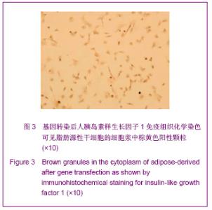
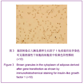
| [1] Fortier LA, Barker JU, Strauss EJ, et al. The role of growth factors in cartilage repair. Clin Orthop Relat Res. 2011; 469(10):2706-2715.[2] Spiller KL, Liu Y, Holloway JL, et al. A novel method for the direct fabrication of growth factor-loaded microspheres within porous nondegradable hydrogels: Controlled release for cartilage tissue engineering. J Control Release. 2012; 157(1): 39-45.[3] Orth P, Kaul G, Cucchiarini M, et al. Transplanted articular chondrocytes co-overexpressing IGF-I and FGF-2 stimulate cartilage repair in vivo. Knee Surg Sports Traumatol Arthrosc. 2011;19(12):2119-2130.[4] Kanazawa I, Yamaguchi T, Sugimoto T. Serum insulin-like growth factor-I is negatively associated with serum adiponectin in type 2 diabetes mellitus. Growth Horm IGF Res. 2011;21(5):268-271.[5] Jansen M, Van Schaik FM, Richer AT, et al. Sequence of cDNA encoding human insulin-like growth factor I precursor. Nature. 1983;306(8):609-611.[6] Panek B, Gacko M, Palka J.Metalloproteinases, insulin-like growth factor-I and its binding proteins in aortic aneurysm. Int J Exp Pathol.2004;85(3):159-164. [7] Ongaro A, Pellati A, Masieri FF, et al. Chondroprotective effects of pulsed electromagnetic fields on human cartilage explants. Bioelectromagnetics. 2011;32(7):543-551.[8] Hayes AJ, Ralphs JR. The response of foetal annulus fibrosus cells to growth factors: modulation of matrix synthesis by TGF-β1 and IGF-1. Histochem Cell Biol. 2011;136(2): 163-175.[9] Gallagher EJ, LeRoith D. Minireview: IGF, Insulin, and Cancer. Endocrinology. 2011;152(7):2546-2551.[10] Ng KW, O'Conor CJ, Kugler LE, et al. Transient supplementation of anabolic growth factors rapidly stimulates matrix synthesis in engineered cartilage. Ann Biomed Eng. 2011;39(10):2491-2500.[11] Chen Y, Ke J, Long X, Meng Q, et al. Insulin-like growth factor-1 boosts the developing process of condylar hyperplasia by stimulating chondrocytes proliferation. Osteoarthritis Cartilage. 2012;20(4):279-287.[12] Xue JX,Gong YY,Zhou GD,et al.Chondrogenic differentiation of bone marrow-derived mesenchymal stem cells induced by acellular cartilage sheets. Biomaterials. 2012;33(24):5832- 5840.[13] Montagnani A, Gonnelli S, Alessandri M,et al. Osteoporosis and risk of fracture in patients with diabetes: an update. Aging Clin Exp Res. 2011;23(2):84-90.[14] Shi S, Mercer S, Eckert GJ, et al. Regulation of articular chondrocyte aggrecan and collagen gene expression by multiple growth factor gene transfer. J Orthop Res. 2012; 30(7):1026-1031.[15] Rodrigues MT, Lee SJ,Gomes ME,et al.Bilayered constructs aimed at osteochondral strategies: The influence of medium supplements in the osteogenic and chondrogenic differentiation of amniotic fluid-derived stem cells. Acta Biomater. 2012;8(7):2795-2806.[16] Zhao R, Ren Y, Sun B, et al. Experimental study on chitosan mediated insulin-like growth factor gene transfection repairing injured articular cartilage in rabbits. Zhongguo Xiu Fu Chong Jian Wai Ke Za Zhi. 2010;24(11):1372-1375.[17] Thrailkill K, Quattrin T, Baker L, et al. Dual hormonal replacement therapy with insulin and recombinant human insulin-like growth factor(IGF)-1 in insulin-dependent diabetes mellitus: effects on the growth hormone/IGF/IGF-binding protein system.J Clin Endocrinol Metab. 1997;82:1181-1187.[18] Thrailkill K, Quattrin T, Baker L, et al. Cotherapy with recombinant human insulin-like growth factor I and insulin improves glycemic control in type 1 diabetes. RhIGF-I in IDDM Study Group. Diabetes Care. 1999;22:585-592.[19] Zuk PA, Zhu M, Mizuno H, et al.Multilineage cells from human adipose tissue: implications for cell-based therapies. Tissue Eng 2001;7: 211-217.[20] Evans CH, Robbins PD, Ghivizzani SC, et al. Clinical trial to assess the safety, feasibility and efficiency of transferring a potentially anti-arthritic cytokine gene to human joints with rheumatoid arthritis. Hum Gene Ther.1996;7:1261-1264. |