Chinese Journal of Tissue Engineering Research ›› 2019, Vol. 23 ›› Issue (17): 2651-2658.doi: 10.3969/j.issn.2095-4344.1715
Previous Articles Next Articles
Isolation and identification of exosomes from human adipose-derived mesenchymal stem cells
Wang Jing1, Cai Xia2, Wang Zhiguo2, Xu Quanchen3, Li Kun4, Hua Cheng5
-
Revised:2019-02-25Online:2019-06-18Published:2019-06-18 -
Contact:Cai Xia, Department of Burn and Plastic Surgery, the Affiliated Hospital of Qingdao University, Qingdao 266021, Shandong Province, China; Wang Zhiguo, Department of Burn and Plastic Surgery, the Affiliated Hospital of Qingdao University, Qingdao 266021, Shandong Province, China -
About author:Wang Jing, Master, Physician, School of Medicine, Qingdao University, Qingdao 266021, Shandong Province, China -
Supported by:the Natural Science Foundation of Shandong Province, No. XR2017MH083 (to WZG); the Shandong Provincial Key Research and Development Foundation, No. 2018GSF118150 (to XQC)
CLC Number:
Cite this article
Wang Jing, Cai Xia, Wang Zhiguo, Xu Quanchen, Li Kun, Hua Cheng. Isolation and identification of exosomes from human adipose-derived mesenchymal stem cells[J]. Chinese Journal of Tissue Engineering Research, 2019, 23(17): 2651-2658.
share this article
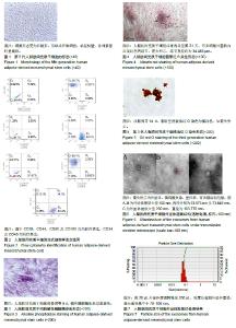
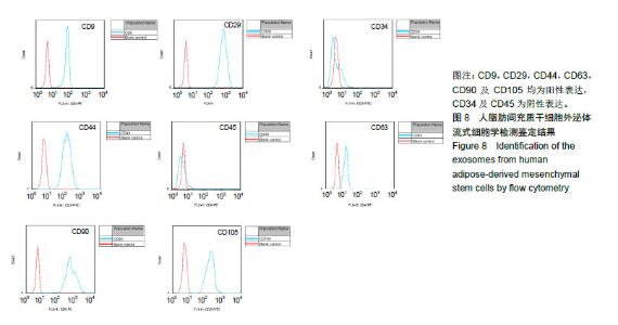
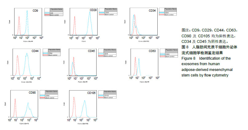
2.1 脂肪间充质干细胞的形态学观察 脂肪间充质干细胞培养24-48 h后贴壁,待5-7 d融合生长至80-90%,原代细胞形态呈梭形、圆形、多边形,大小不一。传代至第5代细胞形态变为长梭形,形似成纤维细胞,单层贴壁,排列紧密时呈巢状生长。见图1。 2.2 脂肪间充质干细胞的流式细胞学检测 CD29,CD44,CD90及CD105均为阳性表达,阳性率分别为99.7%,88.9%,96.1%及83.4%,CD34及CD45为阴性表达,阴性率分别为98.7%和92.5%,见图2。 2.3 成骨诱导细胞形态 成骨细胞诱导至4天时细胞开始变大,形态变为立方形。诱导至第9天,碱性磷酸酶染色为蓝紫色,见图3。诱导至第21天,细胞呈复层生长,结构不清,茜素红S染色可见细胞外基质内出现红色钙结节,见图4。 2.4 成脂诱导细胞形态 成脂肪细胞诱导4天细胞胞浆内开始出现透光性良好的小空泡,随着诱导时间的延长而增加,诱导至14天时出现大量透光性良好的大小不等的空泡,油红O染色为橘红色,见图5。 2.5 外泌体的形态学观察 外泌体外形为杯状,有膜结构,直径为(81.225±22.226) nm,见图6,BCA蛋白浓度测定结果为1.5 g/L,粒度仪鉴定其直径集中分布在70-100 nm,见图7。 2.6 外泌体的流式细胞学鉴定 CD9,CD29,CD44,CD63,CD90及CD105均为阳性表达,阳性率分别为99.5%,100%,99.8%,95.6%,100%及99.9%,CD34及CD45为阴性表达,阴性率分别为97.6%和82.5%。见图8。"

"
| [1] Williams KJ, Picou AA, Kish SL, et al. Isolation and characterization of porcine adipose tissue-derived adult stem cells. Cells Tissues Organs. 2008;(3):251.[2] Liao HT, Chen CT. Osteogenic potential: comparison between bone marrow and adipose-derived mesenchymal stem cells. World J Stem Cells. 2014;6(3):288.[3] De Ugarte DA, Ashjian PH, Elbarbary A, et al. Future of fat as raw material for tissue regeneration. Ann Plas Surg. 2003; 50(2):215-219.[4] Zhang J, Li S, Li L, et al. Exosome and exosomal microrna: trafficking, sorting, and function. Genom Proteom Bioinf. 2015;13(1):17 [5] van der Pol E, Boing AN, Harrison P, et al. Classification, functions, and clinical relevance of extracellular vesicles. Pharmacol Rev. 2012;64(3):676.[6] Richard JS, Hina K, Suresh M. ExoCarta as a resource for exosomal research. J Extracell Vesicles. 2012. doi: 10.3402/jev.v1i0.18374.[7] Zuk PA, Zhu M, Ashjian P. human adipose tissue is a source of multipotent stem cells. Mol Biol Cell. 2002;13(12): 4279-4295.[8] De Ugarte DA, Alfonso Z, Zuk PA, et al. Differential expression of stem cell mobilization-associated molecules on multi-lineage cells from adipose tissue and bone marrow. Immunol Lett. 2003;89(2-3):267. [9] Bunnell BA, Flaat M, Gagliardi C, et al. Adipose-derived stem cells: isolation, expansion and differentiation. Methods. 2008; 45(2):115.[10] Arrigoni E, Lopa S, de Girolamo L, et al. Isolation, characterization and osteogenic differentiation of adipose-derived stem cells: from small to large animal models. Cell Tissue Res. 2009;338(3):401 [11] Palumbo P, Lombardi F, Siragusa G, et al. Methods of isolation, characterization and expansion of human adipose-derived stem cells (ASCs): an overview. Int J Mol Sci. 2018;19(7):1897. [12] Bliley JM, Argenta A, Satish L, et al. Administration of adipose-derived stem cells enhances vascularity, induces collagen deposition, and dermal adipogenesis in burn wounds. Burns. 2016;42(6):1212. [13] Liew L, Ong H, Dilley R. Isolation and culture of adipose-derived stromal cells from subcutaneous fat. Methods Mol Biol. 2017;1627:193.[14] Riis S. Critical steps in the isolation and expansion of adipose-derived stem cells for translational therapy. Expert Rev Mol Med. 2015;17:e11.[15] Jaiswal N, Haynesworth SE, Caplan AI, et al. Osteogenic differentiation of purified, culture-expanded human mesenchymal stem cells in vitro. J Cell Biochem. 1997; 64(2): 295.[16] Baglioni S, Francalanci M, Squecco R, et al. Characterization of human adult stem-cell populations isolated from visceral and subcutaneous adipose tissue. FASEB J. 2009;23(10): 3494.[17] Farshid G, Kristen EL, Hani AA, et al. Clonal analysis of the differentiation potential of human adipose-derived adult stem cells. J Cell Physiol. 2006;206(1):229-237.[18] Jason W, Robert S, Mason MD, et al. Cancer exosomes trigger fibroblast to myofibroblast differentiation. Cancer Res. 2010;70(23):9621 [19] 方硕.干细胞来源的外泌体对成纤维细胞向肌成纤维细胞分化的调控作用及其机制研究[D].上海:第二军医大学,2016.[20] 王伟伟.HiPSC-MSCs来源Exosome促进部分肝切除后肝再生修复作用的研究[D].苏州:苏州大学,2016.[21] 王娟.脂肪干细胞来源的外来体促进皮肤创伤愈合的研究[D].武汉:华中科技大学,2016.[22] Ruenn Chai L, Fatih A, Soon Sim T, et al. Derivation and characterization of human fetal MSCs: an alternative cell source for large-scale production of cardioprotective microparticles. J Mol Cell Cardiol. 2010,48(6):1215-1224.[23] Zuk PA, Zhu M, Mizuno H, et al. Multilineage cells from human adipose tissue: implications for cell-based therapies. Tissue Eng. 2001;7(2):211-218.[24] Toyserkani NM, Christensen ML, Sheikh SP, et al. Adipose-derived stem cells: new treatment for wound healing? Ann Plas Surg. 2015;75(1):117-123.[25] John KF, Isabella W, Zeni A, et al. Fat tissue: an underappreciated source of stem cells for biotechnology. Trends Biotechnol. 2006;24(4):150-154.[26] Zhu Y, Liu T, Song K, et al. Adipose-derived stem cell: a better stem cell than BMSC. Cell Biochem Funct. 2008;26(6):664.[27] Yoshimura K, Shigeura T, Matsumoto D, et al. Characterization of freshly isolated and cultured cells derived from the fatty and fluid portions of liposuction aspirates. J Cell Physiol. 2006;208(1):64.[28] McIntosh K, Zvonic S, Garrett S, et al. The immunogenicity of human adipose-derived cells: temporal changes in vitro. Stem Cells. 2006;24(5):1246-1253.[29] Aust L, Devlin B, Foster SJ, et al. Yield of human adipose- derived adult stem cells from liposuction aspirates. Cytotherapy. 2004;6(1):7. [30] Di Taranto G, Cicione C, Visconti G, et al. Qualitative and quantitative differences of adipose-derived stromal cells from superficial and deep subcutaneous lipoaspirates: a matter of fat. Cytotherapy. 2015;(8):1076 [31] 毛曦媛,程辰,李华,等.不同部位脂肪组织来源干细胞的特性[J].中华整形外科杂志,2015,31(3):234. [32] Mitchell JB, McIntosh K, Zvonic S, et al. Immunophenotype of human adipose-derived cells: temporal changes in stromal-associated and stem cell-associated markers. Stem cells (Dayton, Ohio). 2006;24(2):376-385.[33] 吴尉,梁芳,宋小琴,等.人脂肪干细胞的提取和鉴定[J].中国组织工程研究,2015,19(28):4498. [34] Lee RH, Kim B, Choi I, et al. Characterization and expression analysis of mesenchymal stem cells from human bone marrow and adipose tissue. Cell Physiol Biochem. 2004; 14(4-6):311-324. [35] 田霖,孙筱放,刘海波,等.人脂肪干细胞的分离培养与生物学特性[J].中国组织工程研究,2012,16(32):5946 [36] Matthias S, Christoph P, Florian H, et al. Human mesenchymal stem cells at the single-cell level: simultaneous seven-colour immunofluorescence. J Anat. 2007;210(5):592-599.[37] Katia M, Ivana F, Deborah R, et al. Expansion of mesenchymal stem cells isolated from pediatric and adult donor bone marrow. J Cell Biochem. 2006;97(4):744-754.[38] Wankhade UD, Shen M, Kolhe R, et al. Advances in adipose-derived stem cells isolation, characterization, and application in regenerative tissue engineering. Stem Cells Int. 2016;2016:3206807.[39] Vishnubalaji, R., Al-Nbaheen, et al. Comparative investigation of the differentiation capability of bone-marrow- and adipose-derived mesenchymal stem cells by qualitative and quantitative analysis. Cell Tissue Res. 2012;(2):419.[40] Guilak F, Awad HA, Fermor B, et al. Adipose-derived adult stem cells for cartilage tissue engineering. Biorheology. 2004; (3/4):389. [41] Scott DM, Kent GN, Cohn DV. Collagen synthesis in cultured osteoblast-like cells. Arch Biochem Biophys. 1980;201(2): 384-391.[42] Reger RL, Tucker AH, Wolfe MR.Differentiation and characterization of human MSCs. Methods Mol Biol. 2008; 449:93.[43] Gimble J, Guilak F. Adipose-derived adult stem cells: isolation, characterization, and differentiation potential. Cytotherapy. 2003;5(5):362-369.[44] 李洪超,金银鹏,王皙,等.人脂肪干细胞及其外泌体的分离与鉴定[J].中国组织工程研究,2018,22(13):2033-2038.[45] Johnstone RM, Adam M, Hammond JR, et al. Vesicle formation during reticulocyte maturation. Association of plasma membrane activities with released vesicles (exosomes). J Biol Chem. 1987;262(19):9412.[46] Harding C, Heuser J, Stahl P. Receptor-mediated endocytosis of transferrin and recycling of the transferrin receptor in rat reticulocytes. J Cell Biol. 1983;97(2):329.[47] Pan BT, Teng K, Wu C, et al. Electron microscopic evidence for externalization of the transferrin receptor in vesicular form in sheep reticulocytes. J Cell Biol. 1985;101(3):942.[48] Huang X, Yuan T, Tschannen M, et al. Characterization of human plasma-derived exosomal RNAs by deep sequencing. Bmc Genomics. 2013;14(1):319.[49] Huang C, Fisher KP, Hammer SS, et al. Plasma exosomes contribute to microvascular damage in diabetic retinopathy (DR) by activating classical complement pathway. Diabetes. 2018;67(8):1639-1649.[50] Qin W, Tsukasaki Y, Dasgupta S, et al. Exosomes in human breast milk promote EMT. Clin Cancer Res. 2016;22(17): 4517. [51] Lukasik A, Brzozowska I, Zielenkiewicz U, et al. Detection of plant miRNAs abundance in human breast milk. Int J Mol Sci. 2017;19(1):E37.[52] Street JM, Koritzinsky EH, Glispie DM, et al. Urine Exosome Isolation and Characterization. Methods Mol Biol. 2017; 1641:413-423.[53] Gheinani AH, Vogeli M, Baumgartner U, et al. Improved isolation strategies to increase the yield and purity of human urinary exosomes for biomarker discovery. Sci Rep. 2018; 8(1):3945. [54] Nonaka T, Wong DTW. Saliva-exosomics in cancer: molecular characterization of cancer-derived exosomes in saliva. Enzymes. 2017;42:125. [55] Sun Y, Huo C, Qiao Z, et al. Comparative proteomic analysis of exosomes and microvesicles in human saliva for lung cancer. J Proteome Res. 2018;17(3):1101. [56] Raposo G, Stoorvogel W. Extracellular vesicles: exosomes, microvesicles, and friends. J Cell Biol. 2013;200(4):373.[57] Boon RA, Vickers KC. Intercellular transport of MicroRNAs. Arterioscler Thromb Vasc Biol. 2013;33(2):186.[58] A Aryani BD. Exosomes as a nanodelivery system: a key to the future of neuromedicine? Mol Neurobiol. 2016;53(2): 818-834.[59] Otani K, Mai Y, Kodama T, et al. Plasma exosomes regulate systemic blood pressure in rats. Biochem Biophys Res Commun. 2018;503(2):776-783.[60] Cocucci E, Meldolesi J. Ectosomes and exosomes: shedding the confusion between extracellular vesicles. Trends Cell Biol. 2015;25(6):364. [61] Anders WM, Gunnar R.Role of exosomes in myocardial remodeling. Circ Res. 2014;114(2):315. [62] Hu L, Wang J, Zhou X, et al. Exosomes derived from human adipose mensenchymal stem cells accelerates cutaneous wound healing via optimizing the characteristics of fibroblasts. Sci Rep. 2016;6:32993. [63] 郭子宽,张涤平,陈贤光,等.一种人脐带间充质干细胞源外泌体及其获取方法和应用[P].CN201710262727.0, 2017-04-20. [64] 纪成,费书琴,陈鸣,等.间充质干细胞来源外泌体在肾脏疾病中的研究进展[J].第二军医大学学报,2018,39(7):722-725.[65] Januszyk K, Lima CD. The eukaryotic RNA exosome. Curr Opin Struc Biol. 2014;24(1):132.[66] Gould SJ, Raposo G. As we wait: coping with an imperfect nomenclature for extracellular vesicles. J Extracell Vesicles. 2013. doi: 10.3402/jev.v2i0.20389.[67] Crescitelli R, Lässer C, Szabó TG, et al. Distinct RNA profiles in subpopulations of extracellular vesicles: apoptotic bodies, microvesicles and exosomes. J Extracell Vesicles. 2013. doi: 10.3402/jev.v2i0.20677.[68] van der Pol E, Hoekstra AG, Sturk A, et al. Optical and non-optical methods for detection and characterization of microparticles and exosomes. J Thromb Haemost. 2010; 8(12):2596.[69] 黄春煌,马媛媛,任林,等.脂肪间充质干细胞条件培养基及其外泌体促成骨作用的体外研究[J].中华口腔医学研究杂志(电子版), 2018,12(2):101.[70] Raposo G, Nijman HW, Stoorvogel W, et al. Blymphocytes secrete antigen-presenting vesicles. J Exp Med. 1996;183(3): 1161-1172.[71] Caby MP, Lankar D, Vincendeau-Scherrer C, et al. Exosomal-like vesicles are present in human blood plasma. Int Immunol. 2005;17(7):879-887.[72] 肖漓,黄海燕,毕丽丽,等.HLA-G在肾移植受者外周血外泌体中表达的初步研究[J].中华器官移植杂志,2015,36(9):515.[73] 杨向荣,丁娟,徐正阳,等.人脐带间充质干细胞来源外泌体的生物学特性研究[J].华中科技大学学报(医学版),2016,45(2):154 [74] 李玉静,刁振宇,胡娅莉.胎盘外泌体的生物学功能及分离纯化的研究进展[J].中华围产医学杂志,2015,18(6):446. |
| [1] | Pu Rui, Chen Ziyang, Yuan Lingyan. Characteristics and effects of exosomes from different cell sources in cardioprotection [J]. Chinese Journal of Tissue Engineering Research, 2021, 25(在线): 1-. |
| [2] | Fan Quanbao, Luo Huina, Wang Bingyun, Chen Shengfeng, Cui Lianxu, Jiang Wenkang, Zhao Mingming, Wang Jingjing, Luo Dongzhang, Chen Zhisheng, Bai Yinshan, Liu Canying, Zhang Hui. Biological characteristics of canine adipose-derived mesenchymal stem cells cultured in hypoxia [J]. Chinese Journal of Tissue Engineering Research, 2021, 25(7): 1002-1007. |
| [3] | Zhao Min, Feng Liuxiang, Chen Yao, Gu Xia, Wang Pingyi, Li Yimei, Li Wenhua. Exosomes as a disease marker under hypoxic conditions [J]. Chinese Journal of Tissue Engineering Research, 2021, 25(7): 1104-1108. |
| [4] | Yi Zhishuang, Wang Zhihai, Li Ming, Zhou Qingan, Yang Baosheng. Exosomes from human umbilical cord-derived mesenchymal stem cells suppress the proliferation and promote the apoptosis of medulloblastoma Daoy cells [J]. Chinese Journal of Tissue Engineering Research, 2021, 25(31): 4945-4949. |
| [5] | Ling Huajun, Wang Qiyou, Lin Weiwen, Luo Penggang. Bone marrow mesenchymal stem cell derived exosomes delay the occurrence and development of osteoarthritis through cartilage protection [J]. Chinese Journal of Tissue Engineering Research, 2021, 25(31): 4964-4969. |
| [6] | Chen Ziyang, Pu Rui, Deng Shuang, Yuan Lingyan. Regulatory effect of exosomes on exercise-mediated insulin resistance diseases [J]. Chinese Journal of Tissue Engineering Research, 2021, 25(25): 4089-4094. |
| [7] | Jiang Tao, Ma Lei, Li Zhiqiang, Shou Xi, Duan Mingjun, Wu Shuo, Ma Chuang, Wei Qin. Platelet-derived growth factor BB induces bone marrow mesenchymal stem cells to differentiate into vascular endothelial cells [J]. Chinese Journal of Tissue Engineering Research, 2021, 25(25): 3937-3942. |
| [8] | Chen Yang, Huang Denggao, Gao Yuanhui, Wang Shunlan, Cao Hui, Zheng Linlin, He Haowei, Luo Siqin, Xiao Jingchuan, Zhang Yingai, Zhang Shufang. Low-intensity pulsed ultrasound promotes the proliferation and adhesion of human adipose-derived mesenchymal stem cells [J]. Chinese Journal of Tissue Engineering Research, 2021, 25(25): 3949-3955. |
| [9] | Yu Dan, Xu Guanglan, Zhao Mei, Li Jiao, Li Guosheng, Wang Guangyao, Lin Hao, Zheng Manli, Li Yuanling. Significance and possibility of stem cells and multiple cell derived exosomes in the treatment of chronic respiratory diseases [J]. Chinese Journal of Tissue Engineering Research, 2021, 25(25): 4053-4057. |
| [10] | Gao Kun, Chen Dayu, Zhang Yong, Liu Weidong, Sun Shufen, Lai Wenqiang, Ma Dujun, Wu Yihong, Lin Zhanpeng, Jiang Yinglu, Yu Weiji. Achyranthes bidentata alcohol extract inhibits extracellular matrix degradation of the cartilage by regulating synovial fibroblast exosomes [J]. Chinese Journal of Tissue Engineering Research, 2021, 25(23): 3636-3640. |
| [11] | Wang Kang, Zhi Xiaodong, Wang Wei. Effect of stem cell derived exosomes on repairing peripheral nerve injury [J]. Chinese Journal of Tissue Engineering Research, 2021, 25(19): 3083-3089. |
| [12] | Zhao Shuangdan, Zheng Jiahua, Qi Wenbo, Huang Xianghua. Role and mechanism of exosomes derived from mesenchymal stem cells in reproductive system diseases [J]. Chinese Journal of Tissue Engineering Research, 2021, 25(19): 3097-3102. |
| [13] | Zhang Jianhui, Ma Heran, Tan Yi, Wang Zhihui. Knee injury repair using human adipose-derived mesenchymal stem cells-based scaffold-free three-dimensional gel-like construct in pigs [J]. Chinese Journal of Tissue Engineering Research, 2021, 25(19): 2969-2975. |
| [14] | Liu Feng, Zhang Yu, Wang Yanli, Luo Wei, Han Chaoshan, Li Yangxin. Application of temperature-sensitive chitosan hydrogel encapsulated exosomes in ischemic diseases [J]. Chinese Journal of Tissue Engineering Research, 2021, 25(16): 2479-2487. |
| [15] | Cai Yuan, Deng Chengliang. Therapeutic application of adipose stem cell-free liquid extracts: skin aging, wound healing, scar recovery and nerve regeneration [J]. Chinese Journal of Tissue Engineering Research, 2021, 25(13): 2097-2102. |
| Viewed | ||||||
|
Full text |
|
|||||
|
Abstract |
|
|||||