Chinese Journal of Tissue Engineering Research ›› 2019, Vol. 23 ›› Issue (14): 2147-2155.doi: 10.3969/j.issn.2095-4344.1672
Previous Articles Next Articles
Preparation and characterization of sustained-release levofloxacin bone tissue-engineered three-dimensional silk fibroin/chitosan/nano-hydroxyapatite scaffold
Ye Peng, Luo Fuli, Liu Anping, Duan Haizhen, Hu Quan, Huang Wenjin, Cheng Yun, Yu Anyong
- Department of Trauma Emergence, Affiliated Hospital of Zunyi Medical University, Zunyi 563003, Guizhou Province, China
-
Received:2018-09-29 -
Contact:Yu Anyong, MD, Professor, Chief physician, Master’s supervisor, Department of Trauma Emergence, Affiliated Hospital of Zunyi Medical University, Zunyi 563003, Guizhou Province, China -
About author:Ye Peng, MD, Attending physician, Department of Trauma Emergence, Affiliated Hospital of Zunyi Medical University, Zunyi 563003, Guizhou Province, China -
Supported by:the National Natural Science Foundation of China, No. A304 and A226 (to YAY)
CLC Number:
Cite this article
Ye Peng, Luo Fuli, Liu Anping, Duan Haizhen, Hu Quan, Huang Wenjin, Cheng Yun, Yu Anyong. Preparation and characterization of sustained-release levofloxacin bone tissue-engineered three-dimensional silk fibroin/chitosan/nano-hydroxyapatite scaffold[J]. Chinese Journal of Tissue Engineering Research, 2019, 23(14): 2147-2155.
share this article
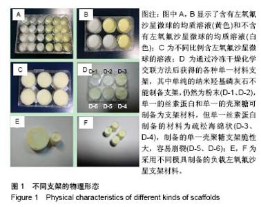
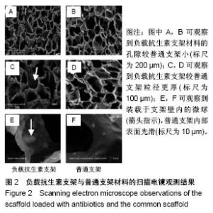
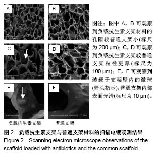
2.1 支架的物理形态观察结果 图1A,B显示了含有左氧氟沙星微球的均质溶液(黄色)和不含有左氧氟沙星微球的均质溶液(白色);图1C为不同比例含左氧氟沙星微球的溶液;图1D为通过冷冻干燥化学交联方法后获得的各种单一材料的支架;图1E,F为采用不同模具制备的负载左氧氟沙星支架材料,可根据实际情况切割制备为所需要的大小。 2.2 负载抗生素支架与普通支架的扫描电镜观察结果 图2为扫描电镜下负载抗生素支架与普通支架的微观结构,其中图2A,B可观察到负载抗生素支架材料的孔隙较普通支架小;图2C,D可观察到负载抗生素支架较普通支架粒径更厚;图2E,F可观察到装载于支架壁内的微球,普通支架内部表面光滑。"
"
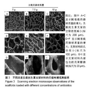
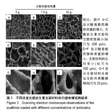
2.3 不同浓度负载抗生素支架的扫描电镜观察结果 图3为不同浓度负载抗生素支架的扫描电镜观察结果,其中图3A-C可随着载药微球质量的增大,支架的空隙密度减小;图3D-F可更典型地观察到支架材料的空隙变化情况,随着载药微球质量的升高,支架材料的孔径变小;图3G-I可以看到支架材料内部包含的壳聚糖微球。"
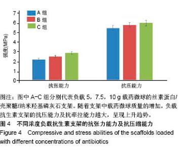
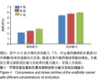
2.4 不同浓度负载抗生素支架的力学性能 根据图4可以看出,随着支架中载药微球质量的增加,负载抗生素支架的抗压能力及抗牵拉能力越大,呈现上升趋势。"
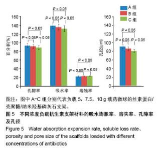
2.5 不同浓度负载抗生素支架的吸水膨胀率、溶失率、孔隙率及孔径 图5显示随着载药微球质量的增高,负载抗生素支架材料的孔隙率、吸水膨胀率、平均孔径呈逐渐减小的趋势,而热水溶失率则表现出相反的趋势。这一现象可能是载药微球的增加填充了内部材料空隙。载药微球的增加虽然能够增加支架的机械性能,但过量会影响其内部孔隙率,而合适的内部孔隙率才能够为细胞生长分化及血管的再生提供充足空间。过多的载药微球会影响支架在体内的代谢速度,影响正常骨组织的生长修复过程。因此认为以1∶1∶1比例加入7.5 g载药微球制作的复合支架最接近骨组织工程需要。"

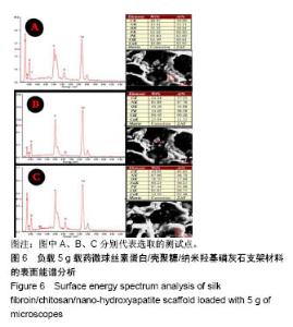
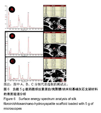
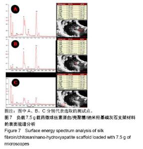
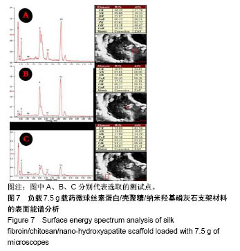
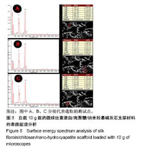
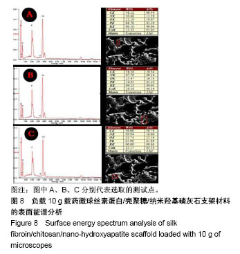
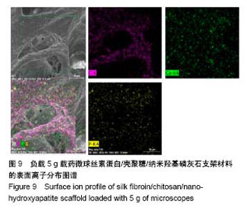
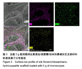
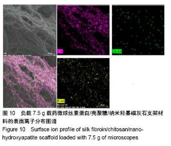
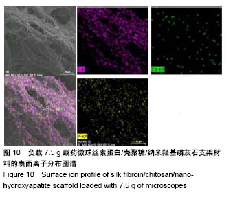
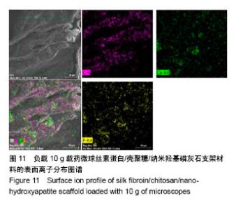
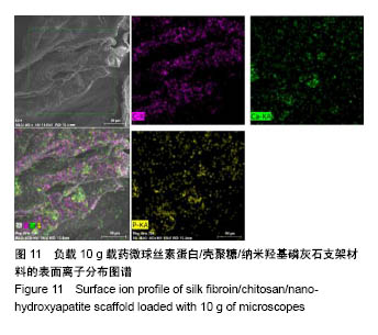
2.6 不同浓度负载抗生素支架的表面能谱分析 图6-8分别为A、B、C组支架选取不同截面进行多次表面元素构成测量的结果,可以看出各种支架材料均含有丰富的钙离子、磷离子,这些离子能为后期骨组织矿化形成提供原材料。 2.7 不同浓度负载抗生素支架的离子分布结果(C、Ca、P离子) 结果显示,各组支架表面都有丰富的钙离子、碳离子及磷离子,且离子分布均匀,其中钙离子可能来源于羟基磷灰石,见图9-11,这与实验预期结果相符合。"
"
"
"
"
"
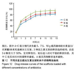
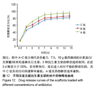
2.8 不同浓度负载抗生素支架的体外药物释放曲线 图12所示,3种抗生素支架的释放趋势相同,在前3 d释放大于50%,呈突释效应,而后进入相对平稳的释放阶段,由缓释曲线可看出浓度最大的C组其相应时间缓释率最低,浓度最小的A组其药物释放速率最快,这一现象与其结构相符合。浓度越大支架的孔径越小,孔隙越小,与溶液接触的相应表面积越小,从而释放速率低,反之浓度小的支架其释放速率快。"
| [1] Howlader D, Vignesh U, Bhutia DP, et al. Hydroxyapatite collagen scaffold with autologous bone marrow aspirate for mandibular condylar reconstruction. J Craniomaxillofac Surg. 2017; 45(9): 1566-1572.[2] Tuukkanen J, Nakamura M. Hydroxyapatite as a Nanomaterial for Advanced Tissue Engineering and Drug Therapy. Curr Pharm Des.2017;23(26):3786-3793.[3] Kamalaldin N, Jaafar M, Zubairi SI, et al. Physico-Mechanical Properties of HA/TCP Pellets and Their Three-Dimensional Biological Evaluation In Vitro.Adv Exp Med Biol.2018. doi: 10.1007/5584_2017_130. [Epub ahead of print][4] Tohamy KM, Mabrouk M, Soliman IE, et al. Novel alginate/hydroxyethyl cellulose/hydroxyapatite composite scaffold for bone regeneration: In vitro cell viability and proliferation of human mesenchymal stem cells. Int J Biol Macromol.2018; 112: 448-460.[5] Tanaka M, Haniu H, Kamanaka T, et al. Physico-Chemical, In Vitro, and In Vivo Evaluation of a 3D Unidirectional Porous Hydroxyapatite Scaffold for Bone Regeneration. Materials(Basel). 2017;10(1). pii: E33. doi: 10.3390/ma10010033.[6] Tamaddon M, Samizadeh S, Wang L, et al. Intrinsic Osteoinductivity of Porous Titanium Scaffold for Bone Tissue Engineering. Int J Biomater.2017;2017:5093063.[7] Huang B, Caetano G, Vyas C, et al. Polymer-Ceramic Composite Scaffolds: The Effect of Hydroxyapatite and beta-tri-Calcium Phosphate. Materials(Basel).2018; 11(1). pii: E129. doi: 10.3390/ ma11010129.[8] Meskinfam M, Bertoldi S, Albanese N, et al. Polyurethane foam/nano hydroxyapatite composite as a suitable scaffold for bone tissue regeneration. Mater Sci Eng C Mater Biol Appl. 2018; 82:130-140.[9] Zang S, Zhu L, Luo K, et al. Chitosan composite scaffold combined with bone marrow-derived mesenchymal stem cells for bone regeneration: in vitro and in vivo evaluation. Oncotarget. 2017;8(67):110890-110903.[10] Varoni EM, Vijayakumar S, Canciani E, et al. Chitosan-Based Trilayer Scaffold for Multitissue Periodontal Regeneration. J Dent Res.2017:1335507775.[11] Li Y, Zhang Z, Zhang Z. Porous Chitosan/ Nano-Hydroxyapatite Composite Scaffolds Incorporating Simvastatin-Loaded PLGA Microspheres for Bone Repair. Cells Tissues Organs. 2018; 205(1):20-31.[12] Zhou T, Wu J, Liu J, et al. Fabrication and characterization of layered chitosan/silk fibroin/nano-hydroxyapatite scaffolds with designed composition and mechanical properties. Biomed Mater. 2015;10(4):45013.[13] Iqbal H, Ali M, Zeeshan R, et al. Chitosan/hydroxyapatite (HA)/hydroxypropylmethyl cellulose (HPMC) spongy scaffolds-synthesis and evaluation as potential alveolar bone substitutes.Colloids Surf B Biointerfaces. 2017;160:553-563.[14] Chen BQ, Kankala RK, Chen AZ, et al. Investigation of silk fibroin nanoparticle-decorated poly(l-lactic acid) composite scaffolds for osteoblast growth and differentiation. Int J Nanomedicine. 2017; 12:1877-1890.[15] Oftadeh MO, Bakhshandeh B, Dehghan MM, et al. Sequential application of mineralized electroconductive scaffold and electrical stimulation for efficient osteogenesis. J Biomed Mater Res A. 2018;106(5):1200-1210.[16] Dhivya S, Keshav NA, Logith KR, et al. Proliferation and differentiation of mesenchymal stem cells on scaffolds containing chitosan, calcium polyphosphate and pigeonite for bone tissue engineering. Cell Prolif. 2018;51(1). doi: 10.1111/cpr.12408. Epub 2017 Nov 21.[17] Shahbazarab Z, Teimouri A, Chermahini AN, et al. Fabrication and characterization of nanobiocomposite scaffold of zein/chitosan/ nanohydroxyapatite prepared by freeze-drying method for bone tissue engineering. Int J Biol Macromol. 2018; 108:1017-1027.[18] Lu Y, Li M, Li L, et al. High-activity chitosan/nano hydroxyapatite/zoledronic acid scaffolds for simultaneous tumor inhibition, bone repair and infection eradication. Mater Sci Eng C Mater Biol Appl.2018;82:225-233.[19] Moraes PC, Marques I, Basso FG, et al. Repair of Bone Defects with Chitosan-Collagen Biomembrane and Scaffold Containing Calcium Aluminate Cement. Braz Dent J. 2017;28(3):287-295.[20] Li J, Wang Q, Gu Y, et al. Production of Composite Scaffold Containing Silk Fibroin, Chitosan, and Gelatin for 3D Cell Culture and Bone Tissue Regeneration. Med Sci Monit. 2017;23: 5311-5320.[21] Kaczmarek B, Sionkowska A, Kozlowska J, et al. New composite materials prepared by calcium phosphate precipitation in chitosan/collagen/hyaluronic acid sponge cross-linked by EDC/NHS. Int J Biol Macromol.2018;107(Pt A):247-253.[22] Lu Y, Li L, Zhu Y, et al. Multifunctional Copper-Containing Carboxymethyl Chitosan/Alginate Scaffolds for Eradicating Clinical Bacterial Infection and Promoting Bone Formation. ACS Appl Mater Interfaces.2018;10(1):127-138.[23] Ruan SQ, Deng J, Yan L, et al. Composite scaffolds loaded with bone mesenchymal stem cells promote the repair of radial bone defects in rabbit model. Biomed Pharmacother. 2018;97:600-606.[24] Shalumon KT, Lai GJ, Chen CH, et al.Modulation of Bone-Specific Tissue Regeneration by Incorporating Bone Morphogenetic Protein and Controlling the Shell Thickness of Silk Fibroin/ Chitosan/Nanohydroxyapatite Core-Shell Nanofibrous Membranes. ACS Appl Mater Interfaces.2015;7(38):21170-21181.[25] Qi X N, Mou Z L, Zhang J, et al. Preparation of chitosan/silk fibroin/hydroxyapatite porous scaffold and its characteristics in comparison to bi-component scaffolds. J Biomed Mater Res A. 2014;102(2):366-372.[26] Ruan SQ, Yan L, Deng J, et al. Preparation of a biphase composite scaffold and its application in tissue engineering for femoral osteochondral defects in rabbits. Int Orthop. 2017;41(9): 1899-1908.[27] Ruan SQ, Deng J, Yan L, et al. Composite scaffolds loaded with bone mesenchymal stem cells promote the repair of radial bone defects in rabbit model. Biomed Pharmacother. 2018;97:600-606.[28] Raina DB, Isaksson H, Teotia AK, et al. Biocomposite macroporous cryogels as potential carrier scaffolds for bone active agents augmenting bone regeneration. J Control Release. 2016;235:365-378.[29] Ran J, Hu J, Sun G, et al. A novel chitosan-tussah silk fibroin/nano-hydroxyapatite composite bone scaffold platform with tunable mechanical strength in a wide range. Int J Biol Macromol.2016;93(Pt A):87-97.[30] Bhuiyan DB, Middleton JC, Tannenbaum R, et al. Bone regeneration from human mesenchymal stem cells on porous hydroxyapatite-PLGA-collagen bioactive polymer scaffolds. Biomed Mater Eng.2017;28(6):671-685.[31] A A, Menon D, T B S, et al. Bioinspired Composite Matrix Containing Hydroxyapatite-Silica Core-Shell Nanorods for Bone Tissue Engineering. ACS Appl Mater Interfaces. 2017;9(32): 26707-26718.[32] Hernandez I, Kumar A, Joddar B. A Bioactive Hydrogel and 3D Printed Polycaprolactone System for Bone Tissue Engineering. Gels.2017;3(3).pii: 26.doi:10.3390/gels3030026. Epub 2017 Jul 6.[33] David N, Nallaiyan R. Biologically anchored chitosan/gelatin-SrHAP scaffold fabricated on Titanium against chronic osteomyelitis infection. Int J Biol Macromol. 2018; 110: 206-214.[34] Chen CK, Chang NJ, Wu YT, et al. Bone Formation Using Cross-Linked Chitosan Scaffolds in Rat Calvarial Defects. Implant Dent.2018;27(1):15-21.[35] Stepniewski M, Martynkiewicz J, Gosk J. Chitosan and its composites: Properties for use in bone substitution. Polim Med. 2017;47(1):49-53.[36] Casagrande S, Tiribuzi R, Cassetti E, et al. Biodegradable composite porous poly(dl-lactide-co-glycolide) scaffold supports mesenchymal stem cell differentiation and calcium phosphate deposition. Artif Cells Nanomed Biotechnol. 2017:1-11.[37] Garcia-Gonzalez CA, Barros J, Rey-Rico A, et al. Antimicrobial Properties and Osteogenicity of Vancomycin-Loaded Synthetic Scaffolds Obtained by Supercritical Foaming. ACS Appl Mater Interfaces. 2018;10(4):3349-3360.[38] Lin HY, Chang TW, Peng TK. 3D plotted alginate fibers embedded with diclofenac and bone cells coated with chitosan for bone regeneration during inflammation. J Biomed Mater Res A. 2018; 106(6):1511-1521.[39] Sommer MR, Vetsch JR, Leemann J, et al. Silk fibroin scaffolds with inverse opal structure for bone tissue engineering. J Biomed Mater Res B Appl Biomater. 2017;105(7):2074-2084.[40] Jo YY, Kim SG, Kwon KJ, et al. Silk Fibroin-Alginate- Hydroxyapatite Composite Particles in Bone Tissue Engineering Applications In Vivo. Int J Mol Sci.2017;18(4). pii: E858. doi: 10.3390/ijms18040858.[41] Farokhi M, Mottaghitalab F, Samani S, et al. Silk fibroin/hydroxyapatite composites for bone tissue engineering. Biotechnol Adv.2018;36(1):68-91.[42] Sangkert S, Kamonmattayakul S, Chai WL, et al. Modified porous scaffolds of silk fibroin with mimicked microenvironment based on decellularized pulp/fibronectin for designed performance biomaterials in maxillofacial bone defect. J Biomed Mater Res A. 2017;105(6):1624-1636.[43] Bhattacharjee P, Naskar D, Maiti TK, et al. Non-mulberry silk fibroin grafted poly (capital JE, Ukrainian-caprolactone)/nano hydroxyapatite nanofibrous scaffold for dual growth factor delivery to promote bone regeneration. J Colloid Interface Sci. 2016;472: 16-33.[44] Mclaren JS, White LJ, Cox HC, et al. A biodegradable antibiotic-impregnated scaffold to prevent osteomyelitis in a contaminated in vivo bone defect model. Eur Cell Mater. 2014;27: 332-349.[45] Lu M, Liao J, Dong J, et al. An effective treatment of experimental osteomyelitis using the antimicrobial titanium/silver-containing nHP66(nano-hydroxyapatite/polyamide-66) nanoscaffold biomaterials. Sci Rep.2016;6:39174.[46] Mostafa AA, El-Sayed M, Mahmoud AA, et al. Bioactive/Natural Polymeric Scaffolds Loaded with Ciprofloxacin for Treatment of Osteomyelitis.AAPS Pharm Sci Tech.2017;18(4):1056-1069.[47] Zhou J, Zhou XG, Wang JW, et al. Treatment of osteomyelitis defects by a vancomycin-loaded gelatin/beta-tricalcium phosphate composite scaffold.Bone Joint Res. 2018;7(1):46-57.[48] Pacheco H, Vedantham K, Aniket, et al. Tissue engineering scaffold for sequential release of vancomycin and rhBMP2 to treat bone infections. J Biomed Mater Res A. 2014;102(12):4213-4223.[49] Dorati R, Detrizio A, Modena T, et al. Biodegradable Scaffolds for Bone Regeneration Combined with Drug-Delivery Systems in Osteomyelitis Therapy. Pharmaceuticals(Basel).2017; 10(4). pii: E96. doi: 10.3390/ph10040096.[50] David N, Nallaiyan R. Biologically anchored chitosan/gelatin-SrHAP scaffold fabricated on Titanium against chronic osteomyelitis infection.Int J Biol Macromol. 2018;110: 206-214.[51] Jr Sanchez CJ, Prieto EM, Krueger CA, et al. Effects of local delivery of D-amino acids from biofilm-dispersive scaffolds on infection in contaminated rat segmental defects. Biomaterials. 2013;34(30):7533-7543.[52] Cao ZD, Jiang DM, Yan L, et al. Biosafety of the Novel Vancomycin-loaded Bone-like Hydroxyapatite/Poly-amino Acid Bony Scaffold. Chin Med J(Engl).2016;129(2):194-199.[53] Hassani B N, Mottaghitalab F, Eslami M, et al. Sustainable Release of Vancomycin from Silk Fibroin Nanoparticles for Treating Severe Bone Infection in Rat Tibia Osteomyelitis Model. ACS Appl Mater Interfaces.2017;9(6):5128-5138.[54] Hess U, Hill S, Treccani L, et al. A mild one-pot process for synthesising hydroxyapatite/biomolecule bone scaffolds for sustained and controlled antibiotic release. Biomed Mater.2015; 10(1):15013.[55] Huang TY, Su WT, Chen PH. Comparing the Effects of Chitosan Scaffolds Containing Various Divalent Metal Phosphates on Osteogenic Differentiation of Stem Cells from Human Exfoliated Deciduous Teeth. Biol Trace Elem Res.2018; 185(2):316-326.[56] Pipattanawarothai A, Suksai C, Srisook K, et al. Non-cytotoxic hybrid bioscaffolds of chitosan-silica: Sol-gel synthesis, characterization and proposed application.Carbohydr Polym. 2017;178:190-199.[57] Drampalos E, Mohammad HR, Kosmidis C, et al. Single stage treatment of diabetic calcaneal osteomyelitis with an absorbable gentamicin-loaded calcium sulphate/hydroxyapatite biocomposite: The Silo technique. Foot(Edinb).2017;34:40-44.[58] Posadowska U, Brzychczy-Wloch M, Pamula E. Gentamicin loaded PLGA nanoparticles as local drug delivery system for the osteomyelitis treatment.Acta Bioeng Biomech. 2015;17(3):41-48.[59] Rumian L, Tiainen H, Cibor U, et al. Ceramic scaffolds enriched with gentamicin loaded poly(lactide-co-glycolide) microparticles for prevention and treatment of bone tissue infections. Mater Sci Eng C Mater Biol Appl.2016;69:856-864.[60] Wang Q, Chen C, Liu W, et al. Levofloxacin loaded mesoporous silica microspheres/nano-hydroxyapatite/ polyurethane composite scaffold for the treatment of chronic osteomyelitis with bone defects. Sci Rep.2017;7:41808. |
| [1] | Zhang Tongtong, Wang Zhonghua, Wen Jie, Song Yuxin, Liu Lin. Application of three-dimensional printing model in surgical resection and reconstruction of cervical tumor [J]. Chinese Journal of Tissue Engineering Research, 2021, 25(9): 1335-1339. |
| [2] | Zeng Yanhua, Hao Yanlei. In vitro culture and purification of Schwann cells: a systematic review [J]. Chinese Journal of Tissue Engineering Research, 2021, 25(7): 1135-1141. |
| [3] | Xu Dongzi, Zhang Ting, Ouyang Zhaolian. The global competitive situation of cardiac tissue engineering based on patent analysis [J]. Chinese Journal of Tissue Engineering Research, 2021, 25(5): 807-812. |
| [4] | Wu Zijian, Hu Zhaoduan, Xie Youqiong, Wang Feng, Li Jia, Li Bocun, Cai Guowei, Peng Rui. Three-dimensional printing technology and bone tissue engineering research: literature metrology and visual analysis of research hotspots [J]. Chinese Journal of Tissue Engineering Research, 2021, 25(4): 564-569. |
| [5] | Li Li, Ma Li. Immobilization of lactase on magnetic chitosan microspheres and its effect on enzymatic properties [J]. Chinese Journal of Tissue Engineering Research, 2021, 25(4): 576-581. |
| [6] | Chang Wenliao, Zhao Jie, Sun Xiaoliang, Wang Kun, Wu Guofeng, Zhou Jian, Li Shuxiang, Sun Han. Material selection, theoretical design and biomimetic function of artificial periosteum [J]. Chinese Journal of Tissue Engineering Research, 2021, 25(4): 600-606. |
| [7] | Liu Fei, Cui Yutao, Liu He. Advantages and problems of local antibiotic delivery system in the treatment of osteomyelitis [J]. Chinese Journal of Tissue Engineering Research, 2021, 25(4): 614-620. |
| [8] | Li Xiaozhuang, Duan Hao, Wang Weizhou, Tang Zhihong, Wang Yanghao, He Fei. Application of bone tissue engineering materials in the treatment of bone defect diseases in vivo [J]. Chinese Journal of Tissue Engineering Research, 2021, 25(4): 626-631. |
| [9] | Zhang Zhenkun, Li Zhe, Li Ya, Wang Yingying, Wang Yaping, Zhou Xinkui, Ma Shanshan, Guan Fangxia. Application of alginate based hydrogels/dressings in wound healing: sustained, dynamic and sequential release [J]. Chinese Journal of Tissue Engineering Research, 2021, 25(4): 638-643. |
| [10] | Chen Jiana, Qiu Yanling, Nie Minhai, Liu Xuqian. Tissue engineering scaffolds in repairing oral and maxillofacial soft tissue defects [J]. Chinese Journal of Tissue Engineering Research, 2021, 25(4): 644-650. |
| [11] | Xing Hao, Zhang Yonghong, Wang Dong. Advantages and disadvantages of repairing large-segment bone defect [J]. Chinese Journal of Tissue Engineering Research, 2021, 25(3): 426-430. |
| [12] | Chen Siqi, Xian Debin, Xu Rongsheng, Qin Zhongjie, Zhang Lei, Xia Delin. Effects of bone marrow mesenchymal stem cells and human umbilical vein endothelial cells combined with hydroxyapatite-tricalcium phosphate scaffolds on early angiogenesis in skull defect repair in rats [J]. Chinese Journal of Tissue Engineering Research, 2021, 25(22): 3458-3465. |
| [13] | Wang Hao, Chen Mingxue, Li Junkang, Luo Xujiang, Peng Liqing, Li Huo, Huang Bo, Tian Guangzhao, Liu Shuyun, Sui Xiang, Huang Jingxiang, Guo Quanyi, Lu Xiaobo. Decellularized porcine skin matrix for tissue-engineered meniscus scaffold [J]. Chinese Journal of Tissue Engineering Research, 2021, 25(22): 3473-3478. |
| [14] | Mo Jianling, He Shaoru, Feng Bowen, Jian Minqiao, Zhang Xiaohui, Liu Caisheng, Liang Yijing, Liu Yumei, Chen Liang, Zhou Haiyu, Liu Yanhui. Forming prevascularized cell sheets and the expression of angiogenesis-related factors [J]. Chinese Journal of Tissue Engineering Research, 2021, 25(22): 3479-3486. |
| [15] | Li Xinping, Cui Qiuju, Zeng Shuguang, Ran Gaoying, Zhang Zhaoqiang, Liu Xianwen, Fang Wei, Xu Shuaimei. Effect of modification of β-tricalcium phosphate/chitosan hydrogel on growth and mineralization of dental pulp stem cells [J]. Chinese Journal of Tissue Engineering Research, 2021, 25(22): 3493-3499. |
| Viewed | ||||||
|
Full text |
|
|||||
|
Abstract |
|
|||||