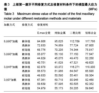
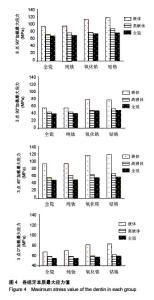
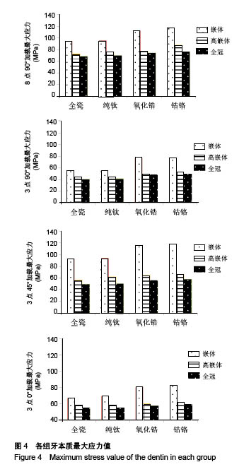
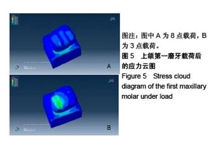
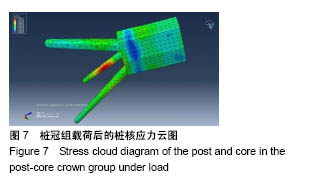
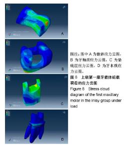
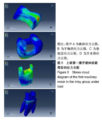
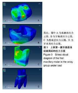
| [1] 冯海兰.口腔修复学[M].北京:北京大学医学出版社,2013.[2] Harsha MS,Praffulla M,Babu MR,et al.The Effect of Cavity Design on Fracture Resistance and Failure Pattern in Monolithic Zirconia Partial Coverage Restorations-An In vitro Study.J Clin Diagn Res. 2017;11(5):ZC45-ZC48.[3] Guo L,Wang XJ,Wan R,et al.Clinical application of gold alloy onlay on restoration of molars with severe attrition.Chin J Prosthodont.2016;17(6):332-335.[4] Haiyue YU,Dandan MA,Lin X,et al.The clinic outcome of CAD/CAM onlay in the restoration of molars with extensive defect after root canal treatment.JPract Stomatol. 2017;33:41-44.[5] 石晓婷,尹新芹,林志勇,等.不同根管预备方法对根管壁应力分布的影响[J].临床口腔医学杂志,2013, 29(2):92-94.[6] Dejak B,M?otkowski A.3D-Finite element analysis of molars restored with endocrowns and posts during masticatory simulation. Dent Mater.2013;29(12):e309.[7] Frankenberger R,ZeilingerI,Krech M,et al.Stability of endodontically treated teeth with differently invasive restorations: Adhesive vs. non-adhesive cusp stabilization. Dent Mater.2015; 31(11):1312-1320.[8] Juloski J,Apicella D,Ferrari M.The effect of ferrule height on stress distribution within a tooth restored with fibre posts and ceramic crown: a finite element analysis.Dent Mater.2014; 30(12):1304-1315.[9] 陈小文,桑卓,古育娣,等.观察三种嵌体对牙体缺损的修复效果[J].中国处方药,2014,12(7):33-34.[10] Lee KS,Shin JH,Kim JE,e tal.Biomechanical Evaluation of a Tooth Restored with High Performance Polymer PEKK Post-Core System: A 3D Finite Element Analysis.Biomed Res Int.2017; 2017 (2):1373127.[11] 王文亚,傅波,罗华,等.不同桩核冠修复上颌中切牙的三维有限元模型建立及应力分析[J].医用生物力学, 2014,29(1):25-30.[12] 赵信义.口腔材料学[M].5版.北京:人民卫生出版社,2013:59-61.[13] 蔡跃,黄英,张慧,等.有限元分析上颌中切牙唇、舌向斜形缺损后纤维桩核冠修复三维模型的应力分布[J]. 中国组织工程研究, 2017, 21(30):4824-4830.[14] Nakamura T,Imanishi A,Kashima H,et al.Stress analysis of metal-free polymer crowns using the three-dimensional finite element method.IntJ Prosthodont.2001;14(5):401-405.[15] Toniollo MB,Macedo AP,Pupim D,et al. Finite Element Analysis of Bone Stress in the Posterior Mandible Using Regular and Short Implants, in the Same Context, with Splinted and Nonsplinted Prostheses.Int J Oral Maxillofac Implants. 2017;32(4):199-206.[16] 刘涛,耿海霞.下颌第一磨牙不同桩核位置数目金合金桩核修复后基牙应力的三维有限元分析[J].中国组织工程研究, 2017,21(22): 3501-3506.[17] 肖丽婷,郑纪伟,林融,等.3种不同方法修复后牙牙体缺损的疗效对比分析[J].中国校医,2014, 28(9):695-696.[18] 顾新华,蔡盛东,M.Kern.不同粘固剂对 IPS-Empress 2后牙全瓷冠边缘适合性的影响[J].实用口腔医学杂志, 2002,18(5):441-443.[19] Hayashi M,Tsuchitani Y,MiuraM,et al.6-year clinical evaluation of fired ceramic inlays.Oper Dent.1999;42(6):318-326.[20] Tortopidis D,Lyons MF,Baxendale RH,et al.The variability of bite force measurement between sessions, in different positions within the dental arch.J Oral Rehabil.1998;25(9):681–686.[21] Bakke M,Holm B,Jensen BL,et al.Unilateral, isometric bite force in 8-68-year-old women and men related to occlusalfactors. Eur J Oral Sci.1990;98(2):149-158.[22] Gibbs CH,Mahan PE,Lundeen HC,et al.Occlusal forces during chewing and swallowing as measured by sound transmission. JProsthet Dent.1981;46(4):443.[23] Koolstra JH, van Eijden TM, Weijs WA, et al. A three-dimensional mathematical model of the human masticatory system predicting maximum possible bite forces. J Biomech.1988;21(7):563-576.[24] 刘利军,缪羽.三种不同材料的带桩高嵌体修复上颌第一前磨牙的三维有限元分析[J].中华老年口腔医学杂志, 2015,13(3):168-172.[25] Durand LB,Guimarães JC,Monteiro JS,et al.Effect of ceramic thickness and composite bases on stress distribution of inlays - a finite element analysis.Braz Dent J.2015;26(2):146-151.[26] 韩梦.四种修复方法对不同程度缺损下颌第一磨牙应力的影响—三维有限元研究[D].郑州:郑州大学,2015.[27] Tek㧠EN,Pala K,Demirci M,et al. Influence of different composite materials and cavity preparation designs on the fracture resistance of mesio-occluso-distal inlay restoration.Dent MaterJ. 2016;35(3): 523-531. [28] Lin C,Chang YP.Multi-factorial analysis of a cusp-replacing adhesive premolar restoration: A finite element study.J Dent. 2008;36(3): 194-203.[29] Guven S,Akdogan M,Oz C,et al.Three-dimensional finite-element analysis of two ceramic inlay restorations with different cavity designs.BiotechnolBiotechnol Eq. 2015;29(3):579-585.[30] 徐红梅,贾静,朱晓英,等.根管治疗后下颌磨牙采用各种充填材料修复的三维有限元分析[J].口腔颌面修复学杂志, 2014,15(2):75-79.[31] Su C,Su Y,Li Z,et al.In situ synthesis of bilayered gradient poly(vinyl alcohol)/hydroxyapatite composite hydrogel by directional freezing-thawing and electrophoresis method. Mater SciEng C Mater Biol Appl.2017;77:76-83.[32] Han SH,Sadr A,Tagami J,et al.Internal adaptation of resin composites at two configurations: Influence of polymerization shrinkage and stress. Dent Mater.2016;32(9):1085-1094.[33] Fronza BM,Rueggeberg FA,Braga RR,et al.Monomer conversion, microhardness, internal marginal adaptation, and shrinkage stress of bulk-fill resin composites.Dent Mater. 2015;31(12):1542-1551.[34] Holberg C,Winterhalder P,Wichelhaus A,et al. Fracture risk of lithium-disilicate ceramic inlays: A finite element analysis.Dent Mater.2013;29(12):1244-1250.[35] 张颜,初晓阳,刘大军,等.三维有限元法分析不同粘接剂层对瓷贴面应力分布的影响[J].口腔颌面修复学杂志,2014,15(2):84-87.[36] 李欢欢.下颌第一磨牙颊牙合缺损不同修复形式的三维有限元分析[D].郑州:郑州大学,2016.[37] 刘利军,缪羽,姚丽英.三种不同材料带桩高嵌体修复上颌第一前磨牙三维有限元分析[J].疾病监测与控制, 2016,10(2):143-144.[38] Ozyoney G,Hayran O.The efficacy of glass-ceramic onlays in the restoration of morphologically compromised and endodontically treated molars.Int J Prosthodont. 2013;26(3):230-234.[39] Morimoto S,Rebello de Sampaio FB,Braga MM,et al.Survival Rate of Resin and Ceramic Inlays, Onlays, and Overlays: A Systematic Review and Meta-analysis.J Dent Res. 2016;95(9):985. [40] 张孝霞,韩丁,朱庆林,等.大面积缺损的下颌第一前磨牙桩核冠与高嵌体修复的三维有限元分析[J].牙体牙髓牙周病学杂志, 2018, 28(5):265-270.[41] 冯广智,蔡婧.上颌中切牙四种不同材料桩核冠的有限元分析[J].临床医药文献电子杂志,2017, 4(6):1046-1047. |