Chinese Journal of Tissue Engineering Research ›› 2019, Vol. 23 ›› Issue (28): 4503-4509.doi: 10.3969/j.issn.2095-4344.1470
Previous Articles Next Articles
Research status and development trend of bibliometrics and visualization analysis in the assessment of Perthes disease
- 1Guangzhou University of Chinese Medicine, Guangzhou 510405, Guangdong Province, China; 2 Third Department of Orthopedics, the First Affiliated Hospital of Guangzhou University of Chinese Medicine, Guangzhou 510405, Guangdong Province, China; 3Research Center for Hip Joint of Guangzhou University of Chinese Medicine, Guangzhou 510405, Guangdong Province, China
-
Online:2019-10-08Published:2019-10-08 -
Contact:Wei Qiushi, MD, Associate chief physician, Third Department of Orthopedics, the First Affiliated Hospital of Guangzhou University of Chinese Medicine, Guangzhou 510405, Guangdong Province, China; Research Center for Hip Joint of Guangzhou University of Chinese Medicine, Guangzhou 510405, Guangdong Province, China -
About author:Shen Yingshan, Master candidate, Guangzhou University of Chinese Medicine, Guangzhou 510405, Guangdong Province, China -
Supported by:the General Program of National Natural Science Foundation of China, No. 81473697 and 81573996 (to HW)| the Department of Science and Technology of Guangdong Province-Guangdong Provincial Academy of Chinese Medicine Combined Research Project, No. 2016A020226028 (to HW)| the Natural Science Foundation of Guangdong Province, No. 2017A030313698 (to HW)| the Guangdong Provincial Strength Province Construction Project of Chinese Medicine for Dominant Disease (Osteonecrosis of Femoral Head), No. [2015]19 (to HW)| the Guangdong Provincial Famous Chinese Medicine (He Wei) Heritage Studio Construction Project, No. [2017]17 (to HW)
CLC Number:
Cite this article
Shen Yingshan, Gong Shuidi, He Xiaoming, Pang Fengxiang, Chen Xiaojun, Li Weifeng, Chen Lixin, Yang Fan, Yang Peng, Chen Zhenqiu, He Wei, Wei Qiushi. Research status and development trend of bibliometrics and visualization analysis in the assessment of Perthes disease[J]. Chinese Journal of Tissue Engineering Research, 2019, 23(28): 4503-4509.
share this article
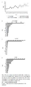
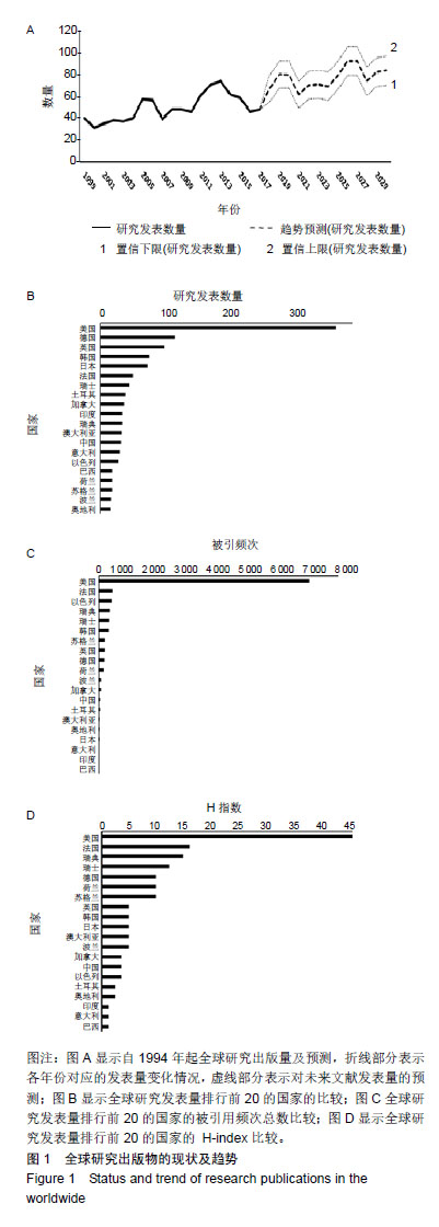
2.1 全球研究出版物的现状及趋势 2.1.1 全球研究出版量及预测 通过检索1994年至 2018年7月30日全球Perthes病的相关文献,共有1 106篇文献被纳入,其中2013年发表的文献最多共74篇,占总文献量的6.7%。图1A折线部分表示从1994到2017年,每年全球文献的发表数量呈波动式的递增。另外,虚线部分表示对未来文献发表数量的预测的置信区间,显示研究发表量会随时间推移而不断增加,预计到2030年的年发表量可达83篇,其中下限为70篇,上限可达96篇左右。 2.1.2 不同国家的研究发表量 图1B显示了在该研究领域的文献发表量在全球排行前20的国家,统计数据显示美国发表相关文章(346篇,31.28%),其次是德国(109篇,9.86%),英国(94篇,8.50%),韩国(72篇,6.639%)和日本(70篇,6.51%)。 2.1.3 不同国家出版物的被引频次 被引频次及H指数是衡量出版文献质量的重要指标,被引频次越高代表文章的价值也越高。图1C显示了该领域的文献发表量在全球排行前20的国家所对应的文献被引频次。统计数据显示,美国在Perthes病的研究领域的被引文频次最高(7 039次),以色列(435次),居第2位,其次是瑞士(351次)、韩国(332次)和英国(210次)。 2.1.4 不同国家出版物的H指数 H指数不仅反映了发表出版文献的次数,还反映其被引用的最低次数。图1D显示了该领域的文献发表量在全球排行前20的国家对应的文献 H指数。统计数据显示美国的相关文献H指数最高(40),其次是瑞士(10)、德国(8)、英国(4)和韩国(4)。"
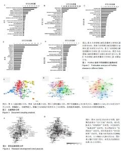
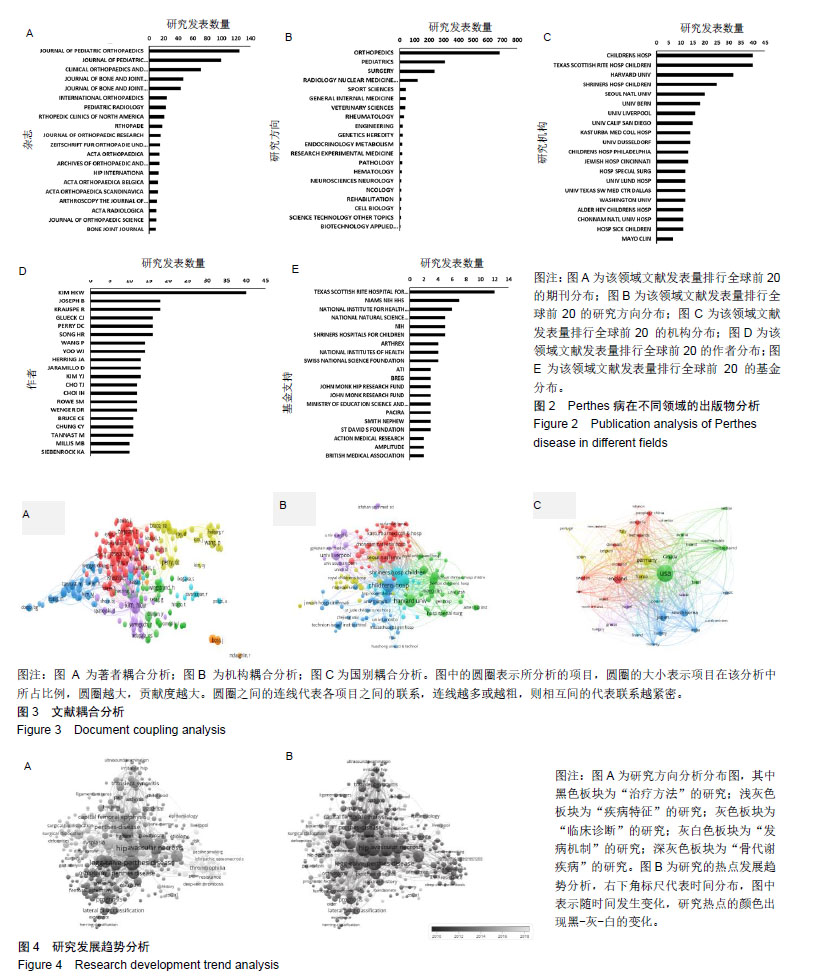
2.2 Perthes病在不同领域的出版物分析 2.2.1 各期刊杂志发表量分析 图2A为该领域文献发表量在全球范围内排行前20的期刊杂志分布情况。根据此次研究所统计的数据显示,在Perthes病领域,《JOURNAL OF PEDIATRIC ORTHOPAEDICS》杂志发表文献最多,共125篇,其次是《JOURNAL OF PEDIATRIC ORTHOPAEDICS PART B》杂志共发表100篇,《CLINICAL ORTHOPAEDICS AND RELATED RESEARCH》杂志共发表72篇,《JOURNAL OF BONE AND JOINT SURGERY AMERICAN VOLUME》杂志共发表47篇,《JOURNAL OF BONE AND JOINT SURGERY BRITISH VOLUME》杂志共发表44篇。 2.2.2 研究方向分析 随着Perthes病的发现与发展,人们对其病因、发病机制、诊治的研究也不断深入。图2B显示了当前Perthes病的研究方向,其中最主要的几个研究领域包括整形手术、儿科学、外科手术、影像学、运动科学。 2.2.3 研究机构文献发表数量分析 图2C为全球对Perthes病进行研究的机构所贡献的文献数量排行前20的机构名称分布。其中“Children Hospital”与“Texas Scottish Rite Hospital For Children”共列第1位,发表文献最多(40篇),“Harvard Univ”排名其次(32篇),“Shriners Hospital for Children”排名第3位(25篇)。 2.2.4 作者文献发表数量分析 图2D显示了全球范围内在该领域文献发表量排行前20的作者排名,其中发表量最多的前3位作者是“KIM HKW”(40篇)、“JOSEPH B”和“KRAUSPE R”(18篇)。 2.2.5 基金支持研究 科学研究离不开研究基金的大力支持,图2E列出了全球范围内提供基金支持产出文献发表量排行前20的基金项目,其中基金项目“Texas Scottish Rite Hospital For Children”(德克萨斯苏格兰礼仪儿童医院)排名第一,共支持发表12篇文献研究。基金项目“NIAMS NIH HHS”(美国国立关节肌肉骨骼及皮肤病研究所)排名第二,共支持发表7篇文献研究。“National Institute For Health Research”(英国国家健康科学研究所)排名第三,共支持发表6篇文献研究。“National Natural Science Foundation Of China”(中国国家自然基金)排名第四,共支持发表 5篇文献研究。“NIH”(美国国立卫生研究院)排名第五,共支持发表5篇文献研究。 2.3 全球研究的可视化分析 2.3.1 文献耦合分析 文献耦合分析图见图3,文献耦合是指文献之间通过参考文献进行的耦合,可分为论文耦合、学科耦合、著者耦合以及期刊耦合等类型。此外,还有文献所属国别耦合、文献语种耦合等。而耦合的强度取决于共同参考文献(被引文献)的数量。该种类型的分析被广泛应用于情报科学、文献计量学、科学学、未来学等领域当中。 (1)著者耦合分析:纳入的全部引文资料共包括3 496 位著者,通过定义每位作者至少有3篇文献相关联,共纳入 296位著者,见图3A,其中在该领域中发表文献的耦合强度排行前五的著者为:Kim HKW(总联系强度为45 097);Josper B(总联系强度为24 116);Wiig O(总联系强度为 22 665);Krauspe R(强度联系次数为21 854);Kim H(总联系强度为19 346)。 (2)机构耦合分析:纳入的全部引文资料共包括1 208个机构,通过定义至少存在3个组织机构的研究文献,纳入163个机构,见图3B,其中该领域发表文献的耦合强度排行前五的机构为:“TEXAS SCOTTISH RITE HOSP CHILDREN”(强度联系次数为25 926),“CHILDRENS HOSP”(强度联系次数为21 214),“HARVARD UNIV”(强度联系次数为15 591),“SHRINERS HOSP CHILDREN” (强度联系次数为13 368),“UNIV BERN”(强度联系次数为13 243)。 (3)国别耦合分析:纳入的全部引文资料共包括57个国家,通过定义至少存在3个国家的研究文献,纳入40个国家,见图3C,其中在该领域中发表文献的耦合强度排行前五的国家为:美国(强度联系次数为117 675)、英国(强度联系次数为42 101)、德国(强度联系次数为40 366)、韩国(强度联系次数为35 611)、日本(强度联系次数为31 669)。 2.3.2 共现分析 “共现分析”是对共现现象的定量研究,以揭示信息的内容关联和特征项所隐含的知识。而对关键词的共现分析是通过研究大量文档中的共现关系,分析共现关键词之间的链接强度,其目的是描述某一学科领域的内部构成关系和结构,揭示该学科的研究前沿[15]。 (1)研究方向分析:通过定义纳入关键词在出版物中使用超过5次的文献,并使用Vos viewer软件进行分析。软件识别出关键词共3 093个,符合条件纳入分析324个,见图4A,根据关键词可把目前全球的研究方向分为5个集群,大致为:疾病评估、疾病特征、疾病诊断、发病机制和坏死情况。 在“治疗方法”研究中,常用的关键词有“手术治疗、保守治疗、控制治疗、骨盆截骨术、内翻截骨术、股骨头截骨术”。在“疾病特征”研究中,常用的关键词有“股骨头骨骺、髋部发育不良、撞击征、盂唇撕裂、骨折、变形”。在“临床诊断”研究中,常用的关键词有“一过性滑膜炎、化脓性关节炎、刺激性髋关节、放射摄影技术、超声检查、MRI”。在“发病机制”研究中,常用的关键词有“血栓形成、被动吸烟、C-反应蛋白、低纤溶的、血管畸形”。在“骨代谢疾病”研究中,常用的关键词有“骨质疏松、特发性关节炎、缺血性坏死、股骨头坏死、塌陷”。 (2)研究发展趋势分析:Vos viewer软件可以通过“Overlay Visualization”(叠加功能)对不同关键词在其对应的文献中所出现的平均时间进行颜色归类,使不同时间内的研究热点一目了然,见图4B。如图中的标尺所示,颜色越深表示关键字出现较早,属于既往的研究热点,随着时间的推移,图中颜色出现黑-灰-白的变化,越靠近白色部分表示近期的研究重点,且连接点的面积越大,表明其相关性越强。 由图4可以分析得出,在2012年之前大多数研究集中在“治疗方法”、“临床诊断”、“发病机制”和“骨代谢疾病”的研究上。2012年之后研究重点集中于“疾病特征”,其中“评分”是该板块近年来提出的研究点。 "
| [1]吴在德,吴肇汉.外科学[M]. 北京:人民卫生出版社,2011:839[2]Wiig O. Perthes' disease in Norway. A prospective study on 425 patients. Acta Orthop Suppl. 2009;80(333):1-44.[3]Perry DC, Bruce CE, Pope D, et al. Comorbidities in Perthes' disease: a case control study using the General Practice Research database. J Bone Joint Surg Br. 2012;94(12):1684.[4]Loder RT, Skopelja EN. The Epidemiology and Demographics of Legg-Calvé-Perthes' Disease. ISRN Orthop. 2011;2011(1): 504393-504393.[5]Tannast M, Macintyre N, Steppacher SD, et al. A systematic approach to analyse the sequelae of LCPD. Hip Int. 2013;23 Suppl 9(Suppl 9): S61-70.[6]Mazloumi SM, Ebrahimzadeh MH, Kachooei AR. Evolution in diagnosis and treatment of Legg-Calve-Perthes disease. Arch Bone Joint Surg. 2014; 2(2):86-92.[7]Rampal V, Clément JL, Solla F. Legg-Calvé-Perthes disease: classifications and prognostic factors. Clin Cases Miner Bone Metab. 2017;14(1):74-82. [8]Waldenström H. The definite form of the coxa plana. Acta Radiol. 1922;4(1): 384-394.[9]Catterall A. The natural history of Perthes’disease. J Bone Joint Surg Br.1971;53B: 37-53.[10]Salter RB, Thompson GH. Legg-Calvé-Perthes disease. The prognostic significance of the subchondral fracture and a two-group classification of the femoral head involvement. J Bone Joint Surg Am. 1984;66(4): 479-489.[11]Herring JA, Neustadt JB, Williams JJ, et al. The lateral pillar classification of Legg-Calve -Perthes disease. J Pediatr Orthop. 1992; 12:143-150.[12]Herring JA, Kim HT, Browne R. Legg-Calve-Perthes disease. Part I: Classification of radiographs with use of the modified lateral pillar and Stulberg classifications. J Bone Joint Surg Am. 2004;86-A(10): 2103.[13]Mose K. Methods of measuring in Legg-Calvé-Perthes disease with special regard to the prognosis. Clin Orthop Relat Res. 1980;(150): 103-109.[14]Mazloumi SM, Ebrahimzadeh MH, Kachooei AR. Evolution in diagnosis and treatment of Legg-Calve-Perthes disease. Arch Bone Joint Surg. 2014; 2(2):86-92.[15]Jamil K, Zacharin M, Foster B, et al. Protocol for a randomised control trial of bisphosphonate (zoledronic acid) treatment in childhood femoral head avascular necrosis due to Perthes disease. BMJ Paediatr Open. 2017;1(1): e000084.[16]吴生康,何鹤皋. 高压氧治疗 38 例儿童股骨头缺血性坏死[J]. 中华航海医学与高气压医学杂志,2005,12(1) : 60-62.[17]俞松,黄辉.Perthes病的病因与临床诊疗现状[J].临床外科杂志, 2017,25 (12):952-954.[18]Perry DC, Skellorn PJ, Bruce CE. The lognormal age of onset distribution in Perthes' disease: an analysis from a large well-defined cohort . Bone Joint J. 2016;98-B (5):710.[19]Sweileh WM, Al-Jabi SW, Zyoud SH, et al. Bibliometric analysis of global publications in medication adherence (1900-2017). Int J Pharm Pract. 2019;27(2):112-120.[20]Xing D, Zhao Y, Dong S, et al. Global research trends in stem cells for osteoarthritis: a bibliometric and visualized study. Int J Rheum Dis. 2018; 21(7):1372-1384.[21]Hirsch JE. An index to quantify an individual's scientific research output Proc Natl Acad Sci U S A. 2005;102(46):16569-16572.[22]Zou X, Yue WL, Vu HL. Visualization and analysis of mapping knowledge domain of road safety studies. Accid Anal Prev. 2018; 118:131-145.[23]Somberg JC. A perspective on the support of scientific research. Am J Ther. 2013;20(3):231. [24]Kim HKW, Kaste S, Dempsey M, et al. A comparison of non-contrast and contrast-enhanced MRI in the initial stage of Legg-Calvé-Perthes disease. Pediatr Radiol. 2013;43(9): 1166-1173.[25]Du J, Lu A, Dempsey M, et al. MR perfusion index as a quantitative method of evaluating epiphyseal perfusion in Legg-Calve-Perthes disease and correlation with short-term radiographic outcome: a preliminary study. J Pediatr Orthop. 2013;33(7):707.[26]Kim HKW, Wiesman KD, Kulkarni V, et al. Perfusion MRI in early stage of Legg-Calvé-Perthes disease to predict lateral pillar involvement: a preliminary study. J Bone Joint Surg. 2014;96(14): 1152-1160.[27]Joseph B. Prognostic factors and outcome measures in Perthes disease. Orthop Clin North Am. 2011;42(3):303-315.[28]Siebenrock KA, Anwander H, Zurmühle CA, et al. Head reduction osteotomy with additional containment surgery improves sphericity and containment and reduces pain in Legg-Calvé-Perthes disease. Clin Orthop Relat Res. 2015;473(4):1274-1283.[29]Aydin BK, Sofu H, Konya MN, et al. Clinical and radiographic outcomes after femoral varusderotation osteotomy for Legg-Calvé-Perthes disease at 25 years follow-up: what are the determinants of outcome in the long term? Hip Int. 2016;26(3): 301-306. [30]Herring J. Legg-Calve-Perthes disease, Part II; multicenter study of the effect of treatment on outcome. J Bone Joint Surg Am. 2014; 86(10): 2121-2134.[31]Daniel AB, Orth HSM, Kamath A, et al. Environmental tobacco and wood smoke increase the risk of Legg-Calvé-Perthes Disease. Clin Orthop Relat Res. 2012;470(9):2369-2375.[32]Joseph B, Varghese G, Mulpuri K, et al. Natural evolution of Perthes disease: a study of 610 children under 12 years of age at disease onset. J Pediatr Orthop. 2003;23(5):590-600. |
| [1] | Wang Mengting, Gu Yanping, Ren Wenbo, Qin Qian, Bai Bingyi, Liao Yuanpeng. Research hotspots of blood flow restriction training for dyskinesia based on visualization analysis [J]. Chinese Journal of Tissue Engineering Research, 2021, 25(8): 1264-1269. |
| [2] | Wu Zijian, Hu Zhaoduan, Xie Youqiong, Wang Feng, Li Jia, Li Bocun, Cai Guowei, Peng Rui. Three-dimensional printing technology and bone tissue engineering research: literature metrology and visual analysis of research hotspots [J]. Chinese Journal of Tissue Engineering Research, 2021, 25(4): 564-569. |
| [3] | Fan Yinuo, Guan Zhiying, Li Weifeng, Chen Lixin, Wei Qiushi, He Wei, Chen Zhenqiu. Research status and development trend of bibliometrics and visualization analysis in the assessment of femoroacetabular impingement [J]. Chinese Journal of Tissue Engineering Research, 2021, 25(3): 414-419. |
| [4] | Lu Yuyun, Huang Mei, Shi Xinlei, Chen Baoyan. Bibliometric and visualization analysis of breast cancer stem cell literature from 2011 to 2020 based on Web of Science database [J]. Chinese Journal of Tissue Engineering Research, 2021, 25(25): 4001-4008. |
| [5] | Wang Yihan, Li Yang, Zhang Ling, Zhang Rui, Xu Ruida, Han Xiaofeng, Cheng Guangqi, Wang Weil. Application of three-dimensional visualization technology for digital orthopedics in the reduction and fixation of intertrochanteric fracture [J]. Chinese Journal of Tissue Engineering Research, 2021, 25(24): 3816-3820. |
| [6] | Fan Jin, Zeng Luyao, Zhong Dongling, Li Yuxi, Tian Yanping, Huang Yijie, Jin Rongjiang. Development of functional near-infrared spectroscopy in recent 10 years: a visual analysis using CiteSpace [J]. Chinese Journal of Tissue Engineering Research, 2021, 25(23): 3711-3717. |
| [7] | Yang Qin, Zhou Honghai, Chen Longhao, Zhong Zhong, Xu Yigao, Huang Zhaozhi. Research status and development trend of pelvic reconstruction techniques: a bibliometric and visual analysis [J]. Chinese Journal of Tissue Engineering Research, 2021, 25(23): 3718-3724. |
| [8] | Huang Maomao, Hu Yue, Wang Binchuan, Zhang Chi, Xie Yujie, Wang Jianxiong, Wang Li, Xu Fangyuan. Bibliometric and visual analysis of international literature addressing ischemic stroke rehabilitation in recent 10 years [J]. Chinese Journal of Tissue Engineering Research, 2021, 25(23): 3725-3733. |
| [9] | Ren Wenbo, Liao Yuanpeng. Visualization analysis of traumatic osteoarthritis research hotspots and content based on CiteSpace [J]. Chinese Journal of Tissue Engineering Research, 2021, 25(21): 3374-3381. |
| [10] | Pan Xuan, Zhao Meng, Zhang Xiumei, Zhao Jie, Zhai Yunkai. Research and application of biological three-dimensional printing technology in the field of precision medicine: analysis of Chinese and English literature [J]. Chinese Journal of Tissue Engineering Research, 2021, 25(21): 3382-3389. |
| [11] | Li Wenhui, Liu Guobin. Knowledge mapping analysis on the international research of diabetic foot: a visual analysis based on CiteSpace [J]. Chinese Journal of Tissue Engineering Research, 2021, 25(20): 3178-3184. |
| [12] | Wei Jinqiang, Huang Dengcheng, Cao Xuewei, Zhou Jianwei, Sun He, Li Zehui. Analysis of researches on TCM treatments for cartilage diseases in recent 20 years by mapping knowledge domains [J]. Chinese Journal of Tissue Engineering Research, 2021, 25(20): 3202-3209. |
| [13] | Zhang Kai, Zhang Xiaobo, Shi Jintao, Wang Keping, Zhou Haiyu. Mesenchymal stem cells for treatment of intervertebral disc degeneration: a bibliometric and visualization analysis based on Web of Science database [J]. Chinese Journal of Tissue Engineering Research, 2021, 25(19): 3031-3038. |
| [14] | Huang Na, Liu Jiayue, Huang Yingjie, Wen Junmao, Wang Haibin, Zhang Qingwen, Zhou Chi . Bibliometric and visualized analysis of research on osteonecrosis of the femoral head from the Web of Science in the last 5 years [J]. Chinese Journal of Tissue Engineering Research, 2021, 25(17): 2711-2718. |
| [15] | Wen Shuaibo, Han Jie, Wu Yukun. Bibliometric and visual analysis of literature on cartilage repair in the Web of Science in recent 15 years [J]. Chinese Journal of Tissue Engineering Research, 2021, 25(17): 2657-2663. |
| Viewed | ||||||
|
Full text |
|
|||||
|
Abstract |
|
|||||