Chinese Journal of Tissue Engineering Research ›› 2019, Vol. 23 ›› Issue (35): 5697-5702.doi: 10.3969/j.issn.2095-4344.1437
Previous Articles Next Articles
Relationship between miRNA and occurrence and development of chondrosarcoma
Wang Jicheng1, 2, Liu Shizhang1, Zhao Song1, 2, Yi Zhi1
- (1Department of Orthopedics, Shaanxi Provincial People’s Hospital, Xi’an 710068, Shaanxi Province, China; 2Xi’an Medical University, Xi’an 710068, Shaanxi Province, China)
-
Received:2019-05-28Online:2019-12-18Published:2019-12-18 -
Contact:Yi Zhi, MD, Professor, Chief physician, Department of Orthopedics, Shaanxi Provincial People’s Hospital, Xi’an 710068, Shaanxi Province, China -
About author:Wang Jicheng, Master candidate, Department of Orthopedics, Shaanxi Provincial People’s Hospital, Xi’an 710068, Shaanxi Province, China; Xi’an Medical University, Xi’an 710068, Shaanxi Province, China -
Supported by:the Key Project of Shaanxi Province, No. 2018ZDXM-SF-054 (to YZ)
CLC Number:
Cite this article
Wang Jicheng1, 2, Liu Shizhang1, Zhao Song1, 2, Yi Zhi1. Relationship between miRNA and occurrence and development of chondrosarcoma[J]. Chinese Journal of Tissue Engineering Research, 2019, 23(35): 5697-5702.
share this article
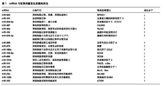
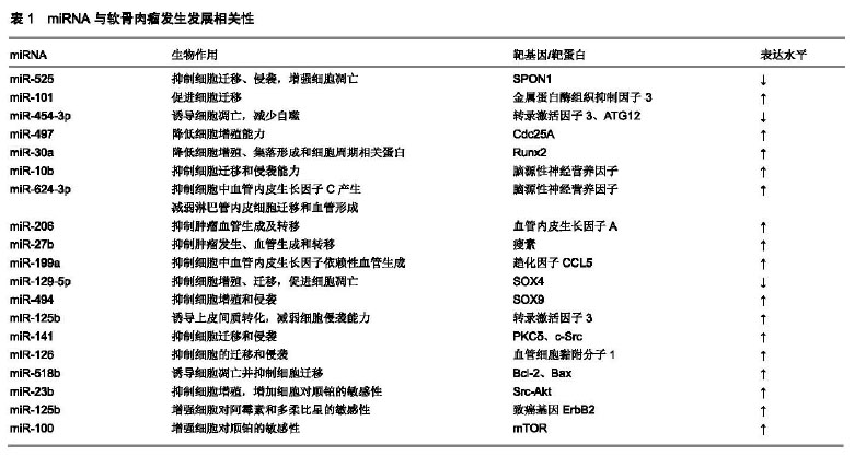
2.1 miRNA的生成及生物学作用 1993年Lee等[11]在秀丽隐杆线虫的研究中首次发现了小RNA的存在,这些分子能够控制生物过程,包括基因表达的调控[12-14]。Reinhart等[15]在秀丽隐杆线虫内发现了另一个具有转录后调节功能的小分子RNA:let-7。随着人们研究的不断深入,至今已发现1 000多种miRNA,每种miRNA调节多种mRNA,从而参与调控不同的生物过程[16]。 miRNA的生成是一个非常复杂的生物过程,其包括胞质合成和胞核合成两个部分,并需要多种酶的参与。首先,编码miRAN的基因在细胞核内经RNA聚合酶Ⅱ转录形成具有特殊发夹结构[多聚腺苷酸尾巴(AAAAA)和7MGpppG]的pri-miRNA,接着核酸酶Drosha(核糖核酸酶Ⅲ)将pri-miRNA进行微切割并处理成具有茎环结构、大小为70-80 nt的miRNA前体,即pre-miRNA。细胞质转运蛋白Exportins-5在Ran-GTP辅助下将pre-miRNA从细胞核内运输到细胞胞质中,然后由核糖核酸酶Ⅲ(Dicer酶)将其剪切成为大小为19-23 nt的miRNA:miRNA*复合结构。miRNA*通常被降解,而miRNA与AGO(argonaute)蛋白结合形成成熟miRNA[17]。成熟的miRNA与RNA诱导的沉默复合物(RNA-induced silencing complex,RISC)结合发挥其生物作用,包括2种方式:①miRNA与靶mRNA完全互补时,miRNA的作用方式是降解靶mRNA;②miRNA与靶mRNA不完全互补时,miRNA与3'UTRs结合抑制靶mRNA的翻译[18]。miRNA参与几个生理过程,如发育、增殖、正常细胞的分化和凋亡[13,19],也参与维护细胞多能性。 2.2 miRNA与肿瘤的关系 大量研究已证明恶性肿瘤细胞的miRNA常表现异常,miRNA对癌症起着正面或负面调控作用,如维持增殖信号传导、抗凋亡、诱导血管生成以及癌细胞侵袭和转移[20]。某些miRNA的异常表达与人类不同类型癌症的诊断、治疗和预后密切相关,因此miRNA成为重要的诊断和预后生物标志物以及癌症的治疗靶点[21]。 2002年Calin等[22]首次描述miRNA失调可能转化为慢性淋巴细胞白血病的表现,有69%的患者出现miR-15a和miR-16表达沉默,这是人们发现的第1个miRNA参与肿瘤发病的证据。在此之后,研究人员就开始了分析miRNA如何导致心血管疾病、肝脏纤维化、糖尿病、神经退行性疾病和癌症[23-27]。关于它们在肿瘤体内的表达和功能,miRNA可以作为癌基因或肿瘤抑制因子起作用,并且它们的功能与miRNA的靶标的作用直接相关[24]。 Volinia等[28]通过对肺癌、前列腺癌、胃癌等中的miRNA表达谱进行分析,发现miRNA-21的表达均显著上调;有研究发现,miRNA-21在肝细胞癌、食管癌、肾癌和宫颈癌中同样表现出过表达状态[29-32]。Wu等[33]研究发现,在非小细胞肺癌中,肺癌细胞的生长增殖是miRNA-25通过下调促凋亡基因MOAP1进行调控的。有研究发现,在神经胶质瘤组织中,过表达的miRNA-25通过抑制周期蛋白依赖激酶抑制因子1C的表达,促进肿瘤的生长[34]。当内源性miRNA-25的作用被解除后,周期蛋白依赖激酶抑制因子1C表达上调,参与正常细胞周期进程。Calin等[35]研究发现,在宫颈癌细胞中miR-199a的表达下调, 能够抑制细胞生长;在胆管癌细胞中,分别下调miR-141、miR-21,同样能够抑制细胞生长。Liu等[36]研究发现,miRNA-144在胃癌患者的癌组织和血清样品中的表达水平显著下调,血清miR-144能够高精度地区分胃癌患者和健康对照,此外,癌组织和血清miR-144水平均与临床分期和淋巴结转移相关,癌组织及血清中低表达miR-144的患者5年总体存活率和无病存活率更差。Li等[37]研究表明,miRNA-144-3p在胰腺癌患者癌组织及PCNA-1细胞系中的表达显著下调。研究人员已经发现,miRNA参与了癌症的发生发展、转移和侵袭[38]。因此,通过对不同miRNA在肿瘤组织中抑癌或促癌作用的进一步研究,可为肿瘤的临床诊断、判断预后及生物治疗提供新思路。 2.3 miRNA在软骨肉瘤中的研究进展 2.3.1 miRNA与软肉瘤病理发展过程 软骨肉瘤的分子发病机制已受到越来越多学者的关注,但miRNAs在软骨肉瘤细胞中的生物学作用仍不甚明了。最近研究表明,在原发性骨肿瘤包括软骨肉瘤、骨肉瘤、尤因氏肉瘤和巨细胞瘤的进展过程中,改变了miRNA的表达模式[39]。 Liu等[40]研究发现,miR-525在软骨肉瘤组织和细胞中表达水平降低,过表达miR-525可抑制SW1353细胞迁移和侵袭,并增强SW1353细胞凋亡;双荧光素酶测定表明F-spondin 1(SPON1)是miR-525的靶基因。SPON1是一种在感觉神经元细胞中高表达的细胞外基质蛋白[41],已证明SPON1可激活黏着斑激酶(FAK)和Src信号传导,从而促进肿瘤的发展;miR-525的过表达显著抑制了软骨肉瘤细胞中FAK/Src/PI3K/Akt信号的激活,降低了软骨肉瘤的恶性进展。有研究表明,miR-101在人软骨肉瘤细胞中显著上调,金属蛋白酶组织抑制因子3是其靶标[42]。鞘氨醇1-磷酸是一种生物活性鞘脂,通过鞘氨醇激酶在细胞内产生,是调节炎症、增殖、血管生成和转移的第二信号分子;该研究发现,鞘氨醇1-磷酸可下调miR-101并通过上调金属蛋白酶组织抑制因子3的表达来抑制细胞迁移和基质金属蛋白酶2表达,减少了软骨肉瘤向肺部的转移。Bao等[43]研究发现,miR-454-3p在软骨肉瘤表达降低,miR-454-3p可通过靶向转录激活因子3和ATG12诱导细胞凋亡并减少自噬。研究表明,miR-497在人软骨肉瘤组织和细胞中的表达显著下调,miR-497过表达通过靶向Cdc25A显著降低JJ012和OUMS-27细胞的增殖能力,miR-497可作为软骨肉瘤中的潜在肿瘤抑制因子[44]。Jiang等[45]通过对软骨肉瘤组织和正常组织比较发现,miR-30a的表达在软骨肉瘤细胞系和人软骨肉瘤组织中显著下调,过表达miR-30a抑制了Runx2,显著降低了软骨肉瘤细胞的增殖、集落形成和细胞周期相关蛋白,延缓了软骨肉瘤的进展。研究表明,miR-10b在JJ012和SW1353软骨肉瘤细胞系中显著下调,过表达miR-10b可抑制JJ012软骨肉瘤细胞的迁移和侵袭能力,脑源性神经营养因子是miR-10b的新靶点[46];该研究还发现,基质金属蛋白酶1参与了miR-10b/脑源性神经营养因子介导的软骨肉瘤细胞在JJ012细胞中的迁移和侵袭。 肿瘤血管生成是癌症发展的基础,可为肿瘤持续生长提供所必需的营养物质,以适应肿瘤微环境内的缺氧环境。血管生成是一个复杂的过程,其特征在于促血管生成因子(如血管内皮生长因子及其受体和血小板衍生因子及其受体)和抗血管生成因子(如TSP-1/TSP-2)的相互作用。Lin等[47]研究发现,miR-624-3p在JJ012(S10)细胞系中显著下调,过表达miR-624-3p可抑制JJ012(S10)细胞中的血管内皮生长因子C产生,减弱淋巴管内皮细胞迁移和血管形成;该研究还发现,脑源性神经营养因子是miR-624-3p的靶标,miR-624-3p通过MEK/ERK/mTOR级联负调控脑源性神经营养因子,抑制血管生成。研究表明,miR-206在软骨肉瘤中表达下调,过表达miR-206可抑制肿瘤血管生成及转移,血管内皮生长因子A是其靶标[48]。Yang等[49]研究发现,miR-27b在软骨肉瘤组织中表达降低。瘦素是一种脂肪细胞因子,可促进肿瘤发生、血管生成和转移,过表达miR-27b可抑制瘦素的产生,并通过FAK/PI3K/Akt级联抑制血管内皮生长因子C生成,瘦素可以作为软骨肉瘤转移和淋巴管生成的治疗靶标。研究发现,miR-199a在软骨肉瘤细胞中下调,过表达miR-199a可靶向趋化因子CCL5,抑制软骨肉瘤细胞中血管内皮生长因子依赖性血管生成[50]。 Zhang等[51]研究发现,与正常组织相比,miR-129-5p在软骨肉瘤组织和细胞中的表达显著下调,SOX4是其直接靶标,miR-129-5p通过靶向SOX4抑制Wnt/β-catenin信号通路,抑制软骨肉瘤细胞增殖、迁移并促进细胞凋亡。SOX4是与SRY相关的HMG盒(SOX)基因家族成员之一,与软骨形成有关,SOX4是软骨肉瘤进展的关键基 因[52]。SOX9是SOX家族的另一个成员,在软骨肉瘤中过度表达且与癌症进展有关[53]。Li等[54]通过对体内和体外的软骨肉瘤细胞研究发现,miR-494显著低表达,上调miR-494课抑制软骨肉瘤细胞增殖和侵袭,SOX9是miR-494的直接靶标。Bao等[55]研究发现,miR-125b在人转移性软骨肉瘤组织和细胞系中显著下调,三氧化二砷通过DNA的去甲基化上调miR-125b的表达,诱导上皮间质转化并减弱软骨肉瘤细胞的侵袭能力,转录激活因子3是miR-125b的直接靶标。有研究表明,miR-141可抑制软骨肉瘤的迁移和侵袭[56]。Tan等[57]研究表明,miR-126可靶向血管细胞黏附分子1,从而抑制软骨肉瘤的迁移和侵袭。有研究表明,miR-518b在软骨肉瘤细胞中低表达,上调SW1353细胞中的miR-518b可诱导细胞凋亡并抑制细胞迁移[58]。 2.3.2 miRNA与软骨肉瘤诊断及预后 基于miRNAs在疾病中的功能的研究,科学家们已经意识到这些小分子不仅可以作为开发新疗法的目标,而且还可以成为疾病的生物标志物。在疾病进展的早期获得正确和及时的诊断是成功治疗疾病的关键,尤其是肿瘤,也是预后良好和抗癌治疗良好反应的基础[59]。迄今为止,癌症的诊断标准通常是通过外科取活组织进行病理学分析,这是一种侵入性的方法且价格昂贵,对患者也有一定危险性。因此,寻找新方法可能有助于改善诊断方式。 已有文献报道,miRNAs在细胞内、血浆、尿液、唾液及乳汁中等都有存在[60-62]。通过对血液循环系统中miRNAs的定性定量分析,可为诊断和监测软骨肉瘤患者的病情变化提供依据。Goudarzi等[63]研究发现,与邻近的正常组织相比,软骨肉瘤骨组织中miR-185的表达显著下降,而miR-218表达水平显著升高;miR-185的低表达和miR-218的高表达与晚期肿瘤分期、肿瘤分级和远处转移显著相关;低miR-185表达组和高miR-218表达组的总生存期明显缩短,总之,miR-185的下调和miR-218的上调可能与软骨肉瘤的进展有关,并且它们两者都可以作为诊断及判断预后的生物标志物。研究表明,过表达的SOX4在软骨肉瘤中与组织学分级和肿瘤复发显著相关,miR-30a在软骨肉瘤患者中低表达[64];SOX4过表达和miR-30a低表达可作为低级别软骨肉瘤患者的预后标志物。 2.3.3 miRNA与软骨肉瘤治疗 软骨肉瘤的治疗大多基于标准治疗方案,包括积极的外科手术切除、全身化疗和靶向放射治疗。虽然已经对化学疗法和放射疗法进行了功效测试,但软骨肉瘤对传统化疗和放疗往往会产生抵抗性[65-66]。因此,降低软骨肉瘤的化学抗性,提高其对化疗及放疗的敏感性尤为重要。 Huang等[67]研究发现,与邻近的正常组织和人原代软骨细胞相比,miR-23b的表达在软骨肉瘤组织和细胞系中下调,过表达miR-23b可抑制Src-Akt途径,进而抑制软骨肉瘤细胞增殖,并增加了软骨肉瘤细胞对顺铂的敏感性。有研究发现,miR-125b在软骨肉瘤组织和细胞系中均下调,过表达的miR-125b通过直接靶向致癌基因ErbB2介导的软骨肉瘤细胞中糖酵解的上调,增强了软骨肉瘤细胞对阿霉素和多柔比星的敏感性[68]。Zhu等[69]研究表明,miR-100在对顺铂耐药的软骨肉瘤细胞中表达降低,过表达miR-100通过靶向mTOR增强软骨肉瘤细胞对顺铂的敏感性。miR-100可作为基于顺铂的联合化疗治疗策略用于治疗临床软骨肉瘤患者。miRNA与软骨肉瘤发生发展的相关性总结见表1。 "
| [1]Stemm M, Beck C, Mannem R, et al. Dedifferentiated chondrosarcoma of bone with prominent rhab-doid component. Ann Diagn Pathol. 2017;28:7-11.[2]Kumar R, Duran C, Amini B, et al. Erratum to: Periosteal mesenchymal chondrosarcoma of the tibia with multifocal bonemetastases: a case report. Skeletal Radiol. 2017; 46(7):1001.[3]Leddy LR, Holmes RE. Chondrosarcoma of bone. Cancer Treat Res, 2014; 162:117-130. [4]Kumar R, Duran C, Amini B, et al. Periosteal mesenchymal chondrosarcoma of the tibia with multifocal bone metastases: A case report. Skeletal Radiol. 2017;46(7):995-1000.[5]Maki D, Mori T, Teshima M, et al. Chondrosarcoma of the hyoid bone-Report of a case and a literature review of the suitable treat-ment strategy. Auris Nasus Larynx. 2017; 44(5):629-634.[6]Palmini G, Marini F, Brandi ML. et al. What Is New in the miRNA World Regarding Osteosarcoma and Chondrosarcoma?. Molecules. 2017; 22(3). pii: E417.[7]Frezza AM, Cesari M, Baumhoer D, et al. Mesenchymal chondrosarcoma: Prognostic factors and outcome in 113 patients. A European Musculoskeletal Oncology Society study. Eur J Cancer. 2015; 51(3):374-381.[8]Lin NY. Signature of circulating microRNAs in osteoarthritis. Ann Rheum Dis. 2015;74(3):e18.[9]Jackson RJ, Standart N. How do microRNAs regulate gene expression? Sci STKE. 2007;2007(367):re1.[10]Ha M, Kim VN. Regulation of microRNA biogenesis. Nat Rev Mol Cell Biol. 2014;15(8):509-524.[11]Lee RC, Feinbaum RL, Ambros V. The C. elegans heterochronic gene lin-4 encodes small RNAs with antisense complementarity to lin-14. Cell.1993;75(5):843-854.[12]Hwang HW, Mendell JT. MicroRNAs in cell proliferation, cell death, and tumorigenesis. Br J Cancer.2006;94(6):776-780.[13]Ebert MS, Sharp PA. Roles for MicroRNAs in conferring robustness to biological processes. Cell.2012;149(3): 515-524.[14]Fire A, Xu S, Montgomery MK, et al. Potent and specific genetic interference by double-stranded RNA in Caenorhabditis elegans. Nature. 1998;391(6669):806-811.[15]Reinhart BJ, Slack FJ, Basson M, et al. The 21-nucleotide let-7 RNA regulates developmental timing in Caenorhabditis elegans. Nature. 2000; 403(6772):901-906.[16]Farh KK, Grimson A, Jan C, et al. The widespread impact of mammalian MicroRNAs on mRNA repression and evolution. Science. 2005; 310(5755): 1817-1821.[17]Jones-Rhoades MW, Bartel DP, Bartel B. MicroRNAs and their regulatory roles in plants. Ann Rev Plant Biol. 2006; 57(1):19-53.[18]Huntzinger E, Izaurralde E. Gene silencing by microRNAs: contributions of translational repression and mRNA decay. Nat Rev Genet. 2011;12(2):99-110.[19]Kosik KS. The neuronal microRNA system. Nat Rev Neurosci. 2006;7(12):911-920.[20]Hanahan D, Weinberg RA. Hallmarks of cancer: the next generation. Cell. 2011;144: 646-674.[21]Reddy KB. MicroRNA (miRNA) in cancer. Cancer Cell Int. 2015;15:38.[22]Calin GA, Dumitru CD, Shimizu M, et al. Frequent deletions and down-regulation of micro-RNA genes miR15 and miR16 at 13q14 in chronic lymphocytic leukemia. Proc Natl Acad Sci USA. 2002; 99(24):15524-15529.[23]Flemming A. Heart Failure: Targeting miRNA pathology in heart disease. Nat Rev Drug Discov. 2014;13(5):336-336.[24]Yu F, Lu Z, Cai J, et al. MALAT1 functions as a competing endogenous RNA to mediate Rac1 expression by sequestering miR-101b in liver fibrosis. Cell Cycle. 2015;14 (24):3885-3896.[25]Farr RJ, Joglekar MV, Hardikar AA.Circulating microRNAs in diabetes progression: Discovery, validation, and research translation. Exp Suppl. 2015;106:215-244.[26]Alaqeel A, Abou Al-Shaar H, Shariff R, et al. The role of RNA metabolism in neurological diseases. Balkan J Med Genet. 2016;18(2):5-14.[27]Di Leva G, Garofalo M, Croce CM.MicroRNAs in cancer.Annu Rev Pathol. 2014;9:287-314.[28]Volinia S, Calin GA, Liu CG, et al. A microRNA expression signature of human solid tumors defines cancer gene targets. Proc Nat Acad Sci. 2006; 103(7):2257-2261.[29]Meng F, Henson R, Wehbe-Janek H, et al. MicroRNA-21 regulates expression of the PTEN tumor suppressor gene in human hepatocellular cancer. Gastroenterology. 2007;133(2): 647-658.[30]Li F, Lv JH, Liang L, et al. Downregulation of microRNA-21 inhibited radiation-resistance of esophageal squamous cell carcinoma. Cancer Cell Int. 2018;18(1):39-50.[31]Chen J, Gu Y, Shen W. MicroRNA-21 functions as an oncogene and promotes cell proliferation and invasion via TIMP3 in renal cancer. Eur Rev Med Pharmacol Sci. 2017; 21(20):4566-4576.[32]Zhang Z, Wang J, Wang X, et al. MicroRNA-21 promotes proliferation, migration, and invasion of cervical cancer through targeting TIMP3. Arch Gynecol Obstet. 2018;297(2):433-442.[33]Wu T, Chen W, Kong D, et al. miR-25 targets the modulator of apoptosis 1 gene in lung cancer. Carcinogenesis. 2015;36(8): 925-935.[34]Zhang J, Gong X, Tian K, et al. miR-25 promotes glioma cell proliferation by targeting CDKN1C. Biomed Pharmacother, 2015;71(3):7-14.[35]Calin GA, Croce CM. MicroRNA signatures in human cancers. Nat Rev Cancer. 2006;6(11):857-866.[36]Liu S, Suo J, Wang C, et al. Prognostic significance of low miR-144 expression in gastric cancer. Cancer Biomark. 2017; 20(4):547-552.[37]Li J, Sun P, Yue Z, et al. miR-144-3p induces cell cycle arrest and apoptosis in pancreatic cancer cells by targeting proline-rich protein 11 expression via the mitogen-activated protein kinase signaling pathway. DNA Cell Biol. 2017;36(8):619-626.[38]Hayes J, Peruzzi PP, Lawler S. MicroRNAs in cancer: biomarkers, functions and therapy. Trends Mol Med. 2014; 20(8):460-469.[39]Nugent M. microRNA and Bone Cancer. Adv Exp Med Biol. 2015;889:201-230.[40]Liu B, Song X, Yan Z, et al. MicroRNA-525 enhances chondrosarcoma malignancy by targeting F-spondin 1. Oncol Lett. 2019; 17(1):781-788.[41]Chang H, Dong T, Ma X, et al. Spondin 1 promotes metastatic progression through Fak and Src dependent pathway in human osteosarcoma. Biochem Biophys Res Commun. 2015; 464(1):45-50. [42]Tsai C, Yang D, Lin C, et al. Sphingosine-1-phosphate suppresses chondrosarcoma metastasis by upregulation of tissue inhibitor of metalloproteinase 3 through suppressing miR-101 expression. Mol Oncol. 2017;11(10):1380-1398.[43]Bao X, Ren T, Huang Y, et al. Knockdown of long non-coding RNA HOTAIR increases miR-454-3p by targeting Stat3 and Atg12 to inhibit chondrosarcoma growth. Cell Death Dis. 2017;8(2):e2605.[44]Lu Y, Li F, Xu T, et al. miRNA-497 Negatively Regulates the Growth and Motility of Chondrosarcoma Cells by Targeting Cdc25A. Oncol Res. 2016;23(4):155-163.[45]Jiang D, Zheng X, Shan W, et al. The overexpression of miR-30a affects cell proliferation of chondrosarcoma via targeting Runx2. Tumour Biol. 2016;37(5):5933-5940.[46]Aili A, Chen Y, Zhang H. MicroRNA 10b suppresses the migration and invasion of chondrosarcoma cells by targeting brain derived neurotrophic factor. Mol Med Rep. 2016;13(1): 441-446.[47]Lin CY, Wang SW, Chen YL, et al. Brain-derived neurotrophic factor promotes VEGF-C-dependent lymphangiogenesis by suppressing miR-624-3p in human chondrosarcoma cells. Cell Death Dis. 2017;8(8):e2964.[48]Wang CQ, Huang YW, Wang SW, et al. Amphiregulin enhances VEGF-A production in human chondrosarcoma cells and promotes angiogenesis by inhibiting miR-206 via FAK/c-Src/PKCδpathway. Cancer Lett. 2017;385:261-270.[49]Yang WH, Chang AC, Wang SW, et al. Leptin promotes VEGF-C production and induces lymphangiogenesis by suppressing miR-27b in human chondrosarcoma cells. Sci Rep. 2016;6:28647.[50]Liu GT, Huang YL, Tzeng HE, et al. CCL5 promotes vascular endothelial growth factor expression and induces angiogenesis by down-regulating miR-199a in human Chondrosarcoma cells. Cancer Lett. 2015; 357(2):476-487.[51]Zhang P, Li J, Song Y, et al. MiR-129-5p inhibits proliferation and invasion of chondrosarcoma cells by regulating SOX4/Wnt/β-Catenin signaling pathway. Cell Physiol Biochem. 2017; 42(1):242-253.[52]Akiyama H, Chaboissier MC, Martin JF, et al. The transcription factor Sox9 has essential roles in successive steps of the chondrocyte differentiation pathway and is required for expression of Sox5 and Sox6. Genes Dev. 2002; 16(21):2813-2828.[53]Kamachi Y, Kondoh H.Sox proteins: regulators of cell fate specification and differentiation. Development. 2013;140(20): 4129-4144.[54]Li J, Wang L, Liu Z, et al. MicroRNA-494 inhibits cell proliferation and invasion of chondrosarcoma cells in vivo and in vitro by directly targeting SOX9. Oncotarget. 2015;6(28): 26216-26229.[55]Bao X, Ren T, Huang Y, et al. Induction of the mesenchymal to epithelial transition by demethylation-activated microRNA-125b is involved in the anti-migration/invasion effects of arsenic trioxide on human chondrosarcoma. J Exp Clin Cancer Res. 2016;35(1):129.[56]Horng CT, Shieh PC, Tan TW, et al. Paeonol suppresses chondrosarcoma metastasis through up-regulation of miR-141 by modulating PKCδ and c-Src signaling pathway. Int J Mol Sci. 2014;15(7):11760-11772.[57]Tan TW, Chou YE, Yang WH, et al. Naringin suppress chondrosarcoma migration through inhibition vascular adhesion molecule-1 expression by modulating miR-126. Int Immunopharmacol. 2014; 22(1):107-114.[58]Liang W, Li X, Li Y, et al. Gallic acid induces apoptosis and inhibits cell migration by upregulating miR-518b in SW1353 human chondrosarcoma cells. Int J Oncol. 2014;44(1):91-98.[59]Hayes J, Peruzzi PP, Lawler S. MicroRNAs in cancer: biomarkers, functions and therapy. Trends Mol Med. 2014; 20(8):460-469.[60]Sampson VB, Yoo S, Kumar A, et al. MicroRNAs and potential targets in osteosarcoma: review. Front Pediatr. 2015;3:69.[61]Zhou G, Shi X, Zhang J, et al. MicroRNAs in osteosarcoma: from biological players to clinical contributors, a review. J Int Med Res. 2013;41(1):1-12.[62]Chang L, Shrestha S, LaChaud G, et al. Review of microRNA in osteosarcoma and chondrosarcoma. Med Oncol.2015; 32(6):613.[63]Goudarzi PK, Taheriazam A, Asghari S, et al. Downregulation of miR-185 and upregulation of miR-218 expression may be potential diagnostic and prognostic biomarkers of human chondrosarcoma. Tumour Biol. 2016; 37(5):5775-5779.[64]Lu N, Lin T, Wang L, et al. Association of SOX4 regulated by tumor suppressor miR-30a with poor prognosis in low-grade chondrosarcoma. Tumour Biol. 2015;36(5):3843-3852.[65]Gelderblom H, Hogendoorn PC, Dijkstra SD, et al. The clinical approach towards chondrosarcoma. Oncologist. 2008; 13(3): 320-329.[66]Onishi AC, Hincker AM, Lee FY. Surmounting chemotherapy and radioresistance in chondrosarcoma: molecular mechanisms and therapeutic targets. Sarcoma. 2011;2011: 381564.[67]Huang K, Chen J, Yang MS, et al. Inhibition of Src by microRNA-23b increases the cisplatin sensitivity of chondrosarcoma cells. Cancer Biomark. 2017; 18(3):231-239.[68]Tang XY, Zheng W, Ding M,et al. miR-125b acts as a tumor suppressor in chondrosarcoma cells by the sensitization to doxorubicin through direct targeting the ErbB2-regulated glucose metabolism. Drug Des Devel Ther. 2016; 10:571-583.[69]Zhu Z, Wang CP, Zhang YF, et al. MicroRNA-100 resensitizes resistant chondrosarcoma cells to cisplatin through direct targeting of mTOR. Asian Pac J Cancer Prev. 2014;15(2): 917-923. |
| [1] | Zhou Jihui, Li Xinzhi, Zhou You, Huang Wei, Chen Wenyao. Multiple problems in the selection of implants for patellar fracture [J]. Chinese Journal of Tissue Engineering Research, 2021, 25(9): 1440-1445. |
| [2] | Liu Cong, Liu Su. Molecular mechanism of miR-17-5p regulation of hypoxia inducible factor-1α mediated adipocyte differentiation and angiogenesis [J]. Chinese Journal of Tissue Engineering Research, 2021, 25(7): 1069-1074. |
| [3] | Hou Jingying, Yu Menglei, Guo Tianzhu, Long Huibao, Wu Hao. Hypoxia preconditioning promotes bone marrow mesenchymal stem cells survival and vascularization through the activation of HIF-1α/MALAT1/VEGFA pathway [J]. Chinese Journal of Tissue Engineering Research, 2021, 25(7): 985-990. |
| [4] | Geng Yao, Yin Zhiliang, Li Xingping, Xiao Dongqin, Hou Weiguang. Role of hsa-miRNA-223-3p in regulating osteogenic differentiation of human bone marrow mesenchymal stem cells [J]. Chinese Journal of Tissue Engineering Research, 2021, 25(7): 1008-1013. |
| [5] | Liu Lihua, Sun Wei, Wang Yunting, Gao Fuqiang, Cheng Liming, Li Zirong, Wang Jiangning. Type L1 steroid-induced osteonecrosis of the femoral head through femoral head and neck junction decompression by fenestration: a single-center prospective clinical study [J]. Chinese Journal of Tissue Engineering Research, 2021, 25(6): 906-911. |
| [6] | Song Shan, Hu Fangyuan, Qiao Jun, Wang Jia, Zhang Shengxiao, Li Xiaofeng. An insight into biomarkers of osteoarthritis synovium based on bioinformatics [J]. Chinese Journal of Tissue Engineering Research, 2021, 25(5): 785-790. |
| [7] | Chen Ziyang, Pu Rui, Deng Shuang, Yuan Lingyan. Regulatory effect of exosomes on exercise-mediated insulin resistance diseases [J]. Chinese Journal of Tissue Engineering Research, 2021, 25(25): 4089-4094. |
| [8] | Cheng Chongjie, Yan Yan, Zhang Qidong, Guo Wanshou. Diagnostic value and accuracy of D-dimer in periprosthetic joint infection: a systematic review and meta-analysis [J]. Chinese Journal of Tissue Engineering Research, 2021, 25(24): 3921-3928. |
| [9] | Liu Zhiwei, Xie Rui, Sun Kai, Li Kaiming, Wang Xiongwei, Zhan Jiawen, Zhu Liguo. Interpretation of diagnostic criteria for cervicogenic headache: challenges and understandings in diagnosis and differential diagnosis [J]. Chinese Journal of Tissue Engineering Research, 2021, 25(23): 3746-3751. |
| [10] | Yuan Changshen, Rong Weiming, Lu Zhixian, Duan Kan, Guo Jinrong, Mei Qijie. Construction of osteosarcoma miRNA-mRNA regulatory network based on bioinformatics [J]. Chinese Journal of Tissue Engineering Research, 2021, 25(17): 2740-2746. |
| [11] | Ma Jinchao, Liu Tiansheng, Liu Aipeng, Wang Hao, Wang Qi, Liang Yongjian, Wang Lin, Di Haiwei. Photodynamic antimicrobial chemotherapy for repairing a rabbit model of osteomyelitis [J]. Chinese Journal of Tissue Engineering Research, 2020, 24(8): 1254-1259. |
| [12] | Xu Changbo, Zhang Yi, Yin Li . Severity of patellofemoral osteoarthritis does not affect the prognosis of total knee arthroplasty with patella retention [J]. Chinese Journal of Tissue Engineering Research, 2020, 24(6): 833-838. |
| [13] | Fang Yi, Zhao Wenzhi, Pan Deyue, Han Xin, Zhang Lu, He Hongtao, Shi Feng, Tian Tingxiao. Acromioclavicular joint dislocation: how to achieve anatomical reduction, sustained stability and micro-motion [J]. Chinese Journal of Tissue Engineering Research, 2020, 24(5): 796-802. |
| [14] | Lin Shu, Hu Jiang, Wan Lun, Tang Liuyi, Wang Yue, Yu Yang, Zhang Wei. Robot-guided percutaneous kyphoplasty in treatment of multi-segmental spinal metastases [J]. Chinese Journal of Tissue Engineering Research, 2020, 24(33): 5249-5254. |
| [15] | Yuan Yiming, Wang Yan, Chen Chengcheng, Zhao Mingyue, Pei Fei. Efficacy of exosomes in peripheral nerve injury [J]. Chinese Journal of Tissue Engineering Research, 2020, 24(31): 5079-5084. |
| Viewed | ||||||
|
Full text |
|
|||||
|
Abstract |
|
|||||