Chinese Journal of Tissue Engineering Research ›› 2018, Vol. 22 ›› Issue (17): 2747-2754.doi: 10.3969/j.issn.2095-4344.0869
Previous Articles Next Articles
Immunomodulation of mesenchymal stem cells in inflammatory microenvironment
Zhou Dian1, 2, Yan Fei1, 2, Zhou Ze-kun1, 2, Li Chen1, 2, Liu Ou-sheng1, 2
- 1Xiangya Stomatological Hospital, Central South University, Changsha 410078, Hunan Province, China; 2Central South University, Changsha 410078, Hunan Province, China
-
Revised:2018-05-10Online:2018-06-18Published:2018-06-18 -
Contact:Liu Ou-sheng, M.D., Associate researcher, Xiangya Stomatological Hospital, Central South University, Changsha 410078, Hunan Province, China; Central South University, Changsha 410078, Hunan Province, China -
About author:Zhou Dian, Master, Xiangya Stomatological Hospital, Central South University, Changsha 410078, Hunan Province, China; Central South University, Changsha 410078, Hunan Province, China -
Supported by:the 2017 Postgraduate Exploration and Innovation Project of Central South University, No. 2017zzts913; Scientific Research Project of Hunan Provincial Health and Family Planning Commission, No. C2016092; Hunan Science and Technology Plan Project, No. 2017JJ2341
CLC Number:
Cite this article
Zhou Dian, Yan Fei, Zhou Ze-kun, Li Chen, Liu Ou-sheng. Immunomodulation of mesenchymal stem cells in inflammatory microenvironment[J]. Chinese Journal of Tissue Engineering Research, 2018, 22(17): 2747-2754.
share this article
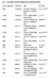
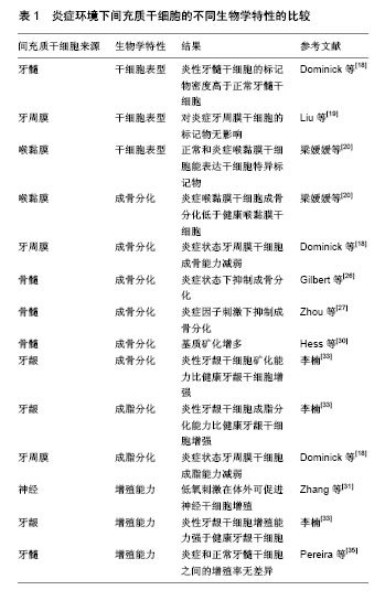
2.1 炎症微环境下MSCs的生物学特性 2.1.1 炎症微环境对MSCs表面标记物的影响 特异性的标记物是鉴别细胞的理想条件,而与MSCs相关的表面标记物现尚未完全明确。迄今为止,已经提出了很多的MSCs表面标记物,较为公认的观点为MSCs表达 CD105,CD73,CD44,CD90,CD71和Stro-1以及黏附分子CD106,CD166,ICAM-1和CD29[14-16],但不表达造血细胞表面标志物(如CD45,CD34,CD14和CD11),同时也不表达CD80,CD86,CD40以及CD31,CD18,CD56。除此之外,CD146也是MSCs的重要标志物[14,17]。 据报道,体外微环境的变化是导致MSCs表型发生变化的最主要因素。Alongi等[18]选择STRO-1,CD90,CD105和CD146这4种用于鉴定MSCs的标记物进行免疫荧光染色,发现炎症牙髓的4种标记物密度明显高于正常牙髓,其可能是由于炎症时血管生成多,而这些标记物都与血管有关。但也有研究表明,炎症微环境并不会影响干细胞的表面标记物[19]。 梁媛媛[20]分离及培养健康喉黏膜MSCs和炎症喉黏膜MSCs,两种细胞均表达MSCs特异性表面细胞分子,说明存在于炎症微环境下的MSCs仍具有干细胞的特性。 2.1.2 炎症微环境对MSCs分化的影响 很多研究表明,炎症环境会对MSCs的生物学行为产生一定程度的影响。梁媛媛[20]实验结果表明,炎症喉黏膜MSCs增殖能力强于健康喉黏膜MSCs,但成骨、成脂分化能力均显著低于健康喉黏膜MSCs,说明炎症状态下MSCs的分化能力受到抑制,这可能是由于炎症微环境中存在的炎细胞、炎性因子和白细胞代谢产物等各种炎性递质能对MSCs产生持续的刺激,从而改变其增殖和分化能力。有报道表明,牙周膜干细胞在炎性环境中会因非经典Wnt/Ca+信号通路的抑制而使其成骨能力受到影响[18]。 在牙周炎环境中,主要致病菌为革兰阴性菌,其细胞壁表面的脂多糖主要激活巨噬细胞而产生肿瘤坏死因子α和白细胞介素1β等,其与牙周炎的病程发展密切相关[21]。牙周膜干细胞是一种成体干细胞,牙周炎状态下脂多糖产生的肿瘤坏死因子α显著抑制牙周膜干细胞的成骨分化,使牙周组织再生能力丧失。在这些炎性因子中,肿瘤坏死因子α被认为是其他因子的上游因子,在炎症发生中起重要作用,其在骨病理生理过程中扮演重要角色,通过抑制转录方式调节Runx2表达[22-24],还有研究表明,肿瘤坏死因子α能抑制成骨细胞分化及矿化结节的形成[25-26]。此外,白细胞介素1和肿瘤坏死因子α在体外可以抑制MSCs的成脂分化能力。这些研究都表明,炎症因子的分泌,可以通过多种复杂分析信号机制影响干细胞的生物学特性。白细胞介素1β和肿瘤坏死因子α是主要的促炎性因子,在炎症组织中高表达,而现在关于肿瘤坏死因子α对干细胞成骨分化的作用有不同的报道,有研究认为其抑制成骨,如Zhou等[27]发现,在炎症反应发生中,肿瘤坏死因子α可激活成骨细胞中的p38 MAPK[28],抑制成骨细胞的分化。也有研究表明肿瘤坏死因子α在干细胞成骨分化中起着积极的作用,通过激活核转录因子κB途径[29],提高成骨相关蛋白的表达,使基质矿化增多[30]。 2.1.3 炎症微环境对MSCs增殖能力的影响 炎症是一个复杂的进程,根据炎症刺激的程度,一些轻度的刺激因子可能会激活MSCs的某些特性。如适当的低氧刺激促进细胞的增殖[31]。此外,当前很多研究都已表明Wnt通路与干细胞的干性息息相关。由于Wnt/β-catenin通路激活后,大量LEF-β-catenin复合体形成激活靶基因Cyclin D1,通过此途径来调控细胞的生长周期以促进干细胞增殖,由此解释了炎性环境中MSCs增殖能力增加的原因[32]。同样,李楠[33-34]通过实验得出,在炎症微环境中,牙龈MSCs的增殖、凋亡、多向分化能力(成骨、成脂能力)、细胞外基质、相关蛋白酶和炎性因子均会有改变。众所周知,克隆形成能力是MSCs的重要特性。通过分析实验结果提示炎症状态下牙龈MSCs提前进入瞬时扩增细胞状态,也就是正常牙龈MSCs在炎症微环境中会提早形成瞬时扩增细胞,但是其多向分化能力降低,在多向分化过程中出现大量的凋亡,尽管此时细胞增殖增强。 然而,也有学者发现,炎性牙髓干细胞的增殖能力与正常牙髓干细胞无明显差异[35]。还有学者将来源于风湿性关节炎患者骨髓的MSCs与正常骨髓的MSCs进行了对比实验,发现来源于患者骨髓的MSCs的增殖能力和克隆形成能力均降低[36]。 炎症环境下间充质干细胞的不同生物学特性的比较,见表1。"
| [1] Chen X, Wang S, Cao W. Mesenchymal stem cell-mediated immunomodulation in cell therapy of neurodegen-erative diseases. Cell Immunol. 2018;326:8-14.[2] Daneshmandi S, Karimi MH, Pourfathollah AA. TGF-β1 Transduced Mesenchymal Stem Cells Have Pro-found Modulatory Effects on DCs and T Cells. Iran J Immunol. 2017;14(1):13-23.[3] Papewalis C, Topolar D, Götz B, et al. Mesenchymal stem cells as cellular immunotherapeutics in allogeneic hematopoietic stem cell transplantation. Adv Biochem Eng Biotechnol. 2013;130:131-162.[4] Shi M, Liu ZW, Wang FS. Immunomodulatory properties and therapeutic application of mesenchymal stem cells. Clin Exp Immunol. 2011;164(1):1-8.[5] Prigione I, Benvenuto F, Bocca P, et al. Reciprocal interactions between human mesenchymal stem cells and gammadelta T cells or invariant natural killer T cells. Stem Cells. 2009;27(3):693-702.[6] Garnett HM, Harigaya K, Cronkite EP. Characterization of a murine cell-line derived from cultured bone marrow stromal cells. Stem Cells. 1982;2(1):11-23.[7] Allakhverdi Z, Comeau MR, Armant M, et al. Mast Cell-Activated Bone Marrow Mesenchymal Stromal Cells Regulate Proliferation and Lineage Commitment of CD34(+) Progenitor Cells. Front Immunol. 2013;4:461.[8] Li N, Liu N, Zhou J, et al. Inflammatory environment induces gingival tissue-specific mesenchymal stem cells to differentiate towards a pro-fibrotic phenotype. Biol Cell. 2013;105(6):261-275.[9] Rustad KC, Gurtner GC. Mesenchymal Stem Cells Home to Sites of Injury and Inflammation. Adv Wound Care (New Rochelle). 2012; 1(4):147-152.[10] Müller I, Lymperi S, Dazzi F. Mesenchymal stem cell therapy for degenerative inflammatory disorders. Curr Opin Organ Transplant. 2008;13(6):639-644.[11] Rameshwar P, Qiu H, Vatner SF. Stem cells in cardiac repair in an inflammatory microenvironment. Mi-nerva Cardioangiol. 2010;58(1): 127-146.[12] Vishnu P, Paulsen A, Roy V, et al. Lenalidomide Enhances Clonogenic Activity, Proliferation and Erythroid Lineage Commitment of CD34+ Progenitor Cells While It Is Cytotoxic to CD34- Accessory Cells. Ash Meeting & Exposition. 2010; 116(21) :509-510.[13] 阮光萍,姚翔,刘菊芬,等. 人脐带间充质干细胞的作用_细胞移植_免疫调节及充当基因治疗靶细胞[J]. 中国组织工程研究, 2014,18(41): 6714-6718.[14] Bartold PM, Mcculloch CA, Narayanan AS, et al. Tissue engineering: a new paradigm for periodontal re-generation based on molecular and cell biology. Periodontology.2000;24(1):253-269.[15] Intini G. Future approaches in periodontal regeneration: gene therapy, stem cells, and RNA interference. Dent Clin North Am. 2010;54(1): 141-155.[16] Yao S, Pan F, Prpic V, et al. Differentiation of stem cells in the dental follicle. J Dent Res. 2008;87(8):767-771.[17] 金岩.口腔颌面组织胚胎学[M].西安:陕西科学技术出版社,2002.[18] Alongi DJ, Yamaza T, Song Y, et al. Stem/progenitor cells from inflamed human dental pulp retain tissue regeneration potential. Regen Med. 2010;5(4):617-631.[19] Liu N, Shi S, Deng M, et al. High levels of β-catenin signaling reduce osteogenic differentiation of stem cells in inflammatory microenvironments through inhibition of the noncanonical Wnt pathway. J Bone Miner Res. 2011;26(9):2082-2095.[20] 梁媛媛.喉黏膜慢性炎症组织来源的间充质干细胞的生物学行为观察[D]. 西安:第四军医大学, 2013.[21] Baud V, Karin M. Signal transduction by tumor necrosis factor and its relatives. Trends Cell Biol. 2001;11(9):372-377.[22] Hla T, Lee MJ, Ancellin N, et al. Lysophospholipids--receptor revelations. Science. 2001;294(5548):1875-1878.[23] Pober JS, Sessa WC. Evolving functions of endothelial cells in inflammation. Nat Rev Immunol. 2007;7(10):803-815.[24] Wei S, Kitaura H, Zhou P, et al. IL-1 mediates TNF-induced osteoclastogenesis. J Clin Invest. 2005;115(2):282-290.[25] Lipsky PE, van der Heijde DM, St Clair EW, et al. Infliximab and methotrexate in the treatment of rheuma-toid arthritis. Anti-Tumor Necrosis Factor Trial in Rheumatoid Arthritis with Concomitant Therapy Study Group. N Engl J Med. 2000;343(22):1594-1602.[26] Gilbert LC, Rubin J, Nanes MS. The p55 TNF receptor mediates TNF inhibition of osteoblast differentiation independently of apoptosis. Am J Physiol Endocrinol Metab. 2005;288(5):E1011-1018.[27] Zhou FH, Foster BK, Zhou XF, et al. TNF-alpha mediates p38 MAP kinase activation and negatively regu-lates bone formation at the injured growth plate in rats. J Bone Miner Res. 2006;21(7):1075-1088.[28] Kitaura H, Sands MS, Aya K, et al. Marrow stromal cells and osteoclast precursors differentially contribute to TNF-alpha-induced osteoclastogenesis in vivo. J Immunol. 2004;173(8):4838-4846.[29] Nanes MS. Tumor necrosis factor-alpha: molecular and cellular mechanisms in skeletal pathology. Gene. 2003;321:1-15.[30] Hess K, Ushmorov A, Fiedler J, et al. TNFalpha promotes osteogenic differentiation of human mesenchymal stem cells by triggering the NF-kappaB signaling pathway. Bone. 2009;45(2):367-376.[31] Zhang XY, Yang YJ, Xu PR, et al. The role of β-catenin signaling pathway on proliferation of rats neural stem cells after hyperbaric oxygen therapy in vitro. Cell Mol Neurobiol. 2011;31(1):101-109.[32] Wang Y, Li YP, Paulson C, et al. Wnt and the Wnt signaling pathway in bone development and disease. Front Biosci (Landmark Ed). 2014;19: 379-407.[33] 李楠.炎症微环境下人牙龈固有层间充质干细胞生物学特性的研究[D]. 西安:第四军医大学, 2011.[34] Harada Y, Nagao S, Nakamura M, et al. Effect of lipopolysaccharide on thymidine salvage as related to macrophage activation. Immunology. 1995;84(2):247-253.[35] Pereira LO, Rubini MR, Silva JR, et al. Comparison of stem cell properties of cells isolated from normal and inflamed dental pulps. Int Endod J. 2012;45(12):1080-1090.[36] 李江.异种基因修饰猪间充质干细胞对人T细胞免疫功能的调节[D].天津:天津医科大学, 2013.[37] Hou C, Peng D, Gao L, et al. Human umbilical cord-derived mesenchymal stem cells protect from hyperoxic lung injury by ameliorating aberrant elastin remodeling in the lung of O2-exposed newborn rat. Biochem Biophys Res Commun. 2018;495(2):1972-1979.[38] 陆晓茜,刘霆,孟文彤,等. 人骨髓间充质干细胞对T淋巴细胞的免疫调节作用[J]. 中国实验血液学杂志, 2005,13(4): 651-655.[39] Fechter K, Dorronsoro A, Jakobsson E, et al. IFNγ Regulates Activated Vδ2+ T Cells through a Feedback Mechanism Mediated by Mesenchymal Stem Cells. PLoS One. 2017;12(1):e0169362.[40] Parekkadan B. Cellular and molecular immunotherapeutics derived from the bone marrow stroma. Massa-chusetts Institute of Technology, 2008.[41] 李建国,颛孙永勋,冉丕鑫,等. 骨髓间充质干细胞对哮喘小鼠CD4+CD25+调节性T细胞及气道炎症的影响[J].中国组织工程研究与临床康复, 2008, 12(47): 9302-9305.[42] Plock JA, Schnider JT, Schweizer R, et al. The Influence of Timing and Frequency of Adipose-Derived Mesenchymal Stem Cell Therapy on Immunomodulation Outcomes After Vascularized Composite Allotransplan-tation. Transplantation. 2017;101(1):e1-e11.[43] Valencia J, Blanco B, Yáñez R, et al. Comparative analysis of the immunomodulatory capacities of human bone marrow- and adipose tissue-derived mesenchymal stromal cells from the same donor. Cytotherapy. 2016;18(10):1297-1311.[44] Ding H, Zhang H, Ding H, et al. Transplantation of placenta-derived mesenchymal stem cells reduces hy-poxic-ischemic brain damage in rats by ameliorating the inflammatory response. Cell Mol Immunol. 2017;14(8):693-701.[45] 房佰俊,宋永平,林全德,等. 成年人脂肪源间充质干细胞治疗急性移植物抗宿主病后CD+8 T细胞亚群的变化[J]. 白血病•淋巴瘤, 2006,15(5): 351-352,355.[46] 杜优优,周胜华,周滔,等. 骨髓间充质干细胞移植对心肌梗死后炎性细胞因子表达的调节[J]. 中国组织工程研究与临床康复,2008,12(8): 1440-1444.[47] 刘元林,江小霞,苏永锋,等. 活化的T细胞促进间充质干细胞分化为成骨细胞[J]. 中国实验血液学杂志, 2009,17(4):974-976.[48] 江小霞,张毅,李秀森,等. 间充质干细胞对T淋巴细胞转化的影响[J]. 解放军医学杂志, 2005,30(2): 130-132.[49] Pinheiro CH, de Queiroz JC, Guimarães-Ferreira L, et al. Local injections of adipose-derived mesenchymal stem cells modulate inflammation and increase angiogenesis ameliorating the dystrophic phenotype in dystrophin-deficient skeletal muscle. Stem Cell Rev. 2012;8(2):363-374.[50] Liu D, Xu J, Liu O, et al. Mesenchymal stem cells derived from inflamed periodontal ligaments exhibit im-paired immunomodulation. J Clin Periodontol. 2012;39(12):1174-1182.[51] Matula Z, Németh A, L?rincz P, et al. The Role of Extracellular Vesicle and Tunneling Nanotube-Mediated Intercellular Cross-Talk Between Mesenchymal Stem Cells and Human Peripheral T Cells. Stem Cells Dev. 2016;25(23):1818-1832.[52] Sivanathan KN, Gronthos S, Grey ST, et al. Immunodepletion and Hypoxia Preconditioning of Mouse Com-pact Bone Cells as a Novel Protocol to Isolate Highly Immunosuppressive Mesenchymal Stem Cells. Stem Cells Dev. 2017;26(7):512-527.[53] Luk F, Carreras-Planella L, Korevaar SS, et al. Inflammatory Conditions Dictate the Effect of Mesenchymal Stem or Stromal Cells on B Cell Function. Front Immunol. 2017;8:1042.[54] Chang TC, Yu CC. Mesenchymal stem cells in inflammatory microenvironment promotes cancer cell migra-tion and epithelialmesenchymal transition through osteopontin. Journal of Cancer Science & Therapy. 2017;9(4): 26.[55] Korkaya H, Liu S, Wicha MS. Regulation of cancer stem cells by cytokine networks: attacking cancer's in-flammatory roots. Clin Cancer Res. 2011;17(19):6125-6129.[56] 王晓宇,侯玲玲,马海滨,等.间充质干细胞对免疫细胞的抑制作用及其机制[J].中国生物化学与分子生物学报, 2011, 27(5): 397-402.[57] 王焕丽,熊兵,陈华德,等.骨髓间充质干细胞对巨噬细胞分泌炎症因子的影响[J].南方医科大学学报, 2014, 34(9): 1259-1264.[58] 靳丽媛,邓子辉,张金英,等.间充质干细胞上清对高糖慢性炎症损伤的巨噬细胞表型极化的影响[J].中华老年多器官疾病杂志, 2017, 16(4): 283-287.[59] Hajkova M, Hermankova B, Javorkova E, et al. Mesenchymal Stem Cells Attenuate the Adverse Effects of Immunosuppressive Drugs on Distinct T Cell Subopulations. Stem Cell Rev. 2017;13(1):104-115.[60] Peng Y, Chen X, Liu Q, et al. Mesenchymal stromal cells infusions improve refractory chronic graft versus host disease through an increase of CD5+ regulatory B cells producing interleukin 10.Leukemia. 2015;29(3):636-646.[61] Meisel R, Zibert A, Laryea M, et al. Human bone marrow stromal cells inhibit allogeneic T-cell responses by indoleamine 2,3-dioxygenase- mediated tryptophan degradation. Blood. 2004;103(12):4619-4621.[62] Luk F, de Witte SF, Korevaar SS, et al. Inactivated Mesenchymal Stem Cells Maintain Immunomodulatory Capacity. Stem Cells Dev. 2016; 25(18):1342-1354.[63] Pezato R, de Almeida DC, Bezerra TF, et al. Immunoregulatory effects of bone marrow-derived mesenchy-mal stem cells in the nasal polyp microenvironment. Mediators Inflamm. 2014;2014:583409.[64] Zachar L, Ba?enková D, Rosocha J. Activation, homing, and role of the mesenchymal stem cells in the in-flammatory environment. J Inflamm Res. 2016;9:231-240.[65] Lu H, Wu X, Wang Z, et al. Erythropoietin-activated mesenchymal stem cells promote healing ulcers by improving microenvironment. J Surg Res. 2016;205(2):464-473.[66] Franquesa M, Hoogduijn MJ, Bestard O, et al. Immunomodulatory effect of mesenchymal stem cells on B cells. Front Immunol. 2012;3: 212.[67] Han Z, Tian Z, Lv G, et al. Immunosuppressive effect of bone marrow-derived mesenchymal stem cells in inflammatory microenvironment favours the growth of B16 melanoma cells. J Cell Mol Med. 2011;15(11):2343-2352.[68] Sun Z, Wang S, Zhao RC. The roles of mesenchymal stem cells in tumor inflammatory microenvironment. J Hematol Oncol. 2014;7:14.[69] 鲁刚,马奎,付小兵,等.脐带间充质干细胞调节炎性微环境干预急性肾损伤的效应研究[J].感染、炎症、修复, 2014,14(2):79-83.[70] Jing Y, Han Z, Liu Y, et al. Mesenchymal stem cells in inflammation microenvironment accelerates hepato-cellular carcinoma metastasis by inducing epithelial-mesenchymal transition. PLoS One. 2012;7(8): e43272.[71] Hogan NM, Dwyer RM, Joyce MR, et al. Mesenchymal stem cells in the colorectal tumor microenvironment: recent progress and implications. Int J Cancer. 2012;131(1):1-7.[72] Brennen WN, Denmeade SR, Isaacs JT. Mesenchymal stem cells as a vector for the inflammatory prostate microenvironment. Endocr Relat Cancer. 2013;20(5):R269-290.[73] Liu J, Wang L, Liu W, et al. Dental follicle cells rescue the regenerative capacity of periodontal ligament stem cells in an inflammatory microenvironment. PLoS One. 2014;9(9):e108752.[74] Munir H, Benjamin A, Allwood JW, et al. A1.18 Mesenchymal stem cells lose their immuno-protective ef-fects upon changes in their local microenvironment.Annals of the Rheumatic Diseases.2016;75(Suppl1): A8.1-A8.[75] Buhrmann C, Mobasheri A, Matis U, et al. Curcumin mediated suppression of nuclear factor-κB promotes chondrogenic differentiation of mesenchymal stem cells in a high-density co-culture microenvironment. Arthritis Res Ther. 2010;12(4):R127.[76] Lerrer S, Liubomirski Y, Bott A, et al. Co-Inflammatory Roles of TGFβ1 in the Presence of TNFα Drive a Pro-inflammatory Fate in Mesenchymal Stem Cells. Front Immunol. 2017;8:479. |
| [1] | Pu Rui, Chen Ziyang, Yuan Lingyan. Characteristics and effects of exosomes from different cell sources in cardioprotection [J]. Chinese Journal of Tissue Engineering Research, 2021, 25(在线): 1-. |
| [2] | Lin Qingfan, Xie Yixin, Chen Wanqing, Ye Zhenzhong, Chen Youfang. Human placenta-derived mesenchymal stem cell conditioned medium can upregulate BeWo cell viability and zonula occludens expression under hypoxia [J]. Chinese Journal of Tissue Engineering Research, 2021, 25(在线): 4970-4975. |
| [3] | Zhang Tongtong, Wang Zhonghua, Wen Jie, Song Yuxin, Liu Lin. Application of three-dimensional printing model in surgical resection and reconstruction of cervical tumor [J]. Chinese Journal of Tissue Engineering Research, 2021, 25(9): 1335-1339. |
| [4] | Geng Qiudong, Ge Haiya, Wang Heming, Li Nan. Role and mechanism of Guilu Erxianjiao in treatment of osteoarthritis based on network pharmacology [J]. Chinese Journal of Tissue Engineering Research, 2021, 25(8): 1229-1236. |
| [5] | Gu Xia, Zhao Min, Wang Pingyi, Li Yimei, Li Wenhua. Relationship between hypoxia inducible factor 1 alpha and hypoxia signaling pathway [J]. Chinese Journal of Tissue Engineering Research, 2021, 25(8): 1284-1289. |
| [6] | Hou Jingying, Yu Menglei, Guo Tianzhu, Long Huibao, Wu Hao. Hypoxia preconditioning promotes bone marrow mesenchymal stem cells survival and vascularization through the activation of HIF-1α/MALAT1/VEGFA pathway [J]. Chinese Journal of Tissue Engineering Research, 2021, 25(7): 985-990. |
| [7] | Shi Yangyang, Qin Yingfei, Wu Fuling, He Xiao, Zhang Xuejing. Pretreatment of placental mesenchymal stem cells to prevent bronchiolitis in mice [J]. Chinese Journal of Tissue Engineering Research, 2021, 25(7): 991-995. |
| [8] | Liang Xueqi, Guo Lijiao, Chen Hejie, Wu Jie, Sun Yaqi, Xing Zhikun, Zou Hailiang, Chen Xueling, Wu Xiangwei. Alveolar echinococcosis protoscolices inhibits the differentiation of bone marrow mesenchymal stem cells into fibroblasts [J]. Chinese Journal of Tissue Engineering Research, 2021, 25(7): 996-1001. |
| [9] | Fan Quanbao, Luo Huina, Wang Bingyun, Chen Shengfeng, Cui Lianxu, Jiang Wenkang, Zhao Mingming, Wang Jingjing, Luo Dongzhang, Chen Zhisheng, Bai Yinshan, Liu Canying, Zhang Hui. Biological characteristics of canine adipose-derived mesenchymal stem cells cultured in hypoxia [J]. Chinese Journal of Tissue Engineering Research, 2021, 25(7): 1002-1007. |
| [10] | Geng Yao, Yin Zhiliang, Li Xingping, Xiao Dongqin, Hou Weiguang. Role of hsa-miRNA-223-3p in regulating osteogenic differentiation of human bone marrow mesenchymal stem cells [J]. Chinese Journal of Tissue Engineering Research, 2021, 25(7): 1008-1013. |
| [11] | Lun Zhigang, Jin Jing, Wang Tianyan, Li Aimin. Effect of peroxiredoxin 6 on proliferation and differentiation of bone marrow mesenchymal stem cells into neural lineage in vitro [J]. Chinese Journal of Tissue Engineering Research, 2021, 25(7): 1014-1018. |
| [12] | Zhu Xuefen, Huang Cheng, Ding Jian, Dai Yongping, Liu Yuanbing, Le Lixiang, Wang Liangliang, Yang Jiandong. Mechanism of bone marrow mesenchymal stem cells differentiation into functional neurons induced by glial cell line derived neurotrophic factor [J]. Chinese Journal of Tissue Engineering Research, 2021, 25(7): 1019-1025. |
| [13] | Duan Liyun, Cao Xiaocang. Human placenta mesenchymal stem cells-derived extracellular vesicles regulate collagen deposition in intestinal mucosa of mice with colitis [J]. Chinese Journal of Tissue Engineering Research, 2021, 25(7): 1026-1031. |
| [14] | Pei Lili, Sun Guicai, Wang Di. Salvianolic acid B inhibits oxidative damage of bone marrow mesenchymal stem cells and promotes differentiation into cardiomyocytes [J]. Chinese Journal of Tissue Engineering Research, 2021, 25(7): 1032-1036. |
| [15] | Wang Zhengdong, Huang Na, Chen Jingxian, Zheng Zuobing, Hu Xinyu, Li Mei, Su Xiao, Su Xuesen, Yan Nan. Inhibitory effects of sodium butyrate on microglial activation and expression of inflammatory factors induced by fluorosis [J]. Chinese Journal of Tissue Engineering Research, 2021, 25(7): 1075-1080. |
| Viewed | ||||||
|
Full text |
|
|||||
|
Abstract |
|
|||||