Chinese Journal of Tissue Engineering Research ›› 2018, Vol. 22 ›› Issue (18): 2836-2842.doi: 10.3969/j.issn.2095-4344.0864
Previous Articles Next Articles
Preparation and characterization of laser microporous acellular osteochondral scaffolds
Liu Xue-jian1, 2, 3, Liu Shi-chen1, Sun Bai-chuan1, 2, 3, Zhang Kai-hong2, 3, Meng Hao-ye2, 3, Wang Yu2, 3, Huang Shao-dai2, 3, Lu Chang-feng2, 3, Wang Chong2, 3, Yu Wen2, 3, Jing Xiao-guang1, 2, 3, Zhao Yue1, Yang Jian-hua1, 4, Peng Jiang2, 3
- 1First Affiliated Hospital of Jiamusi University, Jiamusi 154007, Heilongjiang Province, China; 2Institute of Orthopedics, General Hospital of Chinese PLA, Beijing 100853, China; 3Beijing Key Laboratory of Orthopaedic Regeneration Medicine, Beijing 100853, China; 4People’s Hospital of Longgang District, Shenzhen 518172, Guangdong Province, China
-
Received:2018-03-16Online:2018-06-28Published:2018-06-28 -
Contact:Peng Jiang, Associate professor, Institute of Orthopedics, General Hospital of Chinese PLA, Beijing 100853, China; Beijing Key Laboratory of Orthopaedic Regeneration Medicine, Beijing 100853, China -
About author:Liu Xue-jian, Master candidate, First Affiliated Hospital of Jiamusi University, Jiamusi 154007, Heilongjiang Province, China; Institute of Orthopedics, General Hospital of Chinese PLA, Beijing 100853, China; Beijing Key Laboratory of Orthopaedic Regeneration Medicine, Beijing 100853, China -
Supported by:the Special Scientific Fund of Beijing, No. Z161100005016059; the National Natural Science Foundation of China, No. 81572148; the National Key Research and Development Plan of China, No. 2016YFC1102104; the Key Scientific Program of Jiamusi University (Basic Research), No. 12Z1201507; College Students Innovation and Entrepreneurship Training Project of Heilongjiang Province, No. 201610222058; the Basic Research Program of Heilongjiang Provincial Education Department, No. 2016-KYYWF-0548
CLC Number:
Cite this article
Liu Xue-jian, Liu Shi-chen, Sun Bai-chuan, Zhang Kai-hong, Meng Hao-ye, Wang Yu Huang Shao-dai, Lu Chang-feng, Wang Chong, Yu Wen, Jing Xiao-guang, Zhao Yue, Yang Jian-hua, Peng Jiang . Preparation and characterization of laser microporous acellular osteochondral scaffolds[J]. Chinese Journal of Tissue Engineering Research, 2018, 22(18): 2836-2842.
share this article

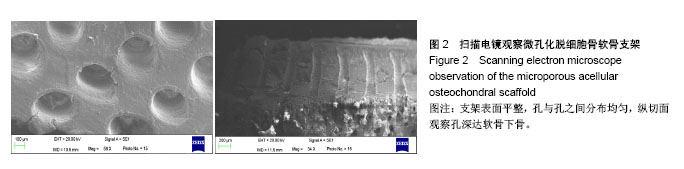
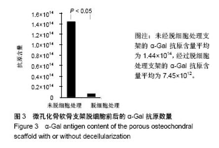
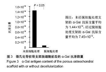
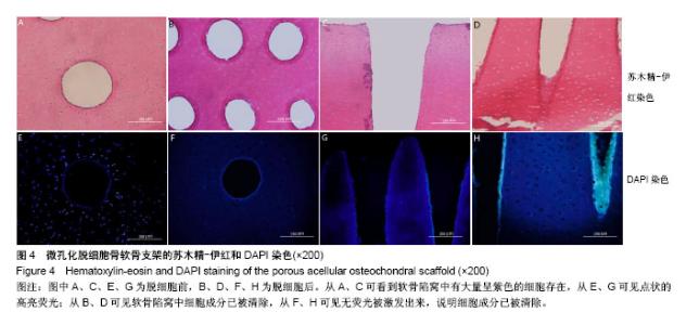
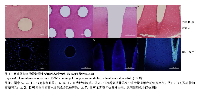
2.1 支架组织学染色和抗原含量检测 扫描电镜下观察支架表面平整,孔与孔之间分布均匀,纵切面孔深达软骨下骨,孔径范围为250-300 μm(图2);α-Gal抗原试剂盒检测多孔骨软骨支架脱细胞前后抗原数量比较差异有显著性意义(P < 0.05),见图3;苏木精-伊红及DAPI染色显示,支架脱细胞后细胞成分及抗原含量去除满意(图4)。"
"
"
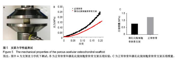
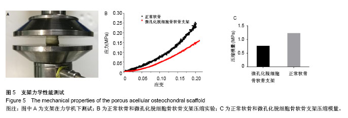
2.2 支架力学性能检测结果 根据计算得到压缩模量,微孔化骨软骨支架的压缩模量平均为0.77 MPa,与正常软骨的1.15 MPa相比较无差异(P > 0.05),见图5。"

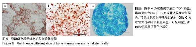
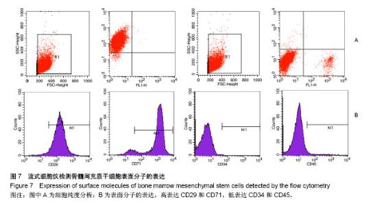
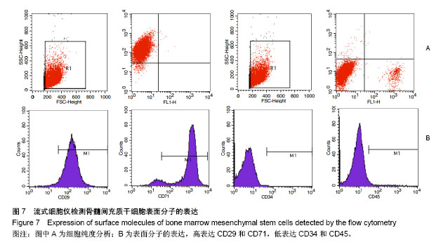
2.3 骨髓间充质干细胞的三系诱导及流式细胞鉴定结果 在脂肪诱导培养基中培养8 d后,可看到大部分细胞中出现了脂滴,经过油红“O”染色后呈红色;在成骨诱导培养基中培养3周,可看到细胞呈团块状生长,茜素红染色呈现红色;在软骨培养基中培养24 h后可看到细胞成球生长,培养28 d后,阿尔新蓝染色可见细胞周围基质呈蓝色(图6)。流式细胞仪检测细胞表面抗原,高表达CD29和CD71,低表达CD34和CD45(图7)。"
"

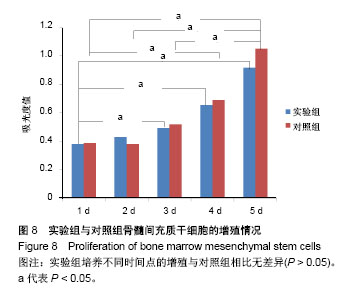
2.4 CCK-8细胞毒性实验结果 实验组培养第1天的吸光度值与第3,4,5天相比差异有显著性意义(P < 0.05);对照组培养第1天的吸光度值与第4,5天相比相比差异有显著性意义(P < 0.05),培养第2,3天的吸光度值与第5天相比差异有显著性意义(P < 0.05);实验组培养不同时间点的吸光度值与对照组相比无差异,见图8。因此,在两种培养基中,骨髓干细胞具有良好的生长状况。"
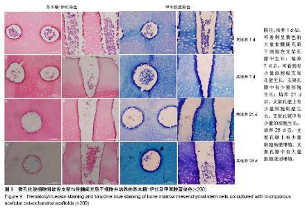
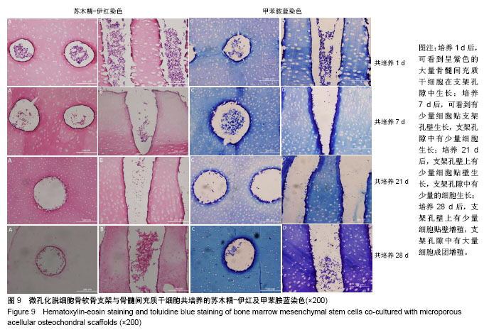
2.5 支架与细胞共培养结果 苏木精-伊红染色和甲苯胺蓝染色显示,共培养1 d后,可看到呈紫色的大量骨髓干细胞在微孔化脱细胞骨软骨支架孔隙中生长;共培养7 d后,可看到有少量细胞贴支架孔壁生长,支架孔隙中有少量细胞生长;共培养21 d后,孔壁上有少量细胞贴壁生长,支架孔隙中有少量细胞生长;共培养28 d后,可看到支架孔壁上有少量细胞贴壁增殖,支架孔隙中有大量细胞成团增殖,见图9。"
| [1] Choi B,Kim S,Lin B,et al.Cartilaginous Extracellular Matrix-Modified Chitosan Hydrogels for Cartilage Tissue Engineering.Acs Appl Mater Interfaces. 2014;6(22):20110-20121.[2] Hunziker EB.Articular cartilage repair: basic science and clinical progress. A review of the current status and prospects. Osteoarthritis Cartilage.2002;10(6):432-463.[3] Redler LH,Caldwell JM,Schulz BM,et al.Management of articular cartilage defects of the knee.Phys Sportsmed. 2010;92(4): 994-1009.[4] Zhou M,Yu D.Cartilage tissue engineering using PHBV and PHBV/Bioglass scaffolds[J].Mol Med Rep. 2014;10(1):508-514.[5] Cheng CW,Solorio LD,Alsberg E.Decellularized tissue and cell-derived extracellular matrices as scaffolds for orthopaedic tissue engineering.Biotechnol Adv.2014;32(2):462-484.[6] Zhang W,Zhu Y,Li J,et al.Cell-Derived Extracellular Matrix: Basic Characteristics and Current Applications in Orthopedic Tissue Engineering.Tissue Eng Part B Rev. 2016;22(3):193-207.[7] Dao TT,Vu NB,Pham LH,et al.In Vitro Production of Cartilage Tissue from Rabbit Bone Marrow-Derived Mesenchymal Stem Cells and Polycaprolactone Scaffold.Adv Exp Med Biol. 2018: 1-16.doi: 10.1007/5584_2017_133.[Epub ahead of print][8] 林建华,陈雷.组织工程软骨生物材料特征与研究现状[J].中国临床康复,2004,8(8):1522-1523.[9] 叶春婷,邹海燕,叶惠贞,等.猪软骨II型胶原的生物相容性研究[J].中国临床康复,2002,6(6):797-797.[10] 谭可.具有不同孔径的三维多孔支架材料的制备以及孔径对细胞体外生长和分化调节作用的研究[D].华东理工大学, 2014:20-43.[11] 徐小龙.组织工程技术治疗股骨头骨坏死的探索性研究[D].中国人民解放军总医院,2015:33-40.[12] 王永成.一体化关节软骨细胞外基质/羟基磷灰石双相支架构建与骨软骨界面组织工程应用[D].中国人民解放军医学院, 2014:40-52.[13] 徐丽娟,张云巍,王淑芳,等.小鼠骨髓间充质干细胞的分离、培养新方法[J].肝脏,2017,26(3):252-255.[14] Schwarz S,Elsaesser AF,Koerber L,et al.Processed xenogenic cartilage as innovative biomatrix for cartilage tissue engineering: effects on chondrocyte differentiation and function.J Tissue Eng Regen Med.2015;9(12):E239-251. [15] Smith BD,Grande DA.The current state of scaffolds for musculoskeletal regenerative applications. Nat Rev Rheumatol. 2015;11(4):213-222.[16] Duda GN,Haisch A,Endres M,et al.Mechanical quality of tissue engineered cartilage: results after 6 and 12 weeks in vivo.J Biomed Mater Res.2000;53(6):673-677.[17] Rotter N,Aigner J,Naumann A,et al.Behavior of tissue-engineered human cartilage after transplantation into nude mice.J Mater Sci Mater Med.1999;10(10/11):689-693.[18] Sutherland AJ,Converse GL,Hopkins RA,et al.The Bioactivity of Cartilage Extracellular Matrix in Articular Cartilage Regeneration. Adv Healthc Mater.2015;4(1):29-39.[19] Chen CH,Lee MY,Shyu VH,et al.Surface modification of polycaprolactone scaffolds fabricated via selective laser sintering for cartilage tissue engineering.Mater Sci Eng C Mater Biol Appl. 2014; 40:389-397.[20] Vinatier C,Guicheux J.Cartilage tissue engineering: From biomaterials and stem cells to osteoarthritis treatments.Ann Phys Rehabil Med.2016;59(3):139-144.[21] Elder BD,Eleswarapu SV,Athanasiou KA.Extraction techniques for the decellularization of tissue engineered articular cartilage constructs.Biomaterials. 2009;30(22):3749-3756.[22] Wu LC,Kuo YJ,Sun FW,et al.Optimized decellularization protocol including α-Gal epitope reduction for fabrication of an acellular porcine annulus fibrosus scaffold.Cell Tissue Bank. 2017; 18(3): 383-396.[23] 李坤,赵艳红,徐晨,等.基于软骨细胞外基质的取向支架的制备及评价[J].华西口腔医学杂志,2017,35(1):51-56.[24] Veronesi F,Maglio M,Tschon M,et al.Adipose-derived mesenchymal stem cells for cartilage tissue engineering: State-of-The-Art in in vivo, studies.J Biomed Mater Res A. 2014; 102(7):2448-2466.[25] Haleem AM,Singergy AA,Sabry D,et al.The Clinical Use of Human Culture-Expanded Autologous Bone Marrow Mesenchymal Stem Cells Transplanted on Platelet-Rich Fibrin Glue in the Treatment of Articular Cartilage Defects: A Pilot Study and Preliminary Results. Cartilage. 2010;1(4):253-261.[26] Vadalà G,Di MA,Tirindelli MC,et al.Use of autologous bone marrow cells concentrate enriched with platelet-rich fibrin on corticocancellous bone allograft for posterolateral multilevel cervical fusion.J Tissue Eng Regen Med.2008;2(8):515-520.[27] Wakitani S,Okabe T,Horibe S,et al.Safety of autologous bone marrow-derived mesenchymal stem cell transplantation for cartilage repair in 41 patients with 45 joints followed for up to 11 years and 5 months.J Tissue Eng Regen Med. 2011;5(2):146-150. [28] Liu X,Meng H,Guo Q,et al.Tissue-derived scaffolds and cells for articular cartilage tissue engineering: characteristics, applications and progress.Cell Tissue Res. 2018;372(1):13-22.[29] Juran CM,Dolwick MF,Mcfetridge PS.Engineered microporosity: enhancing the early regenerative potential of decellularized temporomandibular joint discs.Tissue Eng Part A. 2015;21(3-4): 829-839.[30] Luo L,Eswaramoorthy R,Mulhall KJ,et al.Decellularization of porcine articular cartilage explants and their subsequent repopulation with human chondroprogenitor cells.J Mech Behav Biomed Mater.2015; 55:21-31.[1] Choi B,Kim S,Lin B,et al.Cartilaginous Extracellular Matrix-Modified Chitosan Hydrogels for Cartilage Tissue Engineering.Acs Appl Mater Interfaces. 2014;6(22):20110-20121.[2] Hunziker EB.Articular cartilage repair: basic science and clinical progress. A review of the current status and prospects. Osteoarthritis Cartilage.2002;10(6):432-463.[3] Redler LH,Caldwell JM,Schulz BM,et al.Management of articular cartilage defects of the knee.Phys Sportsmed. 2010;92(4): 994-1009.[4] Zhou M,Yu D.Cartilage tissue engineering using PHBV and PHBV/Bioglass scaffolds[J].Mol Med Rep. 2014;10(1):508-514.[5] Cheng CW,Solorio LD,Alsberg E.Decellularized tissue and cell-derived extracellular matrices as scaffolds for orthopaedic tissue engineering.Biotechnol Adv.2014;32(2):462-484.[6] Zhang W,Zhu Y,Li J,et al.Cell-Derived Extracellular Matrix: Basic Characteristics and Current Applications in Orthopedic Tissue Engineering.Tissue Eng Part B Rev. 2016;22(3):193-207.[7] Dao TT,Vu NB,Pham LH,et al.In Vitro Production of Cartilage Tissue from Rabbit Bone Marrow-Derived Mesenchymal Stem Cells and Polycaprolactone Scaffold.Adv Exp Med Biol. 2018: 1-16.doi: 10.1007/5584_2017_133.[Epub ahead of print][8] 林建华,陈雷.组织工程软骨生物材料特征与研究现状[J].中国临床康复,2004,8(8):1522-1523.[9] 叶春婷,邹海燕,叶惠贞,等.猪软骨II型胶原的生物相容性研究[J].中国临床康复,2002,6(6):797-797.[10] 谭可.具有不同孔径的三维多孔支架材料的制备以及孔径对细胞体外生长和分化调节作用的研究[D].华东理工大学, 2014:20-43.[11] 徐小龙.组织工程技术治疗股骨头骨坏死的探索性研究[D].中国人民解放军总医院,2015:33-40.[12] 王永成.一体化关节软骨细胞外基质/羟基磷灰石双相支架构建与骨软骨界面组织工程应用[D].中国人民解放军医学院, 2014:40-52.[13] 徐丽娟,张云巍,王淑芳,等.小鼠骨髓间充质干细胞的分离、培养新方法[J].肝脏,2017,26(3):252-255.[14] Schwarz S,Elsaesser AF,Koerber L,et al.Processed xenogenic cartilage as innovative biomatrix for cartilage tissue engineering: effects on chondrocyte differentiation and function.J Tissue Eng Regen Med.2015;9(12):E239-251. [15] Smith BD,Grande DA.The current state of scaffolds for musculoskeletal regenerative applications. Nat Rev Rheumatol. 2015;11(4):213-222.[16] Duda GN,Haisch A,Endres M,et al.Mechanical quality of tissue engineered cartilage: results after 6 and 12 weeks in vivo.J Biomed Mater Res.2000;53(6):673-677.[17] Rotter N,Aigner J,Naumann A,et al.Behavior of tissue-engineered human cartilage after transplantation into nude mice.J Mater Sci Mater Med.1999;10(10/11):689-693.[18] Sutherland AJ,Converse GL,Hopkins RA,et al.The Bioactivity of Cartilage Extracellular Matrix in Articular Cartilage Regeneration. Adv Healthc Mater.2015;4(1):29-39.[19] Chen CH,Lee MY,Shyu VH,et al.Surface modification of polycaprolactone scaffolds fabricated via selective laser sintering for cartilage tissue engineering.Mater Sci Eng C Mater Biol Appl. 2014; 40:389-397.[20] Vinatier C,Guicheux J.Cartilage tissue engineering: From biomaterials and stem cells to osteoarthritis treatments.Ann Phys Rehabil Med.2016;59(3):139-144.[21] Elder BD,Eleswarapu SV,Athanasiou KA.Extraction techniques for the decellularization of tissue engineered articular cartilage constructs.Biomaterials. 2009;30(22):3749-3756.[22] Wu LC,Kuo YJ,Sun FW,et al.Optimized decellularization protocol including α-Gal epitope reduction for fabrication of an acellular porcine annulus fibrosus scaffold.Cell Tissue Bank. 2017; 18(3): 383-396.[23] 李坤,赵艳红,徐晨,等.基于软骨细胞外基质的取向支架的制备及评价[J].华西口腔医学杂志,2017,35(1):51-56.[24] Veronesi F,Maglio M,Tschon M,et al.Adipose-derived mesenchymal stem cells for cartilage tissue engineering: State-of-The-Art in in vivo, studies.J Biomed Mater Res A. 2014; 102(7):2448-2466.[25] Haleem AM,Singergy AA,Sabry D,et al.The Clinical Use of Human Culture-Expanded Autologous Bone Marrow Mesenchymal Stem Cells Transplanted on Platelet-Rich Fibrin Glue in the Treatment of Articular Cartilage Defects: A Pilot Study and Preliminary Results. Cartilage. 2010;1(4):253-261.[26] Vadalà G,Di MA,Tirindelli MC,et al.Use of autologous bone marrow cells concentrate enriched with platelet-rich fibrin on corticocancellous bone allograft for posterolateral multilevel cervical fusion.J Tissue Eng Regen Med.2008;2(8):515-520.[27] Wakitani S,Okabe T,Horibe S,et al.Safety of autologous bone marrow-derived mesenchymal stem cell transplantation for cartilage repair in 41 patients with 45 joints followed for up to 11 years and 5 months.J Tissue Eng Regen Med. 2011;5(2):146-150. [28] Liu X,Meng H,Guo Q,et al.Tissue-derived scaffolds and cells for articular cartilage tissue engineering: characteristics, applications and progress.Cell Tissue Res. 2018;372(1):13-22.[29] Juran CM,Dolwick MF,Mcfetridge PS.Engineered microporosity: enhancing the early regenerative potential of decellularized temporomandibular joint discs.Tissue Eng Part A. 2015;21(3-4): 829-839.[30] Luo L,Eswaramoorthy R,Mulhall KJ,et al.Decellularization of porcine articular cartilage explants and their subsequent repopulation with human chondroprogenitor cells.J Mech Behav Biomed Mater.2015; 55:21-31. |
| [1] | Zhang Tongtong, Wang Zhonghua, Wen Jie, Song Yuxin, Liu Lin. Application of three-dimensional printing model in surgical resection and reconstruction of cervical tumor [J]. Chinese Journal of Tissue Engineering Research, 2021, 25(9): 1335-1339. |
| [2] | Li Cai, Zhao Ting, Tan Ge, Zheng Yulin, Zhang Ruonan, Wu Yan, Tang Junming. Platelet-derived growth factor-BB promotes proliferation, differentiation and migration of skeletal muscle myoblast [J]. Chinese Journal of Tissue Engineering Research, 2021, 25(7): 1050-1055. |
| [3] | Liu Cong, Liu Su. Molecular mechanism of miR-17-5p regulation of hypoxia inducible factor-1α mediated adipocyte differentiation and angiogenesis [J]. Chinese Journal of Tissue Engineering Research, 2021, 25(7): 1069-1074. |
| [4] | Zeng Yanhua, Hao Yanlei. In vitro culture and purification of Schwann cells: a systematic review [J]. Chinese Journal of Tissue Engineering Research, 2021, 25(7): 1135-1141. |
| [5] | Ma Zetao, Zeng Hui, Wang Deli, Weng Jian, Feng Song. MicroRNA-138-5p regulates chondrocyte proliferation and autophagy [J]. Chinese Journal of Tissue Engineering Research, 2021, 25(5): 674-678. |
| [6] | Xu Dongzi, Zhang Ting, Ouyang Zhaolian. The global competitive situation of cardiac tissue engineering based on patent analysis [J]. Chinese Journal of Tissue Engineering Research, 2021, 25(5): 807-812. |
| [7] | Wang Yujiao, Liu Dan, Sun Song, Sun Yong. Biphasic calcium phosphate loaded with advanced platelet rich fibrin can promote the activity of rabbit bone marrow mesenchymal stem cells [J]. Chinese Journal of Tissue Engineering Research, 2021, 25(4): 504-509. |
| [8] | Zhou Jihui, Yao Meng, Wang Yansong, Li Xinzhi, Zhou You, Huang Wei, Chen Wenyao. Influence of novel nanoscaffolds on biological behaviors of neural stem cells and the related gene expression [J]. Chinese Journal of Tissue Engineering Research, 2021, 25(4): 532-536. |
| [9] | Wu Zijian, Hu Zhaoduan, Xie Youqiong, Wang Feng, Li Jia, Li Bocun, Cai Guowei, Peng Rui. Three-dimensional printing technology and bone tissue engineering research: literature metrology and visual analysis of research hotspots [J]. Chinese Journal of Tissue Engineering Research, 2021, 25(4): 564-569. |
| [10] | Chang Wenliao, Zhao Jie, Sun Xiaoliang, Wang Kun, Wu Guofeng, Zhou Jian, Li Shuxiang, Sun Han. Material selection, theoretical design and biomimetic function of artificial periosteum [J]. Chinese Journal of Tissue Engineering Research, 2021, 25(4): 600-606. |
| [11] | Liu Fei, Cui Yutao, Liu He. Advantages and problems of local antibiotic delivery system in the treatment of osteomyelitis [J]. Chinese Journal of Tissue Engineering Research, 2021, 25(4): 614-620. |
| [12] | Li Xiaozhuang, Duan Hao, Wang Weizhou, Tang Zhihong, Wang Yanghao, He Fei. Application of bone tissue engineering materials in the treatment of bone defect diseases in vivo [J]. Chinese Journal of Tissue Engineering Research, 2021, 25(4): 626-631. |
| [13] | Zhang Zhenkun, Li Zhe, Li Ya, Wang Yingying, Wang Yaping, Zhou Xinkui, Ma Shanshan, Guan Fangxia. Application of alginate based hydrogels/dressings in wound healing: sustained, dynamic and sequential release [J]. Chinese Journal of Tissue Engineering Research, 2021, 25(4): 638-643. |
| [14] | Chen Jiana, Qiu Yanling, Nie Minhai, Liu Xuqian. Tissue engineering scaffolds in repairing oral and maxillofacial soft tissue defects [J]. Chinese Journal of Tissue Engineering Research, 2021, 25(4): 644-650. |
| [15] | Xing Hao, Zhang Yonghong, Wang Dong. Advantages and disadvantages of repairing large-segment bone defect [J]. Chinese Journal of Tissue Engineering Research, 2021, 25(3): 426-430. |
| Viewed | ||||||
|
Full text |
|
|||||
|
Abstract |
|
|||||