Chinese Journal of Tissue Engineering Research ›› 2018, Vol. 22 ›› Issue (18): 2843-2848.doi: 10.3969/j.issn.2095-4344.0776
Previous Articles Next Articles
Effects of Biodentine on proliferation and osteogenesis of MG-63 cells
Zhai Jiao-yan1, Wang Li-na2, Liu Qi-cheng2, Shi Chun2, Niu Wei-dong2
- 1Hospital of Stomatology, School of Stomatology, Dalian Medical University, Dalian 116023, Liaoning Province, China; 2College of Stomatology, Dalian Medical University, Dalian 116044, Liaoning Province, China
-
Received:2018-01-28Online:2018-06-28Published:2018-06-28 -
Contact:Niu Wei-dong, M.D., Professor, College of Stomatology, Dalian Medical University, Dalian 116044, Liaoning Province, China -
About author:Zhai Jiao-yan, Master, Hospital of Stomatology, School of Stomatology, Dalian Medical University, Dalian 116023, Liaoning Province, China -
Supported by:the National Natural Science Foundation of China, No. 81171538
CLC Number:
Cite this article
Zhai Jiao-yan, Wang Li-na, Liu Qi-cheng, Shi Chun, Niu Wei-dong. Effects of Biodentine on proliferation and osteogenesis of MG-63 cells[J]. Chinese Journal of Tissue Engineering Research, 2018, 22(18): 2843-2848.
share this article

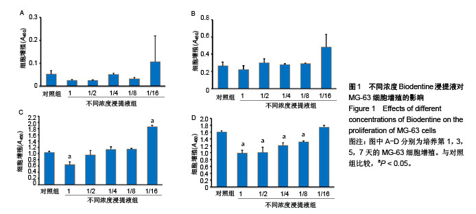
2.1 不同浓度Biodentine对MG-63细胞增殖的影响 如图1A所示,Biodentine作用于MG-63细胞第1天,与对照组比,1浓度Biodentine组、1/2浓度Biodentine组、1/8浓度Biodentine组活细胞数量略有下降,1/4浓度Biodentine组无明显变化,1/16浓度Biodentine组增多,各组间活细胞数比较差异无显著性意义(P > 0.05)。 如图1B所示,Biodentine作用于MG-63细胞第3天,与对照组相比,1浓度Biodentine组活细胞数量略有下降,1/2浓度Biodentine组、1/4浓度Biodentine组和1/8浓度Biodentine组活细胞数量无明显变化,1/16浓度Biodentine组活细胞数量略有增多,各组间活细胞数比较差异无显著性意义(P > 0.05)。 如图1C所示,Biodentine作用于MG-63细胞第5天,与对照组相比,1浓度Biodentine组活细胞数量明显降低(P < 0.05),1/2浓度Biodentine组活细胞数量略降低(P > 0.05),1/4浓度Biodentine组、1/8浓度Biodentine组活细胞数量略有升高(P > 0.05),1/16浓度Biodentine组活细胞数量明显升高(P < 0.05)。 如图1D所示,Biodentine作用于MG-63细胞第7天,与对照组相比,1浓度Biodentine组、1/2浓度Biodentine组活细胞数量明显降低(P < 0.05),1/4浓度Biodentine组、1/8浓度Biodentine组活细胞数量降低(P < 0.05),1/16浓度Biodentine组活细胞数量略有上升(P > 0.05)。"
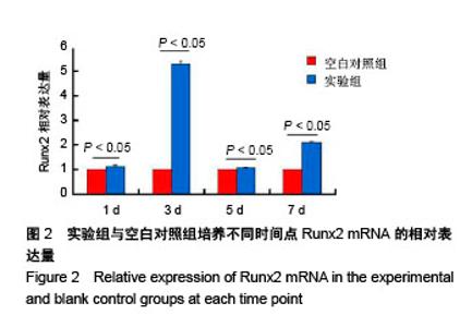
2.2 各组Runx2 mRNA的表达 如图2所示,1/16浓度Biodentine作用于MG-63细胞第1天时,MG-63细胞Runx2 mRNA的表达量为空白对照组的1.14倍;培养第3天时,Runx2 mRNA的表达量为空白对照组的5.29倍;培养第5天时,Runx2 mRNA的表达量为空白对照组的1.08倍;培养第7天时,Runx2 mRNA的表达量为空白对照组的2.11倍,实验组每一个时间点与空白对照组相比差异均有显著性意义(P < 0.05)。"


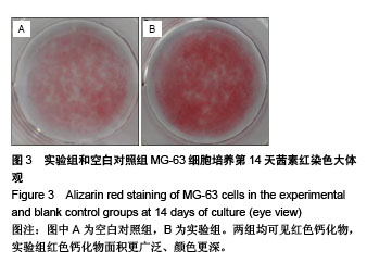
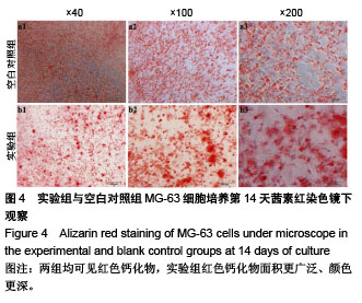
2.3 各组茜素红染色 MG-63细胞按不同处理组加入药物后,于第10天行茜素红染色,大体可见空白对照组无矿 化结节,镜下观察可见二三处小面积红色钙化物;实验组与空白对照组大体和镜下观察结果一致,两组之间未见显著差异。茜素红染色第14天,大体可见空白对照组有大面积红色钙化物;实验组也有红色钙化物,面积比对照组更广泛、颜色更深(图3);显微镜下观察结果与大体观察结果一致(图4)。"

"
| [1] De Rossi A,Fukada SY,De Rossi M,et al.Cementocytes Express Receptor Activator of the Nuclear Factor Kappa-B Ligand in Response to Endodontic Infection in Mice.J Endod. 2016;42(8):1251-1257.[2] Kok SH,Hou KL, Hong CY,et al.Sirtuin 6 Modulates Hypoxia-induced Apoptosis in Osteoblasts via Inhibition of Glycolysis:Implication for Pathogenesis of Periapical Lesions. J Endod. 2015;41(10): 1631-1637. [3] Zhang R,Huang S,Wang L,et al.Histochemical Localization of Dickkopf-1 in Induced Rat Periapical Lesions.J Endod. 2014;40(9): 1394-1399.[4] Parirokh M,Torabinejad M,Dummer PMH.Mineral trioxide aggregate and other bioactive endodontic cements: An updated overview- Part I: Vital pulp therapy.Int Endod J.2017; 24.doi: 10.1111/iej.12841.[5] Huang TH,Ding SJ,Hsu TC,et al.Effects of mineral trioxide aggregate (MTA) extracts on mitogen-activated protein kinase activity in human osteosarcoma cell line(U2OS). Biomaterials. 2003;24 (22):3909-3913.[6] Huang TH,Yang CC,Ding SJ,et al.Inflammatory cytokines reaction elicited by root-end filling materials.J Biomed Mater Res B Appl Biomater.2005;73(1):123-128.[7] Bruserud O,Wendelbo O,Paulsen K.Lipoteichoic acid derived from Enterococcus faecalis modulates the functional characteristics of both normal peripheral blood leukocytes and native human acute myelogenous leukemia blasts.Eur J Haematol.2004;73(5):340-350.[8] Maeda H,Nakano T,Tomokiyo A,et al. Mineral Trioxide Aggregate induces bone morphogenetic protein-2 expression and calcification in human periodontal ligament cells.J Endod. 2010;36(4):647-652.[9] Gomes-Filho JE,Rodrigues G,Watanabe S,et al.Evaluation of the tissue reaction to fast endodontic cement (CER) and Angelus MTA.J Endod.2009;35(10):1377-1380.[10] Camilleri J.Characterization and hydrationkinetics of tricalciumsilicatecement for use as a dentalbiomaterial.Dent Mater. 2011;27(8):836-844.[11] Malkondu Ö,Karapinar Kazanda? M,Kazazo?lu E.A Review on Biodentine, a Contemporary Dentine Replacement and Repair Material.Biomed Res Int.2014;2014:160951.[12] Grech L,Mallia B,Camilleri J.Characterization of set Intermediate Restorative Material, Biodentine, Bioaggregate and a prototype calcium silicate cement for use as root-end filling materials. Int Endod J.2013;46(7):632-641.[13] Kayahan MB,Nekoofar MH,McCann A,et al.Effect of acid etching procedures on the compressive strength of 4 calcium silicate-based endodontic cements.J Endod. 2013;39(12):1646-1648.[14] Grech L,Mallia B,Camilleri J.Investigation of the physical properties of tricalcium silicate cement-based root-end filling materials.Dent Mater. 2013;29(2):e20-28.[15] Koubi G,Colon P,Franquin JC,et al.Clinical evaluation of the performance and safety of a new dentine substitute, Biodentine, in the restoration of posterior teeth—a prospective study. Clin Oral Investig. 2013;17(1):243-249.[16] Guneser MB,Akbulut MB,Eldeniz AU.Effect of various endodontic irrigants on the push-out bond strength of biodentine and conventional root perforation repair materials. J Endod.2013;39(3): 380-384.[17] Camilleri J,Grech L,Galea K,et al.Porosity and root dentine to material interface assessment of calcium silicate-based root-end filling materials.Clin Oral Investig. 2014;18(5):1437-1446.[18] De Souza ET,Nunes Tameirão MD,Roter JM,et al.Tridimensional quantitative porosity characterization of three set calcium silicate-based repair cements for endodontic use.Microsc Res Tech. 2013;76(10): 1093-1098.[19] Camilleri J,Sorrentino F,Damidot D.Investigation of the hydration and bioactivity of radiopacified tricalcium silicate cement,Biodentine and MTA Angelus.Dent Mater. 2013;29(5):580-593.[20] Tanalp J,Karap?nar-Kazanda? M,Döleko?lu S,et al.Comparison of the radiopacities of different root-end filling and repair materials. Scientific World J.2013;23:594950.[21] Raskin A,Eschrich G,Dejou J,et al.In vitro microleakage of Biodentine as a dentin substitute compared to Fuji II LC in cervical lining restorations.J Adhes Dent.2012;14(6):535-542.[22] Villat C,Tran XV,Pradelle-Plasse N,et al.Impedance methodology: A new way to characterize the setting reaction of dental cements.Dent Mater.2010;26(12):1127-1132.[23] Vallés M,Roig M,Duran-Sindreu F,et al.Color Stability of Teeth Restored with Biodentine: A 6-month In Vitro Study.J Endod. 2015; 41(7):1157-1160.[24] Vallés M,Mercadé M,Duran-Sindreu F,et al.Influence of light and oxygen on the color stability of five calcium silicate-based materials.J Endod.2013;39(4):525-528.[25] Han L,Okiji T.Bioactivity evaluation of three calcium silicate-based endodontic materials.Int Endod J. 2013;46(9):808-814.[26] Bortoluzzi EA,Broon NJ,Bramante CM,et al.The influence of calciumchloride on the setting time, solubility, disintegration, and pH of mineral trioxide aggregate and white Portland cementwith a radiopacifier. J Endod.2009;35(4):550-554.[27] Villat C,Tran VX,Pradelle-Plasse N,et al.Impedance methodology: a new way to characterize the setting reaction of dental cements.Dent Mater.2010;26(12):1127-1132.[28] Nowicka A,Lipski M,Parafiniuk M,et al.Response of human dental pulp capped with biodentine and mineral trioxide aggregate.J Endod. 2013; 39(6):743-747.[29] Shayegan A,Jurysta C,Atash R,et al.Biodentine used as a pulp-capping agent in primary pig teeth. Pediatr Dent.2012;34(7):e202-208.[30] Zanini M,Sautier JM,Berdal A,et al.Biodentine induces immortalized murine pulp cell differentiation into odontoblast-like cells and stimulates biomineralization.J Endod.2012;38(9):1220-1226.[31] Girish K,Mandava J,Chandra RR,et al.Effect of obturating materials on fracture resistance of simulated immature teeth.J Conserv Dent. 2017; 20(2):115-119.[32] Villat C,Grosgogeat B,Seux D,et al.Conservative approach of a symptomatic carious immature permanent tooth using a tricalcium silicatecement (Biodentine): a case report.Restor Dent Endod. 2013; 38(4):258-262. [33] Pawar AM,Kokate SR,Shah RA.Management of a large periapical lesion using Biodentine as retrograde restoration with eighteen months evident follow-up.J Conserv Dent. 2013;16(6):573-575.[34] Bakhtiar H,Nekoofar MH,Aminishakib P,et al.Human Pulp Responses to Partial Pulpotomy Treatment with TheraCal as Compared withBiodentine and ProRoot MTA: A Clinical Trial.J Endod.2017;16.pii: S0099-2399(17)30812-9.[35] Goel S,Nawal RR,Talwar S.Management of Dens Invaginatus Type II Associated with Immature Apex and Large Periradicular Lesion Using Platelet-rich Fibrin and Biodentine. J Endod.2017;13.pii: S0099-2399(17)30417-X.[36] Jalan AL,Warhadpande MM,Dakshindas DM.A comparison of human dental pulp response to calcium hydroxide and Biodentine as direct pulp-capping agents.J Conserv Dent. 2017;20(2):129-133.[37] Zanini M,Sautier JM,Berdal A,et al.Biodentine induces immortalized murine pulp cell differentiation into odontoblast-like cells and stimulates biomineralization.J Endod.2012;38(9): 1220-1226.[38] Luo Z,Li D,Kohli MR,et al.Effect of Biodentine on the proliferation, migration and adhesion of human dental pulp stem cells.J Dent. 2014;42(4):490-497.[39] Zhou HM,Shen Y,Wang ZJ,et al.Haapasalo. In vitro cytotoxicity evaluation of a novel root repair material.J Endod. 2013;39(4):478-483.[40] Luo Z,Kohli MR,Yu Q,et al.Biodentine induces human dental pulp stem cell differentiation through mitogen-activated protein kinase and calcium-/calmodulin-dependent protein kinase II pathways.J Endod. 2014;40(7):937-942.[41] Ho CC,Fang HY,Wang B,et al.The effects of Biodentine/polycaprolactone three-dimensional -scaffold with odontogenesis properties on human dental pulp cells. Int Endod J. 2017; 20.doi: 10.1111/iej.12799.[42] Laurent P,Camps J,About I.Biodentine(TM) induces TGF-beta1 release from human pulp cells and early dental pulp mineralization.Int Endod J.2012;45(5):439-448.[43] Jung JY,Woo SM,Lee BN,et al. Effect of Biodentine and Bioaggregate on odontoblastic differentiation via mitogen-activated protein kinase pathway in human dental pulp cells.Int Endod J.2015;48(2): 177-184.[44] Chang SW,Lee SY,Ann HJ,et al.Effects of calcium silicate endodontic cements on biocompatibility and mineralization-inducing potentials in human dental pulp cells.J Endod.2014;40(8):1194-1200.[45] Perard M,Le Clerc J,Watrin T,et al.Spheroid model study comparing the biocompatibility of Biodentine and MTA.J Mater Sci Mater Med. 2013;24(6):1527-1534.[46] Corral Nuñez CM,Bosomworth HJ,Field C,et al.Biodentine and mineral trioxide aggregate induce similar cellular responses in a fibroblast cell line.J Endod. 2014;40(3):406-411.[47] Jang YE,Lee BN,Koh JT,et al.Cytotoxicity and physical properties of tricalcium silicate-based endodontic materials. Restor Dent Endod. 2014;39(2):89-94.[48] Poggio C,Arciola CR,Beltrami R,et al.Cytocompatibility and antibacterial properties of capping materials. ScientificWorldJournal. 2014;2014:181945.[49] Poggio C,Ceci M,Beltrami R,et al. Biocompatibility of a new pulp capping cement.Ann Stomatol (Roma).2014;5(2):69-76.[50] Lee BN,Lee KN,Koh JT,et al.Effects of 3 endodontic bioactive cements on osteogenic differentiation in mesenchymal stem cells.J Endod. 2014;40(8):1217-1222. |
| [1] | Chen Ziyang, Pu Rui, Deng Shuang, Yuan Lingyan. Regulatory effect of exosomes on exercise-mediated insulin resistance diseases [J]. Chinese Journal of Tissue Engineering Research, 2021, 25(25): 4089-4094. |
| [2] | Chen Yang, Huang Denggao, Gao Yuanhui, Wang Shunlan, Cao Hui, Zheng Linlin, He Haowei, Luo Siqin, Xiao Jingchuan, Zhang Yingai, Zhang Shufang. Low-intensity pulsed ultrasound promotes the proliferation and adhesion of human adipose-derived mesenchymal stem cells [J]. Chinese Journal of Tissue Engineering Research, 2021, 25(25): 3949-3955. |
| [3] | Yang Junhui, Luo Jinli, Yuan Xiaoping. Effects of human growth hormone on proliferation and osteogenic differentiation of human periodontal ligament stem cells [J]. Chinese Journal of Tissue Engineering Research, 2021, 25(25): 3956-3961. |
| [4] | Sun Jianwei, Yang Xinming, Zhang Ying. Effect of montelukast combined with bone marrow mesenchymal stem cell transplantation on spinal cord injury in rat models [J]. Chinese Journal of Tissue Engineering Research, 2021, 25(25): 3962-3969. |
| [5] | Gao Shan, Huang Dongjing, Hong Haiman, Jia Jingqiao, Meng Fei. Comparison on the curative effect of human placenta-derived mesenchymal stem cells and induced islet-like cells in gestational diabetes mellitus rats [J]. Chinese Journal of Tissue Engineering Research, 2021, 25(25): 3981-3987. |
| [6] | Hao Xiaona, Zhang Yingjie, Li Yuyun, Xu Tao. Bone marrow mesenchymal stem cells overexpressing prolyl oligopeptidase on the repair of liver fibrosis in rat models [J]. Chinese Journal of Tissue Engineering Research, 2021, 25(25): 3988-3993. |
| [7] | Liu Jianyou, Jia Zhongwei, Niu Jiawei, Cao Xinjie, Zhang Dong, Wei Jie. A new method for measuring the anteversion angle of the femoral neck by constructing the three-dimensional digital model of the femur [J]. Chinese Journal of Tissue Engineering Research, 2021, 25(24): 3779-3783. |
| [8] | Meng Lingjie, Qian Hui, Sheng Xiaolei, Lu Jianfeng, Huang Jianping, Qi Liangang, Liu Zongbao. Application of three-dimensional printing technology combined with bone cement in minimally invasive treatment of the collapsed Sanders III type of calcaneal fractures [J]. Chinese Journal of Tissue Engineering Research, 2021, 25(24): 3784-3789. |
| [9] | Qian Xuankun, Huang Hefei, Wu Chengcong, Liu Keting, Ou Hua, Zhang Jinpeng, Ren Jing, Wan Jianshan. Computer-assisted navigation combined with minimally invasive transforaminal lumbar interbody fusion for lumbar spondylolisthesis [J]. Chinese Journal of Tissue Engineering Research, 2021, 25(24): 3790-3795. |
| [10] | Hu Jing, Xiang Yang, Ye Chuan, Han Ziji. Three-dimensional printing assisted screw placement and freehand pedicle screw fixation in the treatment of thoracolumbar fractures: 1-year follow-up [J]. Chinese Journal of Tissue Engineering Research, 2021, 25(24): 3804-3809. |
| [11] | Shu Qihang, Liao Yijia, Xue Jingbo, Yan Yiguo, Wang Cheng. Three-dimensional finite element analysis of a new three-dimensional printed porous fusion cage for cervical vertebra [J]. Chinese Journal of Tissue Engineering Research, 2021, 25(24): 3810-3815. |
| [12] | Wang Yihan, Li Yang, Zhang Ling, Zhang Rui, Xu Ruida, Han Xiaofeng, Cheng Guangqi, Wang Weil. Application of three-dimensional visualization technology for digital orthopedics in the reduction and fixation of intertrochanteric fracture [J]. Chinese Journal of Tissue Engineering Research, 2021, 25(24): 3816-3820. |
| [13] | Sun Maji, Wang Qiuan, Zhang Xingchen, Guo Chong, Yuan Feng, Guo Kaijin. Development and biomechanical analysis of a new anterior cervical pedicle screw fixation system [J]. Chinese Journal of Tissue Engineering Research, 2021, 25(24): 3821-3825. |
| [14] | Lin Wang, Wang Yingying, Guo Weizhong, Yuan Cuihua, Xu Shenggui, Zhang Shenshen, Lin Chengshou. Adopting expanded lateral approach to enhance the mechanical stability and knee function for treating posterolateral column fracture of tibial plateau [J]. Chinese Journal of Tissue Engineering Research, 2021, 25(24): 3826-3827. |
| [15] | Zhu Yun, Chen Yu, Qiu Hao, Liu Dun, Jin Guorong, Chen Shimou, Weng Zheng. Finite element analysis for treatment of osteoporotic femoral fracture with far cortical locking screw [J]. Chinese Journal of Tissue Engineering Research, 2021, 25(24): 3832-3837. |
| Viewed | ||||||
|
Full text |
|
|||||
|
Abstract |
|
|||||