Chinese Journal of Tissue Engineering Research ›› 2021, Vol. 25 ›› Issue (24): 3810-3815.doi: 10.12307/2021.084
Previous Articles Next Articles
Three-dimensional finite element analysis of a new three-dimensional printed porous fusion cage for cervical vertebra
Shu Qihang, Liao Yijia, Xue Jingbo, Yan Yiguo, Wang Cheng
- Department of Spine Surgery, the First Affiliated Hospital of University of South China, Hengyang 421001, Hunan Province, China
-
Received:2020-10-16Revised:2020-10-21Accepted:2020-11-19Online:2021-08-28Published:2021-03-08 -
Contact:Wang Cheng, MD, Associate chief physician, Department of Spine Surgery, the First Affiliated Hospital of University of South China, Hengyang 421001, Hunan Province, China -
About author:Shu Qihang, Master candidate, Department of Spine Surgery, the First Affiliated Hospital of University of South China, Hengyang 421001, Hunan Province, China -
Supported by:the Scientific Research Project of Hunan Health Committee, No. C2019120, 20201961 (to WC); the Hunan Spinal Minimally Invasive Clinical Research Center, No. 2017SK4004 (to YYG); the Key Scientific Research Project of Hunan Health and Family Planning Commission, No. A2017016 (to YYG); the Science and Technology Development Plan Project of Hengyang, No. 2018KJ115 (to YYG)
CLC Number:
Cite this article
Shu Qihang, Liao Yijia, Xue Jingbo, Yan Yiguo, Wang Cheng. Three-dimensional finite element analysis of a new three-dimensional printed porous fusion cage for cervical vertebra[J]. Chinese Journal of Tissue Engineering Research, 2021, 25(24): 3810-3815.
share this article
Add to citation manager EndNote|Reference Manager|ProCite|BibTeX|RefWorks
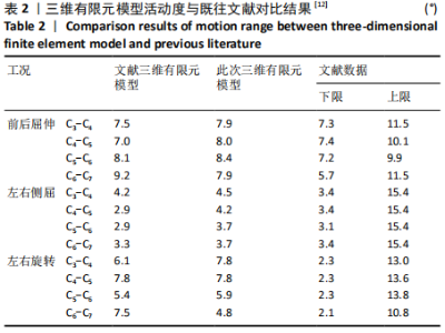
2.1 三维有限元模型有效性验证 模型验证采纳的是C3-C7节段完整模型,参考既往文献,施加1.5 N?m弯矩,查看其活动度,见表2。根据模型测试所得的数据,所得活动度均在文献范围以内[12],故认为该模型满足模型验证要求。"

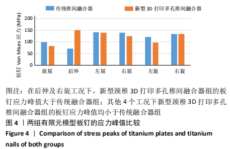
2.2 板钉的应力峰值 两组有限元模型的板钉应力峰值情况见图4。与传统椎间融合器组相比,除在后伸及右旋转这2个工况下,新型3D打印多孔椎间融合器组相对于传统椎间融合器组的板钉的应力峰值分别上升了80.27,0.88 MPa;在前屈、左右侧屈及左旋这4个工况下,新型3D打印多孔椎间融合器组的板钉的应力峰值分别下降了17.84,2.02,14.59,25.05 MPa。 "


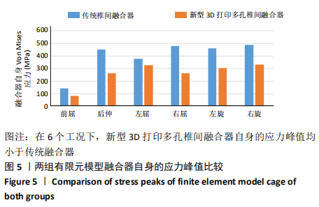
2.3 融合器自身的应力峰值 两组有限元模型融合器自身的应力峰值情况见图5。在前屈、后伸、左右侧屈及左右旋转 这6个工况下,相比于传统融合器,新型3D打印多孔椎间融合器自身的应力峰值分别下降了57.57,189.76,51.51,220.2,155.22,158.51 MPa。 "
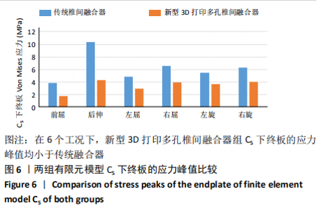
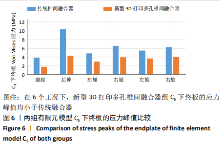
2.4 C5下终板的应力峰值 两组有限元模型C5下终板的应力峰值情况见图6。在前屈、后伸、左右侧屈及左右旋转这6个工况下,新型3D打印多孔椎间融合器组C5下终板的应力峰值相对于传统的椎间融合器组的应力峰值分别下降了2.07,6.07,1.96,2.67,1.79,2.31 MPa。"
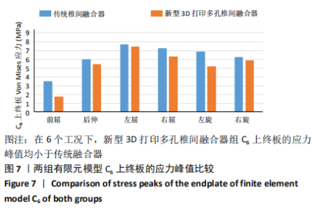
2.5 C6上终板的应力峰值 两组有限元模型C6上终板的应力峰值情况见图7。在前屈、后伸、左右侧屈及左右旋转这6个工况下,新型3D打印多孔椎间融合器组C6上终板的应力峰值相对于传统的椎间融合器组的应力峰值分别下降了1.74,0.60,0.27,0.97,1.70,0.35 MPa。"

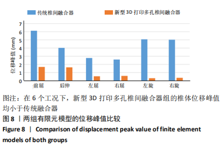
2.6 椎体位移峰值 两组有限元模型位移峰值情况见图8。与传统的椎间融合器组相比,在前屈、后伸、左右侧屈及左右旋转这6个工况下,新型3D打印多孔椎间融合器组的椎体位移峰值分别下降了4.44,2.36,2.27,2.02,4.78,4.69 mm。"
| [1] CLOWARD RB. The anterior approach for removal of ruptured cervical disks. 1958. J Neurosurg Spine. 2007;6(5):496-511. [2] SMITH GW, ROBINSON RA. The treatment of certain cervical-spine disorders by anterior removal of the intervertebral disc and interbody fusion. J Bone Joint Surg Am. 1958;40-A(3):607-624. [3] HEIDECKE V, RAINOV NG, MARX T, et al. Outcome in Cloward anterior fusion for degenerative cervical spinal disease. Acta Neurochir (Wien). 2000;142(3):283-291. [4] BANWART JC, ASHER MA, HASSANEIN RS. Iliac crest bone graft harvest donor site morbidity. A statistical evaluation. Spine (Phila Pa 1976). 1995;20(9):1055-1060. [5] Bagby GW. Arthrodesis by the distraction-compression method using a stainless steel implant. Orthopedics. 1988;11(6):931-934. [6] FARROKHI MR, NIKOO Z, GHOLAMI M, et al. Comparison Between Acrylic Cage and Polyetheretherketone (PEEK) Cage in Single-level Anterior Cervical Discectomy and Fusion: A Randomized Clinical Trial. Clin Spine Surg. 2017;30(1):38-46. [7] PHAN K, PELLETIER MH, RAO PJ, et al. Integral Fixation Titanium/Polyetheretherketone Cages for Cervical Arthrodesis: Evolution of Cage Design and Early Radiological Outcomes and Fusion Rates. Orthop Surg. 2019;11(1):52-59. [8] KIM TH, KIM DH, KIM KH, et al. Can the Zero-Profile Implant Be Used for Anterior Cervical Discectomy and Fusion in Traumatic Subaxial Disc Injury? A Preliminary, Retrospective Study. J Korean Neurosurg Soc. 2018;61(5):574-581. [9] 贾连顺, 魏海峰. 颈椎前路内固定器械的适应证及其进展[J]. 中国脊柱脊髓杂志,2000,10(5):62-64. [10] MALHAM GM, PARKER RM, BLECHER CM, et al. Assessment and classification of subsidence after lateral interbody fusion using serial computed tomography. J Neurosurg Spine. 2015;23(5):589-597. [11] PARK JY, CHOI KY, MOON BJ, et al. Subsidence after single-level anterior cervical fusion with a stand-alone cage. J Clin Neurosci. 2016;33:83-88. [12] LEE SH, IM YJ, KIM KT, et al. Comparison of cervical spine biomechanics after fixed- and mobile-core artificial disc replacement: a finite element analysis. Spine (Phila Pa 1976). 2011;36(9):700-708. [13] CHEUNG J, CHEUNG P, LAW K, et al. Postoperative Rigid Cervical Collar Leads to Less Axial Neck Pain in the Early Stage After Open-Door Laminoplasty-A Single-Blinded Randomized Controlled Trial. Neurosurgery. 2019;85(3):325-334. [14] YANG C, BI Z, YUAN S, et al. [A comparative study of anterior decompression approach by using cervical retractor systems and traditional surgical approach to treat cervical spondylosis]. Zhongguo Xiu Fu Chong Jian Wai Ke Za Zhi. 2008;22(4):394-398. [15] CHONG E, PELLETIER MH, MOBBS RJ, et al. The design evolution of interbody cages in anterior cervical discectomy and fusion: a systematic review. BMC Musculoskelet Disord. 2015;16:99. [16] JAIN S, ELTORAI AE, RUTTIMAN R, et al. Advances in Spinal Interbody Cages. Orthop Surg. 2016;8(3):278-284. [17] IGARASHI H, HOSHINO M, OMORI K, et al. Factors Influencing Interbody Cage Subsidence Following Anterior Cervical Discectomy and Fusion. Clin Spine Surg. 2019;32(7):297-302. [18] LI Z, XU X, WANG W, et al. Modulation of the mesenchymal stem cell migration capacity via preconditioning with topographic microstructure. Clin Hemorheol Microcirc. 2017;67(3-4):267-278. [19] DAENTZER D, WILLBOLD E, KALLA K, et al. Bioabsorbable interbody magnesium-polymer cage: degradation kinetics, biomechanical stiffness, and histological findings from an ovine cervical spine fusion model. Spine (Phila Pa 1976). 2014;39(20):E1220-1227. [20] REN C, SONG Y, XUE Y, et al. Evaluation of Bioabsorbable Multiamino Acid Copolymer/Nanohydroxyapatite/Calcium Sulfate Cage in a Goat Spine Model. World Neurosurg. 2017;103:341-347. [21] LIPPMAN CR, HAJJAR M, ABSHIRE B, et al. Cervical spine fusion with bioabsorbable cages. Neurosurg Focus. 2004;16(3):E4. [22] ZHOU C, SONG Y, LIU H, et al. [Current development of research of biodegradable interbody fusion Cage]. Zhongguo Xiu Fu Chong Jian Wai Ke Za Zhi. 2010;24(12):1500-1505. [23] FROST A, BAGOURI E, BROWN M, et al. Osteolysis following resorbable poly-L-lactide-co-D, L-lactide PLIF cage use: a review of cases. Eur Spine J. 2012;21(3):449-454. [24] ZHANG L, WANG J, TAO Y, et al. Outcome Evaluation of Zero-Profile Implant Compared with an Anterior Plate and Cage Used in Anterior Cervical Discectomy and Fusion: A Two-Year Follow-Up Study. Turk Neurosurg. 2016;26(3):416-422. [25] CHEN Y, LIU Y, CHEN H, et al. Comparison of Curvature Between the Zero-P Spacer and Traditional Cage and Plate After 3-Level Anterior Cervical Discectomy and Fusion: Mid-term Results. Clin Spine Surg. 2017;30(8):E1111-E1116. [26] LI T, YANG JS, WANG XF, et al. Can Zero-Profile Cage Maintain the Cervical Curvature Similar to Plate-Cage Construct for Single-Level Anterior Cervical Diskectomy and Fusion. World Neurosurg. 2020;135: e300-e306. [27] WANG Z, ZHU R, YANG H, et al. Zero-profile implant (Zero-p) versus plate cage benezech implant (PCB) in the treatment of single-level cervical spondylotic myelopathy. BMC Musculoskelet Disord. 2015;16: 290. [28] HAKAŁO J, WROŃSKI J, CIUPIK L. Subsidence and its effect on the anterior plate stabilization in the course of cervical spondylodesis. Part I: definition and review of literature. Neurol Neurochir Pol. 2003;37(4): 903-915. [29] MAYER T, BRÄNDLE G, SCHÖNENBERGER A, et al. Simulation and validation of residual deformations in additive manufacturing of metal parts. Heliyon. 2020;6(5):e03987. [30] HANC M, FOKTER SK, VOGRIN M, et al. Porous tantalum in spinal surgery: an overview. Eur J Orthop Surg Traumatol. 2016;26(1):1-7. |
| [1] | Zhou Jihui, Li Xinzhi, Zhou You, Huang Wei, Chen Wenyao. Multiple problems in the selection of implants for patellar fracture [J]. Chinese Journal of Tissue Engineering Research, 2021, 25(9): 1440-1445. |
| [2] | Chen Junming, Yue Chen, He Peilin, Zhang Juntao, Sun Moyuan, Liu Youwen. Hip arthroplasty versus proximal femoral nail antirotation for intertrochanteric fractures in older adults: a meta-analysis [J]. Chinese Journal of Tissue Engineering Research, 2021, 25(9): 1452-1457. |
| [3] | Hu Kai, Qiao Xiaohong, Zhang Yonghong, Wang Dong, Qin Sihe. Treatment of displaced intra-articular calcaneal fractures with cannulated screws and plates: a meta-analysis of 15 randomized controlled trials [J]. Chinese Journal of Tissue Engineering Research, 2021, 25(9): 1465-1470. |
| [4] | Xu Feng, Kang Hui, Wei Tanjun, Xi Jintao. Biomechanical analysis of different fixation methods of pedicle screws for thoracolumbar fracture [J]. Chinese Journal of Tissue Engineering Research, 2021, 25(9): 1313-1317. |
| [5] | Jiang Yong, Luo Yi, Ding Yongli, Zhou Yong, Min Li, Tang Fan, Zhang Wenli, Duan Hong, Tu Chongqi. Von Mises stress on the influence of pelvic stability by precise sacral resection and clinical validation [J]. Chinese Journal of Tissue Engineering Research, 2021, 25(9): 1318-1323. |
| [6] | Chen Zehua, Ye Xiangling, Chen Weijian, Du Jianping, Liu Wengang, Xu Xuemeng. Effect of pronated foot posture on proprioception and postural stability based on foot posture index [J]. Chinese Journal of Tissue Engineering Research, 2021, 25(9): 1324-1328. |
| [7] | Lu Dezhi, Mei Zhao, Li Xianglei, Wang Caiping, Sun Xin, Wang Xiaowen, Wang Jinwu. Digital design and effect evaluation of three-dimensional printing scoliosis orthosis [J]. Chinese Journal of Tissue Engineering Research, 2021, 25(9): 1329-1334. |
| [8] | Zhang Tongtong, Wang Zhonghua, Wen Jie, Song Yuxin, Liu Lin. Application of three-dimensional printing model in surgical resection and reconstruction of cervical tumor [J]. Chinese Journal of Tissue Engineering Research, 2021, 25(9): 1335-1339. |
| [9] | Du Xiupeng, Yang Zhaohui. Effect of degree of initial deformity of impacted femoral neck fractures under 65 years of age on femoral neck shortening [J]. Chinese Journal of Tissue Engineering Research, 2021, 25(9): 1410-1416. |
| [10] | Zhang Shangpu, Ju Xiaodong, Song Hengyi, Dong Zhi, Wang Chen, Sun Guodong. Arthroscopic suture bridge technique with suture anchor in the treatment of acromioclavicular dislocation [J]. Chinese Journal of Tissue Engineering Research, 2021, 25(9): 1417-1422. |
| [11] | Pei Lili, Sun Guicai, Wang Di. Salvianolic acid B inhibits oxidative damage of bone marrow mesenchymal stem cells and promotes differentiation into cardiomyocytes [J]. Chinese Journal of Tissue Engineering Research, 2021, 25(7): 1032-1036. |
| [12] | Liu Zhengpeng, Wang Yahui, Zhang Yilong, Ming Ying, Sun Zhijie, Sun He. Application of 3D printed interbody fusion cage for cervical spondylosis of spinal cord type: half-year follow-up of recovery of cervical curvature and intervertebral height [J]. Chinese Journal of Tissue Engineering Research, 2021, 25(6): 849-853. |
| [13] | Hou Guangyuan, Zhang Jixue, Zhang Zhijun, Meng Xianghui, Duan Wen, Gao Weilu. Bone cement pedicle screw fixation and fusion in the treatment of degenerative spinal disease with osteoporosis: one-year follow-up [J]. Chinese Journal of Tissue Engineering Research, 2021, 25(6): 878-883. |
| [14] | He Li, Tian Wei, Xu Song, Zhao Xiaoyu, Miao Jun, Jia Jian. Factors influencing the efficacy of lumbopelvic internal fixation in the treatment of traumatic spinopelvic dissociation [J]. Chinese Journal of Tissue Engineering Research, 2021, 25(6): 884-889. |
| [15] | Yang Weiqiang, Ding Tong, Yang Weike, Jiang Zhengang. Combined variable stress plate internal fixation affects changes of bone histiocyte function and bone mineral density at the fractured end of goat femur [J]. Chinese Journal of Tissue Engineering Research, 2021, 25(6): 890-894. |
| Viewed | ||||||
|
Full text |
|
|||||
|
Abstract |
|
|||||