Chinese Journal of Tissue Engineering Research ›› 2018, Vol. 22 ›› Issue (22): 3506-3512.doi: 10.3969/j.issn.2095-4344.0847
Previous Articles Next Articles
Topical usage of nerve growth factor-insulin gel on deep II scald wound increases microvessel density and proliferating cell nuclear antigen expression
Xu Hai-tao1, Wang Min2, Xiang Xi-juan3
- 1Department of Orthopedics, Yongchuan Hospital of Chongqing Medical University, Chongqing 402160, China; 2Department of Burn, Central Hospital of Zibo, Zibo 255000, Shandong Province, China; 3Department of Ophthalmology, Yongchuan District Hospital of Traditional Chinese Medicine, Chongqing 402160, China
-
Received:2018-02-20Online:2018-08-08Published:2018-08-08 -
Contact:Xiang Xi-juan, Department of Ophthalmology, Yongchuan District Hospital of Traditional Chinese Medicine, Chongqing 402160, China -
About author:Xu Hai-tao, Master, Attending physician, Department of Orthopedics, Yongchuan Hospital of Chongqing Medical University, Chongqing 402160, China -
Supported by:the Funded Project of Yongchuan Hospital Affiliated to Chongqing Medical University, No. YJLCX201540
CLC Number:
Cite this article
Xu Hai-tao, Wang Min, Xiang Xi-juan. Topical usage of nerve growth factor-insulin gel on deep II scald wound increases microvessel density and proliferating cell nuclear antigen expression[J]. Chinese Journal of Tissue Engineering Research, 2018, 22(22): 3506-3512.
share this article
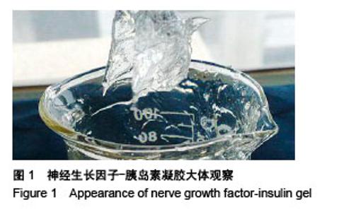
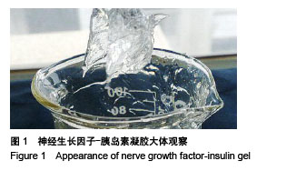
2.1 实验动物数量分析 75只大鼠均进入结果分析。 2.2 神经生长因子-胰岛素凝胶性状 神经生长因子-胰岛素凝胶呈清亮透明的果冻状(图1),延展性、保湿性良好,方便使用、易于清洗。"
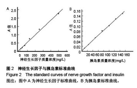
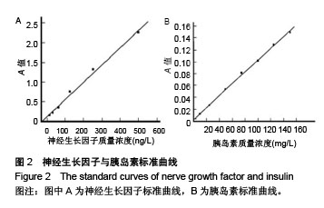
2.3 神经生长因子-胰岛素凝胶体外药物释放实验 2.3.1 神经生长因子、胰岛素标准曲线及回归方程 神经生长因子:y=0.004 4x+0.120 7,胰岛素:y=0.001x+0.002 6,标准曲线见图2。"
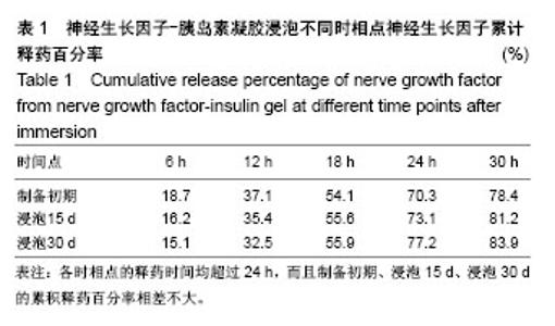
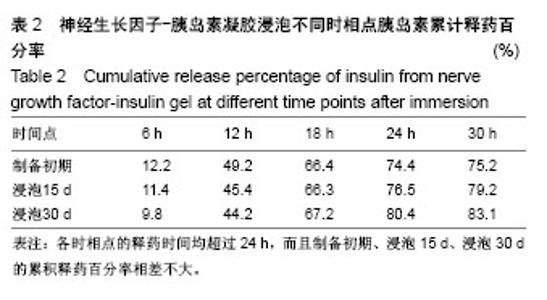
2.3.2 神经生长因子-胰岛素凝胶体外药物释放检测 神经生长因子及胰岛素在各时相点的释药时间均超过24 h,而且制备初期、15 d、30 d的累积释药百分率相差不大,表明制备的凝胶剂稳定性良好,具体见表1,2。"

"
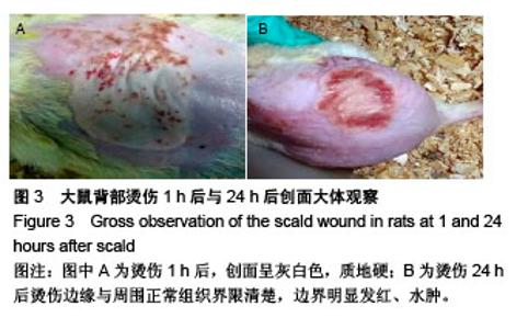
2.4 各组创面观察 初始建立的烫伤模型创面呈灰白色,质地硬;伤后24 h,烫伤边缘与周围正常组织界限清楚,边界明显发红、水肿(图3);经不同凝胶制剂局部外用后,各组烫伤创面随着时间逐渐缩小,联合组在同一观察时相点缩小更为明显。伤后3 d,各组创面均可见不同程度的红肿及炎性渗出,创面未见明显缩小。伤后7 d,除糖尿病对照组外,其余各组创面无明显红肿、渗出,均形成痂皮,而联合组创面干燥、边缘收缩明显,痂皮与基底紧密相连。伤后11 d,各组创面干燥、无红肿,神经生长因子、胰岛素组、联合组可见新生上皮组织移行,以联合组最为显著。伤后15 d,正常对照组、糖尿病对照组也见明显新生上皮移行,而联合组大部分创面痂皮脱落、已愈合。伤后21 d,联合组创面无明显瘢痕形成、完全愈合,而神经生长因子、胰岛素组大部分创面愈合,正常对照组、糖尿病对照组仅部分创面愈合。"

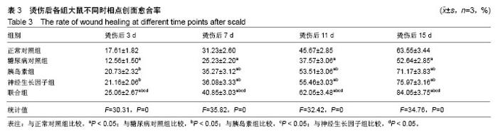
2.5 各组创面愈合率 经不同用药方式治疗后,各组创面随时间延长而逐渐缩小,联合组创面愈合率最高,明显高于胰岛素组、神经生长因子组。烫伤后7,11,15,21 d创面愈合率:联合组>神经生长因子组>胰岛素组>正常对照组>糖尿病对照组,联合组与其余各组比较差异均有显著性意义(P < 0.05),见表3。"
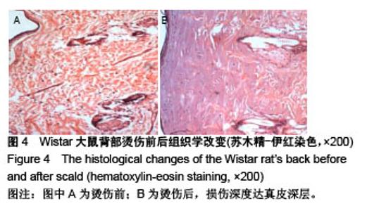
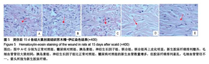
2.6 各组创面组织学观察 糖尿病大鼠烫伤后创面组织苏木精-伊红染色后,观察创面符合深Ⅱ度烫伤,损伤深度可达真皮深层(图4)。烫伤后1 d,各组表现为不同程度的组织结构破坏、血管扩张、炎性细胞浸润;烫伤后3 d可见少量新生毛细血管,联合组最为明显。烫伤后7-11 d,各组毛细血管及炎性细胞逐渐增加。烫伤后15 d,联合组再上皮化明显,新生胶原纤维排列整齐,毛细血管管径大致相同;胰岛素组、神经生长因子组比正常对照组、糖尿病对照组的新生血管数量增多,但胶原纤维排列紊乱,毛细血管管径不一。烫伤后21 d,联合组创面被上皮细胞覆盖,胶原纤维排列整齐;其他各组均有不同程度的上皮化,但正常对照组、糖尿病对照组不及胰岛素组、神经生长因子组,见图5。"
"

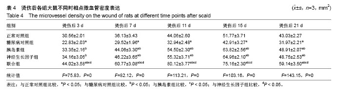
2.7 各组创面微血管密度检测 各组创面CD34在烫伤后3 d均呈阳性表达,阳性细胞随着时间逐渐增多,其中联合组与其他各组分别于11,15 d时达高峰(图6)。联合组微血管密度在不同时相点最高,糖尿病对照组最低,联合组、糖尿病对照组不同时相点比较或与其他组比较均有显著差异(P < 0.05),见表4。"


"
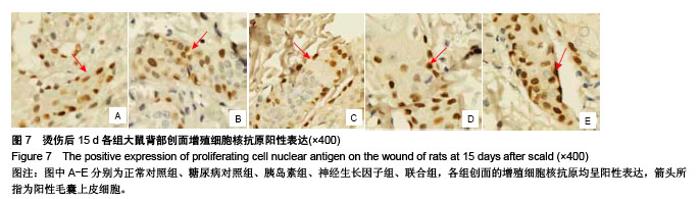
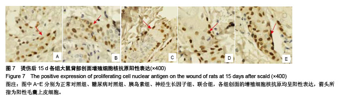
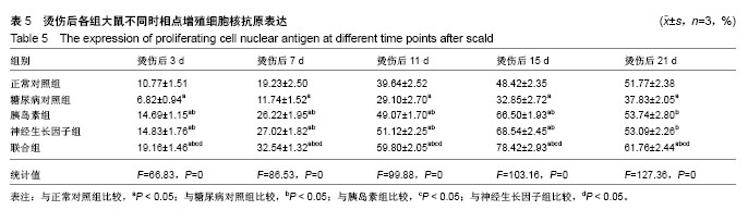
2.8 各组创面增殖细胞核抗原检测 糖尿病大鼠各组创面的增殖细胞核抗原在烫伤后3 d均呈阳性表达,阳性细胞随着时间逐渐增多,神经生长因子组、胰岛素组、联合组及正常对照组、糖尿病对照组分别于15,21 d达高峰(图7)。联合组增殖细胞核抗原在不同时相点最高,糖尿病对照组最低,联合组、糖尿病对照组不同时相点比较或与其他组比较均有差异(P < 0.05),见表5。"

"
| [1] Chen JC,Lin BB,Hu HW,et al.NGF accelerates cutaneous wound healing by promoting the migration of dermal fibroblasts via the PI3K/Akt-Rac1-JNK and ERK pathways. Biomed Res Int. 2014;2014: 547187.[2] Schenck K,Schreurs O,Hayashi K,et al.The Role of Nerve Growth Factor (NGF) and Its Precursor Forms in Oral Wound Healing.Int J Mol Sci.2017;18(2). pii: E386. doi: 10.3390/ijms18020386.[3] Salvinelli F,Frari V,Rocco ML,et al.Enhanced presence of NGF and mast cells number in nasal cavity after autologous stimulation: relation with sensorineural hearing deficit.Eur Rev Med Pharmacol Sci. 2015; 19(3):381-91.[4] 熊华花,李泉水,范锟.一种新型ngf缓释系统的制备[J].实用临床医学, 2008,9(2):3-6.[5] Okon UA,Umoren IU.Comparison of antioxidant activity of insulin, Ocimum gratissimum L. and Vernonia amygdalina L.in type 1 diabetic rat model.J Integr Med.2017;15(4):302-309. [6] 赵亚男,刘明,张玥,等.糖尿病难愈性创面大鼠模型的建立[J].山东医药, 2016,56(14):31-32.[7] 段强,王冰水,牟翔,等.低功率氦氖激光对小鼠烫伤创面血管内皮细胞因子和细胞凋亡因子基因表达的影响[J].中国康复医学杂志, 2016,31(2): 199-200,214.[8] Pareek G,Shevchuk M,Armenakas NA,et al.The effect of finasteride on the expression of vascular endothelial growth factor and microvessel density: a possible mechanism for decreased prostatic bleeding in treated patients.J Urol. 2003;169(1):20-23.[9] 张雨.烧伤创面修复技术的研究进展[J].医疗装备, 2017,30(10):191-192.[10] 孙晓芳,谢挺.创面修复中生长因子研究及临床干预新进展[J].创伤外科杂志,2016,18(3):190-193.[11] 常清璇,杨沛瑯,刘琰,等.局部应用胰岛素调控基质金属蛋白酶对糖尿病创面血管新生的影响[J].上海交通大学学报(医学版), 2015,35(9):1258.[12] 雷雨.小剂量局部胰岛素联合应用重组人表皮生长因子对烧伤大鼠创面MIP-2和MCP-1表达的影响[J].中国现代医学杂志, 2015,25(6):28-33.[13] 杨沛瑯,常清璇,章雄,等.局部应用胰岛素对糖尿病大鼠烫伤创面愈合的影响[J].中华损伤与修复杂志:电子版,2014,9(4):12-15.[14] 王敏,薛晓东,谢沛霖,等.局部应用NGF-胰岛素复合凝胶对糖尿病大鼠深Ⅱ度烫伤创面修复的影响[J].中国修复重建外科杂志, 2013,27(2):182-188.[15] 杨宗城.烧伤治巧学[M].3版.北京:人民卫生出版化,2006:77-116.[16] Achi NK,Ohaeri OC,Ijeh II,et al.Eleazu C(3).Modulation of the lipid profile and insulin levels of streptozotocin induced diabetic rats by ethanol extract of Cnidoscolus aconitifolius leaves and some fractions: Effect on the oral glucose tolerance of normoglycemic rats.Biomed Pharmacother.2016; 86:562-569.[17] 吴健,薛晓东,刘俊玲,等.胰岛素局部应用对老龄糖尿病大鼠烫伤创面愈合的实验研究[J].中国修复重建外科杂志, 2009,23(12):1482-1486.[18] 李超飞,刘琰,陈雪莲,等.局部应用胰岛素对糖尿病小鼠创面愈合的影响[J].中华损伤与修复杂志:电子版, 2012,7(1):54-61.[19] 肖瑶, 吴基良. 糖尿病引发内皮细胞功能紊乱的研究进展[J].湖北科技学报(医学版),2015, 29(2):177-180.[20] 宋振强,王润秀,于德民,等. 晚期糖基化终末产物修饰人血清白蛋白对离体角质形成细胞迁移功能的影响[J].中华医学杂志, 2008,88(38): 2690-2694.[21] 杨秀颖,张莉,陈熙,等.2型糖尿病周围神经病变机制研究进展[J].中国药理学通报,2016,32(5):598-602.[22] 聂发传,石英.糖尿病周围神经病变发生机制研究进展[J].重庆医学, 2015, 44(1):122-125.[23] 徐江平,郭海彪,汪海涛,等.鼠神经生长因子对糖尿病大鼠皮肤损伤的促愈合作用[J].湖北理工学院学报,2015,31(3):51-56.[24] 李政懋,吕辰,杨选鑫,等.外源NGF促进大鼠皮肤创面模型损伤修复的机制研究[J].上海医药, 2014,35(17):51-53.[25] 曾勇,简华刚,高兵,等.胰岛素外用对糖尿病大鼠深Ⅱ度烧伤创面HIF-1α及VEGF表达的影响[J].重庆医科大学学报, 2010,35(6):860-864.[26] 刘波,王甲汉,韩兆峰.局部胰岛素湿敷对兔深Ⅱ度烫伤创面愈合影响的实验研究[J].南方医科大学学报, 2008,28(4):561-563.[27] 张冀北,薛晓东,谢沛霖,等.局部联合应用NGF及胰岛素对糖尿病大鼠烫伤创面血管形成及Bcl-2、Bax表达的影响[J].中国修复重建外科杂志, 2011,25(3):354-360.[28] 倪向梅.卡波姆980流变学性质及影响因素初探[J].中国科技纵横, 2015, 14(15):201-203.[29] Tseng SY,Chao TH,Li YH,et al.Cilostazol improves high glucose-induced impaired angiogenesis in human endothelial progenitor cells and vascular endothelial cells as well as enhances vasculoangiogenesis in hyperglycemic mice mediated by the adenosine monophosphate-activated protein kina.J Vas Surg. 2016; 63(4):1051-1062.e3.[30] Tseng SY,Chao TH,Li YH,et al.Cilostazol improves high glucose-induced impaired angiogenesis in human endothelial progenitor cells and vascular endothelial cells as well as enhances vas. J Vas Surg.2015;63(4):1051.[31] 王珂,李荣贵,程殿林.增殖细胞核抗原(PCNA)功能及晶体结构研究进展[J].山东轻工业学院学报(自然科学版), 2013,27(3):25-31.[32] 宋楠萌,桑建利,徐恒.增殖细胞核抗原(PCNA)的分子结构及其生物学功能研究进展[J].自然科学进展, 2006,16(10):1201-1209.[33] Lu S.Burn Wound Healing[M]// Chinese Burn Surgery. Springer Netherlands,2015:207-248.[34] Graiani G,Emanueli C,Desortes E,et al.Nerve growth factor promotes reparative angiogenesis and inhibits endothelial apoptosis in cutaneous wounds of Type 1 diabetic mice. Diabetologia. 2004;47(6):1047-1054.[35] Zeng M,Zhi Y,Liu W,et al.Clinical study on local application of low-dose insulin for promoting wound healing after operation for deep burns.Exp Ther Med.2016;12(5):3221-3226.[36] Wang C,Wang J,Feng J.Local application of low-dose insulin in improving wound healing after deep burn surgery.Exp Ther Med. 2016; 12(4):2527-2530.[37] Zhang XJ,Wu X,Wolf SE,et al.Local insulin-zinc injection accelerates skin donor site wound healing.J Surg Res. 2007;142(1):90-96. [38] 谢晓繁,贾赤宇.胰岛素在烧伤创面愈合中的作用[J].中华创伤杂志, 2006, 22(6):475-477.[39] 刘虎仙,贾赤宇.胰岛素及血糖调控与烧伤创面愈合[J].中华烧伤杂志, 2005,21(3):232-234 |
| [1] | Luo Xuanxiang, Jing Li, Pan Bin, Feng Hu. Effect of mecobalamine combined with mouse nerve growth factor on nerve function recovery after cervical spondylotic myelopathy surgery [J]. Chinese Journal of Tissue Engineering Research, 2021, 25(5): 719-722. |
| [2] | Chen Ziyang, Pu Rui, Deng Shuang, Yuan Lingyan. Regulatory effect of exosomes on exercise-mediated insulin resistance diseases [J]. Chinese Journal of Tissue Engineering Research, 2021, 25(25): 4089-4094. |
| [3] | Gao Shan, Huang Dongjing, Hong Haiman, Jia Jingqiao, Meng Fei. Comparison on the curative effect of human placenta-derived mesenchymal stem cells and induced islet-like cells in gestational diabetes mellitus rats [J]. Chinese Journal of Tissue Engineering Research, 2021, 25(25): 3981-3987. |
| [4] | Xie Yang, Lü Zhiyu, Zhang Shujiang, Long Ting, Li Zuoxiao. Effects of recombinant adeno-associated virus mediated nerve growth factor gene transfection on oligodendrocyte apoptosis and myelination in experimental autoimmune encephalomyelitis mice [J]. Chinese Journal of Tissue Engineering Research, 2021, 25(23): 3678-3683. |
| [5] | Liu Yulin, Li Guotai. Combined effects of hyperbaric oxygen, vibration training and astaxanthin on bone mineral density, glucose metabolism and oxidative stress in diabetic osteoporosis rats [J]. Chinese Journal of Tissue Engineering Research, 2021, 25(20): 3117-3124. |
| [6] | Tian Lin, Shi Xiaoqing, Duan Zhenglan, Wang Kuan, Zhang Li, Wang Peimin. Efficacy and safety of transverse tibial bone transport technique in the treatment of diabetic foot:a meta-analysis#br# [J]. Chinese Journal of Tissue Engineering Research, 2021, 25(20): 3275-3280. |
| [7] | Xia Wenshen, He Renjiao, Ai Jinwei, Wang Jun, Li Desheng, Pei Bin. Stem cell transplantation for diabetic patients with lower-extremity arterial disease: a meta-analysis [J]. Chinese Journal of Tissue Engineering Research, 2021, 25(19): 3110-3116. |
| [8] | Luo Yicai, Li Hao. Effect of enhanced aryl hydrocarbon receptor expression on inflammatory response and healing of alveolar bone defects in diabetic rats [J]. Chinese Journal of Tissue Engineering Research, 2021, 25(14): 2166-2171. |
| [9] | Zhang Chuanhui, Li Jianjun, Yang Jun. Effects of dynamic pressure on the proliferation and mechanical properties of rabbit adipose mesenchymal stem cells transfected with insulin-like growth factor-1 [J]. Chinese Journal of Tissue Engineering Research, 2021, 25(13): 1982-1987. |
| [10] | Yan Nan, Si Xiaofeng, Zeng Liang, Tian Wei, Shan Guangdong, Xiong Lishuo, Yang Weijie, Wang Zhengdong. Nerve growth factor interferes with proliferation and alpha-actin expression of skeletal muscle satellite cells in rats [J]. Chinese Journal of Tissue Engineering Research, 2021, 25(13): 2030-2035. |
| [11] | Guo Xuan, Xie Jun, Suo Jinrong, Li Yingrui, Huang Lei, Ma Munan, Li Jingjing, Fu Songtao. Transplantation of islet-like cells induced by human umbilical cord mesenchymal stem cells via different ways for the treatment of type 1 diabetic mice [J]. Chinese Journal of Tissue Engineering Research, 2021, 25(1): 78-83. |
| [12] | Cao Guolong, Tian Faming, Liu Jiayin. Lovastatin combined with insulin effects on fracture healing in rat models of bilateral ovariectomized type 2 diabetic mellitus [J]. Chinese Journal of Tissue Engineering Research, 2020, 24(5): 673-681. |
| [13] | Chen Yongjia, Li Yanlin, Liu Dejian, He Yinghong, Yang Xiao. Sustained-release effect and clinical application of thermosensitive gels [J]. Chinese Journal of Tissue Engineering Research, 2020, 24(34): 5428-5433. |
| [14] | Yu Xingge, Lin Kaili. Application of nanocomposite hydrogels in bone tissue engineering [J]. Chinese Journal of Tissue Engineering Research, 2020, 24(34): 5441-5446. |
| [15] | Liu Guoming, Wang Qinfen, Lin Kefeng, Zhou Shiguo, Chen Zuxing, Lin Shishui. Whole body application of nerve growth factor promotes early healing of tibial shaft fracture and improves expression of bone morphogenetic protein-2 and vascular endothelial growth factor in rats [J]. Chinese Journal of Tissue Engineering Research, 2020, 24(29): 4680-4685. |
| Viewed | ||||||
|
Full text |
|
|||||
|
Abstract |
|
|||||