Chinese Journal of Tissue Engineering Research ›› 2018, Vol. 22 ›› Issue (12): 1883-1888.doi: 10.3969/j.issn.2095-4344.0807
Previous Articles Next Articles
Interleukin-17A facilitates optic nerve regeneration in a mouse model of optic nerve crush
Li Yan-fen, Ye Xiao-yang, Chen Xiao-fan, Zhang Wei
- Shenzhen PKU-HKUST Medical Center, Shenzhen 518036, Guangdong Province, China
-
Received:2017-11-16Online:2018-04-28Published:2018-04-28 -
Contact:Zhang Wei, Ph.D., Associate researcher, Master’s supervisor, Shenzhen PKU-HKUST Medical Center, Shenzhen 518036, Guangdong Province, China -
About author:Li Yan-fen, Studying for master’s degree, Shenzhen PKU-HKUST Medical Center, Shenzhen 518036, Guangdong Province, China -
Supported by:the National Natural Science Foundation of China for the Youth, No. 81402600; the Knowledge Innovation and Basic Research Program of Shenzhen Science and Technology Research & Development Foundation, No. JCYJ20150403110829622
CLC Number:
Cite this article
Li Yan-fen, Ye Xiao-yang, Chen Xiao-fan, Zhang Wei . Interleukin-17A facilitates optic nerve regeneration in a mouse model of optic nerve crush[J]. Chinese Journal of Tissue Engineering Research, 2018, 22(12): 1883-1888.
share this article
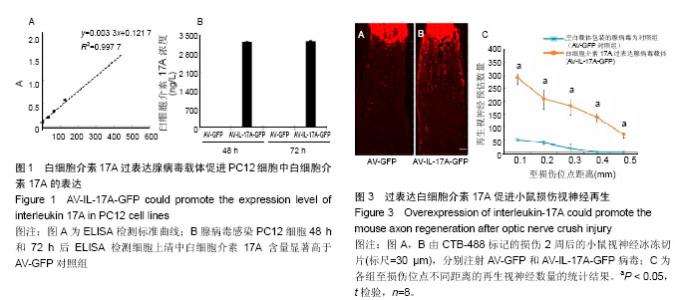
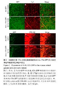
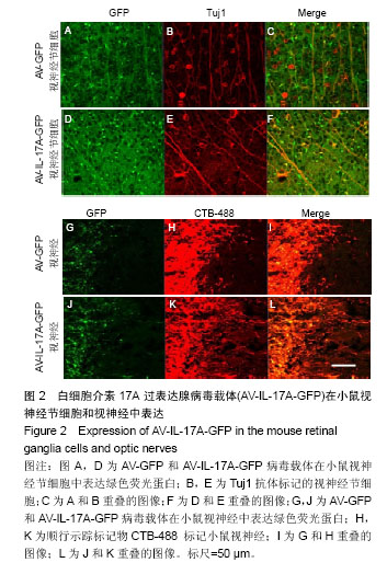
2.1 实验动物数量分析 实验选用C57BL/6J小鼠16只,分为2组,实验过程无脱失,全部进入结果分析。 2.2 PC12细胞中白细胞介素17A的表达 为明确白细胞介素17A在小鼠视神经损伤模型中所发挥的作用,构建了白细胞介素17A过表达腺病毒载体(AV-IL-17A-GFP)。用AV-IL-17A-GFP感染体外培养的PC12细胞系,病毒感染细胞48 h和72 h后,收集细胞上清,采用ELISA检测白细胞介素17A的表达。结果如图1所示,AV-IL-17A-GFP感染的细胞上清中可检测到的白细胞介素17A水平显著高于AV-GFP对照组。 2.3 白细胞介素17A过表达腺病毒载体在小鼠视神经节细胞和视神经中表达 用微量注射器向小鼠左眼玻璃体内注入2 μL腺病毒载体,病毒表达2周后给予小鼠视神经挤压手术,手术2周后进行心脏灌流固定,制备视网膜或视神经切片并进行免疫荧光染色,封固后用荧光显微镜观察。由图2可知,腺病毒载体能够有效地侵染视神经节细胞,并且能够顺行至视神经,表达绿色荧光蛋白。 2.4 小鼠损伤视神经再生结果 用CTB-488抗体对视神经切片进行免疫荧光染色,结果如图3所示,AV-GFP对照组中小鼠视神经在损伤后几乎没有长过损伤点,即使有少数几根神经纤维跨过损伤位点,生长的长度也非常有限。与AV-GFP对照组相比,过表达白细胞介素17A的AV-IL-17A-GFP组中有大量的视神经纤维跨过损伤位点,且长度显著长于对照组(P < 0.05),可以观察到明显的视神经纤维再生的现象。"

"
| [1] Kreutzberg GW. Acute neural reaction to injury//Repair and regeneration of the nervous system. Springer Berlin Heidelberg.1982:57-69.[2] Aguayo AJ, David S, Bray GM.Influences of the glial environment on the elongation of axons after injury: transplantation studies in adult rodents.J Exp Biol. 1981;95: 231-240.[3] David S, Aguayo AJ.Axonal elongation into peripheral nervous system "bridges" after central nervous system injury in adult rats.Science.1981;214:931-933.[4] Richardson PM, Issa VM.Peripheral injury enhances central regeneration of primary sensory neurones, Nature.1984;309: 791-793.[5] Case LC, Tessier-Lavigne M. Regeneration of the adult central nervous system. Current biology.2005;15(18): R749-R753.[6] Filbin MT. Recapitulate development to promote axonal regeneration:good or bad approach? Philos Trans R Soc Lond B Biol Sci. 2006;361(1473):1565-1574.[7] Fitch MT, Silver J. CNS injury, glial scars, and inflammation:Inhibitory extracellular matrices and regeneration failure. Exp Neurol.2008;209(2):294-301.[8] Harel NY, Strittmatter SM. Can regenerating axons recapitulate developmental guidance during recovery from spinal cord injury?. Nat Rev Neurosci.2006;7(8):603-616.[9] Schwab ME, Bartholdi D. Degeneration and regeneration of axons in the lesioned spinal cord. Physiol Rev. 1996;76(2): 319-370.[10] Yiu G, He Z. Glial inhibition of CNS axon regeneration. Nat Rev Neurosci. 2006 Aug;7(8):617-627.[11] Bregman BS, Kunkel-Bagden E, McAtee M, et al.Extension of the critical period for developmental plasticity of the corticospinal pathway.J Comp Neurol.1989;282 :355-370.[12] Cua DJ, Tato CM. Innate IL-17-producing cells:the sentinels of the immune system, Nat Rev Immunol.2010;10:479-489.[13] Iwakura Y,Ishigame H,Saijo S,et al.Functional specialization of interleukin-17 family members.Immunity.2011;34:149-162.[14] Pappu R, Rutz S, Ouyang W.Regulation of epithelial immunity by IL-17 family cytokines, Trends Immunol. 2012;33:343-349.[15] Song X,He X, Li X, et al.The roles and functional mechanisms of interleukin-17 family cytokines in mucosal immunity, Cell Mol Immunol.2016;13:418-431.[16] Fujie H, Niu K, Ohba M, et al.A distinct regulatory role of Th17 cytokines IL-17A and IL-17F in chemokine secretion from lung microvascular endothelial cells.Inflammation.2012;35: 1119-1131.[17] Song X, Qian Y.IL-17 family cytokines mediated signaling in the pathogenesis of inflammatory diseases.Cell Signal.2013; 25:2335-2347.[18] Song X,Qian Y.The activation and regulation of IL-17 receptor mediated signaling, Cytokine.2013;62:175-182.[19] Wang K,Karin M.The IL-23 to IL-17 cascade inflammation-related cancers, Clin Exp Rheumatol.2015; 33: S87-90.[20] Wang K, Kim MK, Di Caro G,et al.Interleukin-17 receptor a signaling in transformed enterocytes promotes early colorectal tumorigenesis.Immunity.2014;41:1052-1063.[21] Grivennikov SI,Wang K,Mucida D,et al.Adenoma-linked barrier defects and microbial products drive IL-23/IL-17-mediated tumour growth.Nature.2012;491: 254-258.[22] Sun C,Zhang J,Chen L,et al.IL-17 contributed to the neuropathic pain following peripheral nerve injury by promoting astrocyte proliferation and secretion of proinflammatory cytokines, Mol Med Rep.2017;15:89-96.[23] Waisman A,Hauptmann J,Regen T.The role of IL-17 in CNS diseases.Acta Neuropathol.2015;129:625-637.[24] Zepp J,Wu L, Li X.IL-17 receptor signaling and T helper 17-mediated autoimmune demyelinating disease.Trends Immunol.2011;32:232-239.[25] Li Z, Burns AR, Han L, et al.IL-17 and VEGF are necessary for efficient corneal nerve regeneration.Am J Pathol.2011; 178:1106-1116.[26] Chisholm SP,Cervi AL, Nagpal S,et al.Interleukin-17A increases neurite outgrowth from adult postganglionic sympathetic neurons.J Neurosci.2012;32(4):1146-155.[27] Zheng B, Atwal J, Ho C, et al. Genetic deletion of the Nogo receptor does not reduce neurite inhibition in vitro or promote corticospinal tract regeneration in vivo. Proc Natl Acad Sci U S A. 2005;102(4):1205-1210.[28] Park KK, Liu K, Hu Y,et al.Promoting axon regeneration in the adult CNS by modulation of the PTEN/mTOR pathway, Science.2008;322:963-966.[29] Smith PD, Sun F, Park KK,et al.SOCS3 deletion promotes optic nerve regeneration in vivo.Neuron.2009;64:617-623.[30] Sun F, Park KK, Belin S, et al.Sustained axon regeneration induced by co-deletion of PTEN and SOCS3, Nature. 2011; 480:372-375.[31] Heuss ND, Pierson MJ, Montaniel KR,et al.Retinal dendritic cell recruitment, but not function, was inhibited in MyD88 and TRIF deficient mice.J Neuroinflammation.2014;11:143.[32] Shum JW, Liu K, So KF.The progress in optic nerve regeneration, where are we?Neural Regen Res. 2016;11(1): 32-36.[33] Fischer D, Heiduschka P, Thanos S.Lens-injury-stimulated axonal regeneration throughout the optic pathway of adult rats, Experimental neurology.2001;172:257-272.[34] Leon S, Yin Y, Nguyen J, et al.Lens injury stimulates axon regeneration in the mature rat optic nerve,J Neurosci. 2000;20(12):4615-4626.[35] Müller A, Hauk TG, Fischer D. Astrocyte-derived CNTF switches mature RGCs to a regenerative state following inflammatory stimulation.Brain.2007;130:3308-3320.[36] Yin DP, Chen QY, Liu L.Synergetic effects of ciliary neurotrophic factor and olfactory ensheathing cells on optic nerve reparation (complete translation).Neural Regen Res. 2016;11(6):1006-1012.[37] Cafferty WB,Gardiner NJ, Das P,et al.Conditioning injury-induced spinal axon regeneration fails in interleukin-6 knock-out mice.J Neurosci.2004;24(18):4432-4432. |
| [1] | Zhang Tongtong, Wang Zhonghua, Wen Jie, Song Yuxin, Liu Lin. Application of three-dimensional printing model in surgical resection and reconstruction of cervical tumor [J]. Chinese Journal of Tissue Engineering Research, 2021, 25(9): 1335-1339. |
| [2] | Wan Ran, Shi Xu, Liu Jingsong, Wang Yansong. Research progress in the treatment of spinal cord injury with mesenchymal stem cell secretome [J]. Chinese Journal of Tissue Engineering Research, 2021, 25(7): 1088-1095. |
| [3] | Zeng Yanhua, Hao Yanlei. In vitro culture and purification of Schwann cells: a systematic review [J]. Chinese Journal of Tissue Engineering Research, 2021, 25(7): 1135-1141. |
| [4] | Xu Dongzi, Zhang Ting, Ouyang Zhaolian. The global competitive situation of cardiac tissue engineering based on patent analysis [J]. Chinese Journal of Tissue Engineering Research, 2021, 25(5): 807-812. |
| [5] | Wu Zijian, Hu Zhaoduan, Xie Youqiong, Wang Feng, Li Jia, Li Bocun, Cai Guowei, Peng Rui. Three-dimensional printing technology and bone tissue engineering research: literature metrology and visual analysis of research hotspots [J]. Chinese Journal of Tissue Engineering Research, 2021, 25(4): 564-569. |
| [6] | Chang Wenliao, Zhao Jie, Sun Xiaoliang, Wang Kun, Wu Guofeng, Zhou Jian, Li Shuxiang, Sun Han. Material selection, theoretical design and biomimetic function of artificial periosteum [J]. Chinese Journal of Tissue Engineering Research, 2021, 25(4): 600-606. |
| [7] | Liu Fei, Cui Yutao, Liu He. Advantages and problems of local antibiotic delivery system in the treatment of osteomyelitis [J]. Chinese Journal of Tissue Engineering Research, 2021, 25(4): 614-620. |
| [8] | Li Xiaozhuang, Duan Hao, Wang Weizhou, Tang Zhihong, Wang Yanghao, He Fei. Application of bone tissue engineering materials in the treatment of bone defect diseases in vivo [J]. Chinese Journal of Tissue Engineering Research, 2021, 25(4): 626-631. |
| [9] | Zhang Zhenkun, Li Zhe, Li Ya, Wang Yingying, Wang Yaping, Zhou Xinkui, Ma Shanshan, Guan Fangxia. Application of alginate based hydrogels/dressings in wound healing: sustained, dynamic and sequential release [J]. Chinese Journal of Tissue Engineering Research, 2021, 25(4): 638-643. |
| [10] | Chen Jiana, Qiu Yanling, Nie Minhai, Liu Xuqian. Tissue engineering scaffolds in repairing oral and maxillofacial soft tissue defects [J]. Chinese Journal of Tissue Engineering Research, 2021, 25(4): 644-650. |
| [11] | Xing Hao, Zhang Yonghong, Wang Dong. Advantages and disadvantages of repairing large-segment bone defect [J]. Chinese Journal of Tissue Engineering Research, 2021, 25(3): 426-430. |
| [12] | Chen Siqi, Xian Debin, Xu Rongsheng, Qin Zhongjie, Zhang Lei, Xia Delin. Effects of bone marrow mesenchymal stem cells and human umbilical vein endothelial cells combined with hydroxyapatite-tricalcium phosphate scaffolds on early angiogenesis in skull defect repair in rats [J]. Chinese Journal of Tissue Engineering Research, 2021, 25(22): 3458-3465. |
| [13] | Wang Hao, Chen Mingxue, Li Junkang, Luo Xujiang, Peng Liqing, Li Huo, Huang Bo, Tian Guangzhao, Liu Shuyun, Sui Xiang, Huang Jingxiang, Guo Quanyi, Lu Xiaobo. Decellularized porcine skin matrix for tissue-engineered meniscus scaffold [J]. Chinese Journal of Tissue Engineering Research, 2021, 25(22): 3473-3478. |
| [14] | Mo Jianling, He Shaoru, Feng Bowen, Jian Minqiao, Zhang Xiaohui, Liu Caisheng, Liang Yijing, Liu Yumei, Chen Liang, Zhou Haiyu, Liu Yanhui. Forming prevascularized cell sheets and the expression of angiogenesis-related factors [J]. Chinese Journal of Tissue Engineering Research, 2021, 25(22): 3479-3486. |
| [15] | Liu Chang, Li Datong, Liu Yuan, Kong Lingbo, Guo Rui, Yang Lixue, Hao Dingjun, He Baorong. Poor efficacy after vertebral augmentation surgery of acute symptomatic thoracolumbar osteoporotic compression fracture: relationship with bone cement, bone mineral density, and adjacent fractures [J]. Chinese Journal of Tissue Engineering Research, 2021, 25(22): 3510-3516. |
| Viewed | ||||||
|
Full text |
|
|||||
|
Abstract |
|
|||||