Chinese Journal of Tissue Engineering Research ›› 2018, Vol. 22 ›› Issue (12): 1823-1828.doi: 10.3969/j.issn.2095-4344.0198
Previous Articles Next Articles
Chondrogenic progenitor cells isolated from the fascia of skeletal muscle
Xiang Yong, Li Guang-heng
- Department of Orthopedics, the First Affiliated Hospital of Zhengzhou University, Zhengzhou 450000, Henan Province, China
-
Received:2017-12-20Online:2018-04-28Published:2018-04-28 -
Contact:Li Guang-heng, M.D., Professor, Doctoral supervisor, Department of Orthopedics, the First Affiliated Hospital of Zhengzhou University, Zhengzhou 450000, Henan Province, China -
About author:Xiang Yong, Master candidate, Department of Orthopedics, the First Affiliated Hospital of Zhengzhou University, Zhengzhou 450000, Henan Province, China -
Supported by:the National Natural Science Foundation of China, No. 81472136
CLC Number:
Cite this article
Xiang Yong, Li Guang-heng. Chondrogenic progenitor cells isolated from the fascia of skeletal muscle[J]. Chinese Journal of Tissue Engineering Research, 2018, 22(12): 1823-1828.
share this article
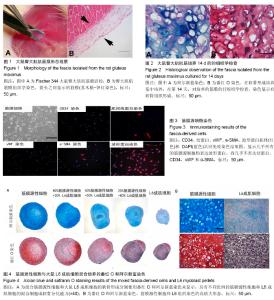
2.1 臀大肌筋膜组织染色和其软骨细胞分化能力的分析 取Fischer 344大鼠臀大肌肌筋膜进行活检,苏木精-伊红染色结果显示,筋膜组织主要是由包裹骨骼肌的纤维和结缔组织组成(图1);筋膜组织块在软骨细胞培养基中培养,在软骨形成培养基中培养14 d之后,番红O和阿尔新蓝染色后可观察到软骨细胞的典型圆形形态和典型的细胞外基质,表明有软骨组织形成(图2)。 2.2 细胞表面标记物分析 筋膜组织分离细胞的免疫组织化学染色显示,细胞核被染成蓝色,结蛋白被染成红色;在软骨形成基质培养之后,筋膜源性细胞获得成纤维细胞样外观(图3)。事实上,当结蛋白(肌原细胞标志物)和波形蛋白(成纤维细胞标志物)染色时,几乎所有的筋膜源性细胞均表达波形蛋白,而几乎不表达结蛋白、CD34,v-WF(内皮标记)和α平滑肌肌动蛋白(周细胞标记),支持这些细胞的成纤维细胞的特性[18-19]。 2.3 筋膜源性细胞与大鼠L6成肌细胞混合培养分析 不同比例的筋膜源性细胞和L6成肌细胞混合群在软骨培养检测结果表明,其成软骨能力随着L6成肌细胞比例的增加而逐渐下降。完全由筋膜源性细胞组成的细胞团表现出最强的成软骨能力,其软骨细胞形态,及细胞外基质形成方面符合体外培养软骨细胞的形态学改变,而完全由L6成肌细胞组成的细胞团表现出最弱的软骨形成潜力,其软骨细胞形态,大小及细胞基质形成方面较差(图4)。在较高放大倍率下,理想的软骨形成分化(筋膜源性细胞)和低分化(L6)的比较,在番红O阳性染色(红色)中,纯筋膜源性细胞组可以见到典型的软骨基质内占据空隙的软骨细胞,而在纯L6组中没有发现典型的软骨细胞。番红O和阿尔新蓝染色后表明其筋膜源性细胞是主要的成软骨细胞,而L6成肌细胞表现出的成软骨能力十分有限。"

| [1] Borakati A, Mafi R, Mafi P, et al. A systematic review and meta-analysis of clinical trials of mesenchymal stem cell therapy for cartilage repair. Curr Stem Cell Res Ther. 2018; 13(3):215-225.[2] Cucchiarini M, Thurn T, Weimer A, et al. Restoration of the extracellular matrix in human osteoarthritic articular cartilage by overexpression of the transcription factor SOX9. Arthritis Rheum.2007;56(1):158-167.[3] Jo CH, Lee YG, Shin WH, et al. Intra-articular injection of mesenchymal stem cells for the treatment of osteoarthritis of the knee: a proof-of-concept clinical trial. Stem Cells. 2014; 32(5):1254-1266.[4] Van Manen MD, Nace J, Mont MA. Management of primary knee osteoarthritis and indications for total knee arthroplasty for general practitioners. J Am Osteopath Assoc. 2012; 112(11):709-715.[5] Cucchiarini M, Madry H. The potential of gene transfer for the treatment of osteoarthritis. Regen Med. 2014;9(1): 5-8.[6] Park BW, Kang EJ, Byun JH, et al. In vitro and in vivo osteogenesis of human mesenchymal stem cells derived from skin, bone marrow and dental follicle tissues. Differentiation. 2012; 83(5): p. 249-59.[7] Wakitani S, Imoto K, Yamamoto T, et al. Human autologous culture expanded bone marrow mesenchymal cell transplantation for repair of cartilage defects in osteoarthritic knees. Osteoarthritis Cartilage. 2002;10(3):199-206.[8] Chew E, Prakash R, Khan W. Mesenchymal stem cells in human meniscal regeneration: A systematic review. Ann Med Surg (Lond). 2017; 24:3-7.[9] Kaul G, Cucchiarini M, Arntzen D, et al. Local stimulation of articular cartilage repair by transplantation of encapsulated chondrocytes overexpressing human fibroblast growth factor 2 (FGF-2) in vivo. J Gene Med. 2006; 8(1):100-111.[10] Chamberlain D, Fox GJ, Ashton B, et al. Concise review: mesenchymal stem cells: their phenotype, differentiation capacity, immunological features, and potential for homing. Stem Cells.2007; 25(11):2739-2749.[11] Sakaguchi Y, Sekiya I, Yagishita K, et al. Comparison of human stem cells derived from various mesenchymal tissues: superiority of synovium as a cell source. Arthritis Rheum. 2005;52(8): 2521-2529.[12] Li G, Corsi-Payne K, Zheng B, et al. The dose of growth factors influences the synergistic effect of vascular endothelial growth factor on bone morphogenetic protein 4-induced ectopic bone formation. Tissue Eng Part A. 2009;15(8): 2123-2133.[13] Mamidi MK, Das AK, Zakaria Z, et al. Mesenchymal stromal cells for cartilage repair in osteoarthritis. Osteoarthritis Cartilage. 2016; 24(8):1307-16.[14] Kuroda R, Usas A, Kubo S, et al. Cartilage repair using bone morphogenetic protein 4 and muscle-derived stem cells. Arthritis Rheum. 2006; 54(2):433-442.[15] Gharaibeh B, Lu A, Tebbets J, et al. Isolation of a slowly adhering cell fraction containing stem cells from murine skeletal muscle by the preplate technique. Nat Protoc. 2008; 3(9): 1501-1509.[16] Peng H, Wright V, Usas A, et al. Synergistic enhancement of bone formation and healing by stem cell-expressed VEGF and bone morphogenetic protein-4. J Clin Invest. 2002; 110(6): 751-759.[17] Kristjansson B, Honsawek S. Mesenchymal stem cells for cartilage regeneration in osteoarthritis. World J Orthop. 2017; 8(9): 674-680.[18] Neuhuber B, Swanger SA, Howard L, et al., Effects of plating density and culture time on bone marrow stromal cell characteristics. Exp Hematol. 2008;36(9): 1176-1185.[19] Crisan M, Yap S, Casteilla L, et al. A perivascular origin for mesenchymal stem cells in multiple human organs. Cell Stem Cell. 2008; 3(3): 301-313.[20] Li G, Zheng B, Meszaros LB, et al., Identification and characterization of chondrogenic progenitor cells in the fascia of postnatal skeletal muscle. J Mol Cell Biol. 2011;3(6): 369-377.[21] Davatchi F, Abdollahi BS, Mohyeddin M, et al., Mesenchymal stem cell therapy for knee osteoarthritis. Preliminary report of four patients. Int J Rheum Dis. 2011; 14(2): 211-215.[22] Crema MD, Roemer FW, Marra MD, et al. Articular cartilage in the knee: current MR imaging techniques and applications in clinical practice and research. Radiographics. 2011; 31(1): 37-61.[23] Battaglia M, Rimondi E, Monti C, et al. Validity of T2 mapping in characterization of the regeneration tissue by bone marrow derived cell transplantation in osteochondral lesions of the ankle. Eur J Radiol. 2011;80(2): e132-139.[24] Shimomura K, Ando W, Tateishi K, et al. The influence of skeletal maturity on allogenic synovial mesenchymal stem cell-based repair of cartilage in a large animal model. Biomaterials. 2010;31(31): 8004-8011.[25] Wang Y, Yu D, Liu Z, et al. Exosomes from embryonic mesenchymal stem cells alleviate osteoarthritis through balancing synthesis and degradation of cartilage extracellular matrix. Stem Cell Res Ther. 2017;8(1):189.[26] Yu HC, Peng H, He XS, et al. Comparison of short- and long-term outcomes after extralevator abdominoperineal excision and standard abdominoperineal excision for rectal cancer: a systematic review and meta-analysis. Int J Colorectal Dis.2014;29(2):183-191. [27] Noth U, Steinert AF, Tuan RS. Technology insight: adult mesenchymal stem cells for osteoarthritis therapy. Nat Clin Pract Rheumatol. 2008;4(7):371-380.[28] Kragh JF, Svoboda SJ, Wenke JC, et al. Epimysium and perimysium in suturing in skeletal muscle lacerations. J Trauma. 2005;59(1):209-212.[29] Kurose T, Asai Y, Mori E, et al. Distribution and change of collagen types I and III and elastin in developing leg muscle in rat. Hiroshima J Med Sci. 2006; 55(3): 85-91.[30] Grande DA, Pitman MI, Peterson L, et al. The repair of experimentally produced defects in rabbit articular cartilage by autologous chondrocyte transplantation. J Orthop Res. 1989;7(2): 208-218.[31] Evans CH, Ghivizzani SC, Robbins PD. Arthritis gene therapy and its tortuous path into the clinic. Transl Res. 2013;161(4): 205-216. |
| [1] | Zhang Tongtong, Wang Zhonghua, Wen Jie, Song Yuxin, Liu Lin. Application of three-dimensional printing model in surgical resection and reconstruction of cervical tumor [J]. Chinese Journal of Tissue Engineering Research, 2021, 25(9): 1335-1339. |
| [2] | Zeng Yanhua, Hao Yanlei. In vitro culture and purification of Schwann cells: a systematic review [J]. Chinese Journal of Tissue Engineering Research, 2021, 25(7): 1135-1141. |
| [3] | Ma Zetao, Zeng Hui, Wang Deli, Weng Jian, Feng Song. MicroRNA-138-5p regulates chondrocyte proliferation and autophagy [J]. Chinese Journal of Tissue Engineering Research, 2021, 25(5): 674-678. |
| [4] | Xie Chongxin, Zhang Lei. Comparison of knee degeneration after anterior cruciate ligament reconstruction with or without remnant preservation [J]. Chinese Journal of Tissue Engineering Research, 2021, 25(5): 735-740. |
| [5] | Xu Dongzi, Zhang Ting, Ouyang Zhaolian. The global competitive situation of cardiac tissue engineering based on patent analysis [J]. Chinese Journal of Tissue Engineering Research, 2021, 25(5): 807-812. |
| [6] | Wu Zijian, Hu Zhaoduan, Xie Youqiong, Wang Feng, Li Jia, Li Bocun, Cai Guowei, Peng Rui. Three-dimensional printing technology and bone tissue engineering research: literature metrology and visual analysis of research hotspots [J]. Chinese Journal of Tissue Engineering Research, 2021, 25(4): 564-569. |
| [7] | Chang Wenliao, Zhao Jie, Sun Xiaoliang, Wang Kun, Wu Guofeng, Zhou Jian, Li Shuxiang, Sun Han. Material selection, theoretical design and biomimetic function of artificial periosteum [J]. Chinese Journal of Tissue Engineering Research, 2021, 25(4): 600-606. |
| [8] | Liu Fei, Cui Yutao, Liu He. Advantages and problems of local antibiotic delivery system in the treatment of osteomyelitis [J]. Chinese Journal of Tissue Engineering Research, 2021, 25(4): 614-620. |
| [9] | Li Xiaozhuang, Duan Hao, Wang Weizhou, Tang Zhihong, Wang Yanghao, He Fei. Application of bone tissue engineering materials in the treatment of bone defect diseases in vivo [J]. Chinese Journal of Tissue Engineering Research, 2021, 25(4): 626-631. |
| [10] | Zhang Zhenkun, Li Zhe, Li Ya, Wang Yingying, Wang Yaping, Zhou Xinkui, Ma Shanshan, Guan Fangxia. Application of alginate based hydrogels/dressings in wound healing: sustained, dynamic and sequential release [J]. Chinese Journal of Tissue Engineering Research, 2021, 25(4): 638-643. |
| [11] | Chen Jiana, Qiu Yanling, Nie Minhai, Liu Xuqian. Tissue engineering scaffolds in repairing oral and maxillofacial soft tissue defects [J]. Chinese Journal of Tissue Engineering Research, 2021, 25(4): 644-650. |
| [12] | Xing Hao, Zhang Yonghong, Wang Dong. Advantages and disadvantages of repairing large-segment bone defect [J]. Chinese Journal of Tissue Engineering Research, 2021, 25(3): 426-430. |
| [13] | Chen Siqi, Xian Debin, Xu Rongsheng, Qin Zhongjie, Zhang Lei, Xia Delin. Effects of bone marrow mesenchymal stem cells and human umbilical vein endothelial cells combined with hydroxyapatite-tricalcium phosphate scaffolds on early angiogenesis in skull defect repair in rats [J]. Chinese Journal of Tissue Engineering Research, 2021, 25(22): 3458-3465. |
| [14] | Wang Hao, Chen Mingxue, Li Junkang, Luo Xujiang, Peng Liqing, Li Huo, Huang Bo, Tian Guangzhao, Liu Shuyun, Sui Xiang, Huang Jingxiang, Guo Quanyi, Lu Xiaobo. Decellularized porcine skin matrix for tissue-engineered meniscus scaffold [J]. Chinese Journal of Tissue Engineering Research, 2021, 25(22): 3473-3478. |
| [15] | Mo Jianling, He Shaoru, Feng Bowen, Jian Minqiao, Zhang Xiaohui, Liu Caisheng, Liang Yijing, Liu Yumei, Chen Liang, Zhou Haiyu, Liu Yanhui. Forming prevascularized cell sheets and the expression of angiogenesis-related factors [J]. Chinese Journal of Tissue Engineering Research, 2021, 25(22): 3479-3486. |
| Viewed | ||||||
|
Full text |
|
|||||
|
Abstract |
|
|||||