Chinese Journal of Tissue Engineering Research ›› 2018, Vol. 22 ›› Issue (7): 1126-1132.doi: 10.3969/j.issn.2095-4344.0128
Previous Articles Next Articles
Intramedullary nailing for proximal tibial fractures: anatomy, biomechanics and design principles of the nail
Zhao Zhi-hui, Ren Lin-hui, Li Yi, Lu Feng-cheng, Kifayat Ullah, Basanta Sapkota, Wang Yong-qing
- Tianjin Fourth Central Hospital, Tianjin 300140, China
-
Online:2018-03-08Published:2018-03-08 -
Contact:Wang Yong-qing, M.D., Chief physician, Professor, Tianjin Fourth Central Hospital, Tianjin 300140, China -
About author:Zhao Zhi-hui, Master, Attending physician, Tianjin Fourth Central Hospital, Tianjin 300140, China -
Supported by:the Key Research Project in Health Industry of Tianjin City, No. 15KG122
CLC Number:
Cite this article
Zhao Zhi-hui, Ren Lin-hui, Li Yi, Lu Feng-cheng, Kifayat Ullah, Basanta Sapkota, Wang Yong-qing . Intramedullary nailing for proximal tibial fractures: anatomy, biomechanics and design principles of the nail [J]. Chinese Journal of Tissue Engineering Research, 2018, 22(7): 1126-1132.
share this article

2.1 传统髓内钉技术治疗胫骨近端骨折失败的原因分析 2.1.1 解剖因素 胫骨近端解剖特殊,越靠近关节面,髓腔越宽大,且髓腔类似漏斗状,骨皮质较薄[17]。髓内钉在安装进近端宽松的髓腔时,由于匹配不佳,不能达到坚强的固定效果。 髌韧带远端固定于胫骨结节部位,在膝关节屈曲时,伸膝装置紧张,牵拉近端骨折块向前移位,而腘绳肌腱和腓肠肌腱同时将远端骨折向后牵拉,引起向后移位。尤其是在插入髓内钉时需要过度屈曲膝关节,这一移位趋势更加明显。鹅足肌腱附着在胫骨前内侧,它的牵拉容易导致骨折端向外成角移位[18]。 2.1.2 技术因素 传统的胫骨髓内钉进钉点,在冠状位上,位于胫骨平台中点,关节面的前下缘位置。胫骨近端骨折偏后或者靠近近端时,髓内钉将很难达到牢固固定的效果。从冠状面看,胫骨干髓腔中轴与胫骨近端中线没有在同一个轴线上。在确定髓内钉进钉点时,若定位不准,将导致骨折断端出现外翻或内翻畸形。Lembcke等[19]选用22例甲醛固定的胫骨,在近端1/5和2/5交界部位截骨,制作骨折模型,应用AO不扩髓胫骨髓内钉固定,研究进钉点与骨折移位的关系。发现主钉插入点偏内侧时将导致骨折断端出现外翻畸形,远端骨折块近端向内侧移位;相反,主钉插入点偏外时将导致骨折断端内翻畸形,远端骨折块近端向外侧移位。所以在应用髓内钉治疗胫骨骨折时,为了有利于复位骨折,应根据骨折的移位方向,选择不同的主钉插入点。 传统的髓内钉近端仅有两枚横行锁钉,其对近端骨折块的把持能力较差,不能达到理想的固定强度。Lang等[1]报道的一组应用普通胫骨髓内钉治疗的胫骨近端骨折病例,其中出现大于5°外翻畸形的病例占到了56%,大于10°向前成角畸形的病例占到了22%,25%的病例出现内固定松动。在这些松动的病例中,有一半近端只安装了1枚锁钉。 传统胫骨髓内钉的近端向前弯曲(Herzog弯)位置较靠下。如果骨折线位置较Herzog弯更靠近胫骨近端,穿入髓内钉将导致远端骨折块向后移位,近端骨折块向前移位,甚至插入过程中,有时会穿出后方皮质,导致髓内钉近端与髓腔不对称[20]。 2.2 髓内钉固定胫骨近端骨折的生物力学研究 胫骨是非常重要的下肢骨,在正常行走时需担负全部的身体质量,只有坚强内固定才能满足早期功能锻炼的需要。髓内钉在抗肢体旋转、以及抗肢体短缩方面具有明显的优势,经生物力学测试,在用于胫骨近端骨折的治疗时表现良好。 周星衡等[21]介绍了一种“多功能带锁髓内钉”,其近端增加了2枚交叉锁定钉。通过有限元分析方法研究发现,该种髓内钉在抗压缩力时,应力分布较均匀,抗疲劳性好,固定坚强可靠,其固定胫骨近端骨折时,与锁定加压接骨板相比稳定性更好。Högel等[22]应用16例尸体胫骨模拟胫骨近端C2型骨折,8例应用髓内钉并辅助2枚空心螺钉固定(图1),另8例应用5孔锁定加压钢板并辅助2枚空心螺钉固定(图2),加载纵向循环载荷,结果显示髓内钉组固定更加牢固,承受的力更大,并且认为在软组织损伤较重时,选择髓内钉作为内固定装置更好。"
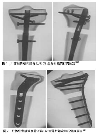

冯卫等[23]应用尸体胫骨模拟近端斜行骨折,分别用带锁髓内钉、锁定加压钢板及外固定支架固定,与正常胫骨做对照,各组分别加载400 N的应力,然后测量应变,并进行了垂直压缩和三点弯曲的力学加载。结果显示:髓内钉固定的胫骨近端骨折压缩应变接近正常对照组。髓内钉组的抗压刚度最强,钢板组的抗弯刚度最强,外固定支架组抗压缩及抗弯曲性能均明显差于其他各组。Hansen等[24]通过比较近端增加交叉锁定钉的“新型胫骨带锁髓内钉”、传统双钢板、微创内固定钢板、胫骨近端非扩髓髓内钉联合T型钢板、外固定架治疗胫骨近端关节外骨折的生物力学特性,发现“新型胫骨带锁髓内钉”在承受轴向载荷和抗扭刚度方面与其他组比较具有明显的优势。Lee等[25]通过比较不同的内固定方式在治疗胫骨近端骨折时,轴向载荷下的力学性能,发现髓内钉的固定强度明显高于单钢板或双钢板。通过对体外胫骨近端粉碎骨折双钢板固定和髓内钉固定的模型进行检测,Kandemir等[26]测定了髓内钉和双钢板固定治疗胫骨近端骨折的疲劳强度,发现二者没有明显差异。但髓内钉可微创置入,对周围组织干扰小,当胫骨近端骨折合并较严重软组织损伤时,他们推荐应用髓内钉固定。 2.3 髓内钉固定胫骨近端骨折的优点 髓内钉可微创置入、稳定性好,术后能较早的开始肢体功能锻炼,可防止因长时间固定而导致的关节强直、深静脉血栓形成等并发症,有利于肢体的功能康复。如果胫骨近端骨折合并了较重的软组织损伤时,髓内钉固定将更加适合。带锁髓内钉可看做是中央型内夹板,在起到固定作用的同时,对骨折部位血运影响较小,骨折不愈合及骨感染等并发症的发生率较低[27]。 李哲等[28]在治疗胫骨近端骨折时,分别选用了交锁髓内钉固定和经皮钢板内固定两种方式,并对治疗结果进行了比较,他们发现髓内钉治疗组,手术中的出血量、操作时间、术后负重时间及骨折临床愈合时间等指标均明显优于钢板组。 对于胫骨近端骨折同时合并胫骨干骨折的病例进行治疗时,髓内钉内固定是比较不错的选择。孙业清 等[29]在治疗胫骨平台骨折合并胫骨干骨折时,选择髓内钉联合钢板的内固定方式,临床结果良好。首先应用钢板将复位的关节内骨折固定,将近端固定为一个整体,然后应用髓内钉固定骨干部位,这样不仅关节内骨折得到了复位及牢固的固定,骨干的骨折也能得到复位,并牢靠固定[30]。 2.4 髓内钉治疗胫骨近端骨折的技术改进 2.4.1 联合钢板内固定 部分临床医生在应用髓内钉治疗胫骨近端、胫骨干多段骨折时,选用联合钢板固定。这种联合固定方式能够为早期功能锻炼提供足够坚强的稳定性。 孙效虎等[31]在治疗胫骨干、胫骨近端多段骨折时,就采用了联合应用髓内钉固定和锁定钢板的方法。在置入髓内钉前,首先应用锁定钢板固定骨折近端,为带锁髓内钉的近端入口提供稳定结构及支撑点,骨干骨折尽量采用闭合复位穿钉技术,这样能够减少对骨折断端血供的破坏。髓内钉并锁定钢板一起内固定的治疗方法,能够取得坚强的固定效果,使患者早期即可进行膝关节屈伸活动及部分负重功能锻炼,提高了骨折的愈合概率。丁强等[32]治疗胫骨骨折不愈合时,也选用了髓内钉联合钢板固定的方法,所有病例均获得了愈合。也有部分作者推荐在对胫骨近端骨折进行治疗时,选用单皮质钢板起到临时复位固定作用[33-34]。陈金亮等[35]总结了8例应用交锁髓内钉连同内侧单皮质钢板固定治疗胫骨近端斜形骨折的经验,认为该方法并没有增加操作的复杂性,而且达到了创伤小、固定稳定的目的,该方法适用于胫骨近端斜形骨折。在髓内钉固定牢固后可选择去除临时固定的单皮质钢板,或保留以增加固定强度。 2.4.2 阻挡钉技术 因胫骨近端髓腔宽大,主钉直径所限,使得主钉与髓腔不完全匹配,这样主钉在髓腔内容易产生偏移,从而导致骨折复位不佳,由此产生了阻挡钉技术,阻挡钉又称作Poller钉,该技术由Krettek等[10]在1999年推广。 在安装主钉前,先安装阻挡钉。阻挡钉可以安装在冠状面上、也可以安装在矢状面上,起到骨皮质的作用,可引导主钉按照正确的方向插入。郭伟等[36]应用髓内钉联合阻挡钉技术,用于治疗胫骨干骺端骨折,取得良好效果。他总结认为:主钉在髓腔内通过与长骨的峡部、阻挡钉、远端的锚点等接触形成稳定的三点接触固定。如果远端锚点不能实现稳定时,可在远端加用1枚阻挡钉增加稳定性。通过选择不同的位置安装阻挡钉,可以对抗软组织牵拉导致的移位趋势。为了建立稳定的三点固定,术前应充分的准备,应详细分析肌肉牵拉导致骨折移位的力的方向,并且应测量健侧胫骨长度及髓腔直径,根据测量分析结果,选择髓内钉及决定置入阻挡钉的位置。通过应用阻挡钉技术,使髓内钉治疗胫骨骨折的范围向两侧延伸,扩大了手术适应证,使得干骺端骨折亦可选用髓内钉技术固定。 Ricci等[37]报道了应用阻挡钉技术治疗的12例胫骨近端骨折的随访结果。认为阻挡钉在恢复、保持下肢力线时非常有效。国内多名作者报道交锁髓内钉辅助阻挡钉技术,用于治疗胫骨近端骨折[38-39],认为该技术可实现微创置入,对组织的损伤小,而且固定牢固可靠。 2.4.3 髓内钉设计的改进 近端锁钉的数量对固定的稳定性有较大的影响,Sayana等[40]的生物力学实验证明,增加近端锁定钉的数量,将明显增加髓内钉对胫骨近端骨折固定的稳定性。也有学者生物力学试验证明,当增加斜向锁定钉时,与单纯横向锁定比,明显增加固定的稳定性[41]。 角稳定锁定技术是另一项技术改进,Horn等[42]在尸体胫骨模拟了角稳定锁定胫骨近端骨折的受力,与普通锁定比较,其抗载荷能力更强。张庆杰等[13]报道锁定多向带锁髓内钉治疗胫骨平台骨折38列,优良率达到94.7%,原因是其应用的锁定多向带锁髓内钉主钉近端80 mm处向前弯曲8°,距近端18 mm和24 mm处分别增加了相互交叉成90°的、带有内螺纹的锁定孔,其固定后能够形成角稳定结构。 孙晓亮等[43]自行改良胫骨交锁髓内钉,并作为治疗胫骨近端骨折的内固定物,取得了很好的临床效果。他选用不锈钢作为制作材料,改进、增加了近端锁孔,将其设计成前后、前内、前外方向,并尽量使得锁定孔与尾端距离缩小,3个锁定孔到尾端距离分别为1.2,1.8,2.4 cm,这样的设计能够增加固定近端骨折块的稳定性。黄涛等[14]分别选择“多功能带锁髓内钉”、双钢板用于胫骨近端骨折的治疗。他应用的“多功能带锁髓内钉”近端增加了从前内、前外两个方向置入的松质骨螺钉。比较了2组间患者的手术时间长短,出血量多少,以及术中平均透视次数等,结果差异无显著性意义。但在比较骨折愈合时间、完全负重时间、骨折愈合优良率等指标时,髓内钉组明显好于钢板组。他认为这两种内固定置入的复杂程度无明显差异,但多功能带锁髓内钉组的稳定性更高,固定牢固的前提下,患者可早期下地活动锻炼。 林成寿等[44]介绍了AO解剖型髓内钉(Expert Tibial Nail,ETN),其近端锁钉孔增加为5个,距离尾端最近的锁钉孔仅1.4 cm,该型髓内钉实现了胫骨近端骨折的多向锁钉固定,明显增加了固定的稳定性。Attal等[15]选择应用ETN治疗胫骨干骨折时,在骨折的再移位率、延迟愈合率、内置物相关并发症发生率等方面低于钢板固定组,但在用于胫骨近端骨折的治疗时,术后的再移位率要明显增高。Trlica等[16]认为ETN能够为胫骨近端骨折,甚至包括部分累及膝关节面的骨折提供足够的固定强度,但在部分患者需加用阻挡钉技术。 2.4.4 入路改进 恰当的进钉点能够有效的纠正力线,使复位更加顺利,简化手术操作。多位学者的研究发现,胫骨髓腔轴线在胫骨近端冠状面中点偏内部位[45-46],建议选择主钉进钉点时,应选择胫骨棘稍偏内侧部位,这样能够保证在冠状面上的主钉方向与胫骨髓腔轴线一致,避免出现内、外翻畸形。陈震宇等[47]应用尸体胫骨模拟了胫骨近端不同方向走形的斜行骨折线,并研究了进钉点位置与骨折对位的关系。进钉点偏内时,骨折线自内上斜向外下标本组中移位最大;进钉点偏外时,骨折线自外上斜向内下标本组移位最大。根据实验结果,他们认为胫骨近端骨折为斜行骨折线时,选用交锁髓内钉固定应根据骨折线的走形选择不同位置的进钉点,以便达到更好的固定强度。除外进钉点在冠状面上的位置对骨折固定的稳定性有影响外,在矢状面上进钉点的高低亦非常重要。有学者介绍进钉点更加靠近近端,髓内钉不仅顺利置入,还能避免膝关节内结构损伤[48]。也有学者在选用髓内钉作为内固定物治疗胫骨近端骨折时,将进钉点选在胫骨平台前缘稍下,约0.5 cm处,与传统进钉点相比上移了1.0-1.5 cm,这样就能够防止近端锁钉不能固定近端骨折块的情况发生[49]。 尽管胫骨髓内钉内固定技术不断进步,但其手术入路仍采用髌韧带旁或劈开髌韧带入路,基本没有变 化[50]。标准的胫骨髓内钉技术置入主钉时,需将膝关节过度屈曲,超过120°,如果为胫骨近端骨折,在过度屈曲时将导致骨折断端向前成角移位。髌上入路置入胫骨髓内钉固定技术,由Tornetta等[51]最早介绍,当时设计也是用于胫骨近端骨折治疗。该技术在治疗胫骨近端骨折时有明显的优势,它半伸直体位,使得骨折复位更容易保持;髓内钉中轴线与胫骨髓腔中轴线共线更容易实现,避免了穿出后侧皮质;通过滑车定位,降低了内外翻发生的风险;术中透视更加容易,避免了加重对胫骨近端软组织进一步的损伤[52-53]。解冰等[54]报道采用膝关节半伸直位髌上入路胫骨髓内钉内固定,治疗胫骨近端骨折16例,术后膝关节目测类比评分为0-1分,证实该手术入路可以很好地降低术后膝前疼痛的发生率。Sanders等[55]随访了经髌上入路置入胫骨髓内钉的13例患者,并对患者进行了关节镜检查及MR检查,证实术后没有对髌股关节软骨造成损伤,也没有发生关节内游离体。他认为该技术具有术中出血量小、手术时间短、对骨膜及软组织血运影响小、骨折愈合率高、术后并发症少等优点。有学者通过尸体标本研究证实髌上入路与髌前入路比较,其关节内软骨、半月板等重要结构损伤的发生率并不增加[56-57]。 "

| [1] Lang GJ, Cohen BE, Bosse MJ, et al. Proximal third tibial shaft fractures. Should they be nailed? Clin Orthop Relat Res. 1995;315(315):64-74.[2] Seyhan M, Unay K, Sener N. Intramedullary nailing versus percutaneous locked plating of distal extra-articular tibial fractures: a retrospective study. Eur J Orthop Surg Traumatol Orthop Traumatol. 2013;23(5):595-601.[3] Decoster TA, Nepola JV, Elkhoury GY. Cast brace treatment of proximal tibia fractures. A ten-year follow-up study. Clin Orthop Relat Res. 1988; 231(231):196-204.[4] Milner SA, Davis TR, Muir KR, et al. Long-term outcome after tibial shaft fracture: is malunion important? J Bone Joint Surg Am. 2002;84-A(6):971–980.[5] Meena RC, Meena UK, Gupta GL, et al. Intramedullary nailing versus proximal plating in the management of closed extra-articular proximal tibial fracture: a randomized controlled trial. J Orthop Traumatol. 2015;16(3):203-208.[6] Pairon P, Ossendorf C, Kuhn S, et al. Intramedullary nailing after external fixation of the femur and tibia: a review of advantages and limits. Eur J Trauma Emerg Surg. 2015;41(1):25-38. [7] Lindvall E, Sanders RT. Intramedullary nailing versus percutaneous locked plating of extra-articular proximal tibial fractures: comparison of 56 cases. J Orthop Trauma. 2009; 23(7):485-492.[8] Tytherleigh-Strong GM, Keating JF, Court-Brown CM. Extra-articular fractures of the proximal tibial diaphysis: their epidemiology, management and outcome. J R Coll Surg Edinb. 1997;42(5):334-338.[9] Freedman EL, Johnson EE. Radiographic analysis of tibial fracture malalignment following intramedullary nailing. Clin Orthop Relat Res. 1995;(315):25-33.[10] Krettek C, Stephan C, Schandelmaier P, et al. The use of Poller screws as blocking screws in stabilising tibial fractures treated with small diameter intramedullary nails. J Bone Joint Surg Br. 1999;81(6):963-968. [11] Vidyadhara S, Sharath KR. Prospective study of the clinico-radiological outcome of interlocked nailing in proximal third tibial shaft fractures. Injury. 2006;37(6):536-542.[12] Josten C, Marquass B, Schwarz C, et al. Intramedullary nailing of proximal tibial fractures. Complications and risk factors. Der Unfallchirurg. 2010;113(1):21-28.[13] 张庆杰,王永清,熊庆广,等. 锁定多向带锁髓内钉治疗胫骨平台骨折[J]. 中华骨科杂志,2016,36(13):833-840.[14] 黄涛,马凯. 多功能带锁髓内钉与双钢板治疗胫骨近端关节外骨折的临床效果比较[J]. 创伤外科杂志, 2017,19(2):126-129.[15] Attal R, Hansen M, Kirjavainen M, et al. A multicentre case series of tibia fractures treated with the Expert Tibia Nail (ETN). Arch Orthop Trauma Surg. 2012;132(7):975-984. [16] Trlica J, Dedek T, Smejkal K, et al. Expert Tibial Nail (ETN) for treatment of diaphyseal tibial fractures in current and extended indications: technique and clinical results. Acta Chirurgiae Orthopaedicae Et Traumatologiae Cechoslovaca. 2010;77(3):235-241.[17] 王永清,罗先正,刘长贵. 国人股骨胫骨髓腔的形态学研究及带锁髓内钉的改进[J]. 中华骨科杂志,1998,18(4):215-218. [18] Hiesterman TG, Shafiq BX, Cole PA. Intramedullary nailing of extra-articular proximal tibia fractures. J Am Acad Orthop Surg. 2011;19(11):690-700.[19] Lembcke O, Rüter A, Beck A. The nail-insertion point in unreamed tibial nailing and its influence on the axial malalignment in proximal tibial fractures. Arch Orthop Trauma Surg. 2001;121(4):197-200.[20] Liporace FA, Stadler CM, Yoon R S. Problems, tricks, and pearls in intramedullary nailing of proximal third tibial fractures. J Orthop Trauma. 2013;27(1):56-62.[21] 周星衡,董黎敏,叶金铎,等. 带锁髓内钉与锁定加压接骨板固定胫骨近端骨折的有限元分析[J]. 实用骨科杂志, 2014,20(11): 995-998.[22] Högel F, Hoffmann S, Panzer S, et al. Biomechanical comparison of intramedullar versus extramedullar stabilization of intra-articular tibial plateau fractures. Arch Orthop Trauma Surg. 2013;133(1):59-64.[23] 冯卫,刘建国,裴福兴.带锁髓内钉在胫骨近端骨折的生物力学研究及临床应用[J].中国生物医学工程学报,2005,24(6):728-732.[24] Hansen M, Mehler D, Hessmann MH, et al. Intramedullary stabilization of extraarticular proximal tibial fractures: a biomechanical comparison of intramedullary and extramedullary implants including a new proximal tibia nail (PTN). J Orthop Trauma. 2007;21(10):701-709.[25] Lee SM, Oh CW, Oh JK, et al. Biomechanical analysis of operative methods in the treatment of extra-articular fracture of the proximal tibia. Clin Orthop Surg. 2014; 6(3):312-317.[26] Kandemir U, Herfat S, Herzog M, et al. Fatigue failure in extra-articular proximal tibia fractures: locking intramedullary nail versus double locking plates-a biomechanical study. J Orthop Trauma. 2017;31(2):e49-e54.[27] 罗先正,邱贵兴. 髓内钉内固定[M]. 2版.北京:人民卫生出版社, 2008:31-36.[28] 李哲,孙贺,付世杰,等. 交锁髓内钉治疗胫骨近端骨折的临床分析[J]. 临床医药文献电子杂志, 2015,2(7):1247-1247.[29] 孙业青,吴卫平,楼列名,等. 胫骨、股骨干骨折合并同侧膝关节内骨折的髓内钉治疗[J]. 中国骨与关节损伤杂志, 2004,19(6): 369-371. [30] 张宏其,龙文荣,李康华,等. 半圆形动态交锁髓内钉治疗胫骨平台伴胫骨上段骨折[J]. 中华创伤杂志, 2001, 17(3):148-150.[31] 孙效虎,袁景. 锁定钢板联合带锁髓内钉治疗胫骨多段骨折[J]. 临床骨科杂志, 2016, 19(1):126-126.[32] 丁强,李超,刘又文,等.交锁髓内钉附加钛板固定治疗胫骨骨折不愈合疗效观察[J].中国骨与关节损伤杂志,2014,29(4):402-403.[33] Kim KC, Lee JK, Hwang DS, et al. Provisional unicortical plating with reamed intramedullary nailing in segmental tibial fractures involving the high proximal metaphysis. Orthopedics. 2007;30(3):189-192.[34] Archdeacon MT, Wyrick JD. Reduction plating for provisional fracture fixation. J Orthop Trauma. 2006;20(3):206-211.[35] 陈金亮,江起庭,朱东波,等. 交锁髓内钉联合钢板在胫骨近端斜形骨折治疗中的应用[J]. 实用手外科杂志, 2015, 29(2): 186-187.[36] 郭伟,杜静德. 胫骨髓内钉联合阻挡钉治疗胫骨干骺端骨折[J]. 实用骨科杂志, 2014,20(9):852-854.[37] Ricci WM, O'Boyle M, Borrelli J, et al. Fractures of the proximal third of the tibial shaft treated with intramedullary nails and blocking screws. J Orthop Trauma. 2001;15(4): 264-270.[38] 王庆,罗建宁,李欣玻.阻挡螺钉技术在胫骨交锁髓内钉治疗胫骨近端骨折中的应用[J].中国骨与关节损伤杂志,2014,29(1):76-77.[39] 祝海炳,武理国,方智松,等.阻挡钉结合生根技术在带锁髓内钉治疗胫骨远端骨折中的应用[J].中国骨伤,2012,25(7):569-571.[40] Sayana MK, Davis BJ, Kapoor B, et al. Fracture strain and stability with additional locking screws in intramedullary nailing: a biomechanical study. J Trauma Acute Care Surg. 2006;60(5):1053-1057.[41] Laflamme GY, Heimlich D, Stephen D, et al. Proximal tibial fracture stability with intramedullary nail fixation using oblique interlocking screws. J Orthop Trauma. 2003;17(7):496-502.[42] Horn J, Linke B, Höntzsch D, et al. Angle stable interlocking screws improve construct stability of intramedullary nailing of distal tibia fractures: a biomechanical study. Injury. 2009; 40(7):767-771. [43] 孙晓亮, 孙有声, 刘志伟,等. 交锁髓内钉治疗胫骨近端骨折(附46例报告)[J].中国骨与关节损伤杂志, 2000, 15(6):451-452.[44] 林成寿, 刘寿坤, 吴锦清,等. AO解剖型胫骨髓内钉微创治疗胫骨远端骨折[J].创伤外科杂志, 2010, 12(6):520-522.[45] Song SJ, Jeong BO. Three-dimensional analysis of the intramedullary canal axis of tibia: clinical relevance to tibia intramedullary nailing. Arch Orthop Trauma Surg. 2010; 130(7):903-907.[46] Schmidt AH, Templeman DC, Tornetta P, et al. Anatomic assessment of the proper insertion site for a tibial intramedullary nail. J Orthop Trauma. 2002;16(1):23-25.[47] 陈震宇,任栋. 胫骨近端骨折不同进钉点交锁髓内钉固定的生物力学比较[J].河北医科大学学报, 2014, 35(3):269-271.[48] Mcconnell T, Rd TP, Tilzey J, et al. Tibial portal placement: the radiographic correlate of the anatomic safe zone. J Orthop Trauma. 2001; 15(3):207-209.[49] 韦阁. 交锁髓内钉治疗胫骨Ⅱ区骨折43例分析[J]. 华夏医学, 2004,17(4):577-578.[50] Morandi M, Banka T, Gaiarsa GP, et al. Intramedullary nailing of tibial fractures: review of surgical techniques and description of a percutaneous lateral suprapatellar approach. Orthopedics. 2010;33(3):172.-179.[51] Tornetta P 3rd, Collins E. Semiextended position of intramedullary nailing of the proximal tibia. Clin Orthop Relat Res.1996;(328):185-189.[52] Jones M, Parry M, Whitehouse M, et al. Radiologic outcome and patient-reported function after intramedullary nailing: a comparison of the retropatellar and infrapatellar approach. J Orthop Trauma. 2014;28(5):256-262. [53] Franke J, Hohendorff B, Alt V, et al. Suprapatellar nailing of tibial fractures-Indications and technique. Injury. 2016;47(2): 495-501.[54] 解冰,杨超,田竞,等. 髌上入路胫骨髓内钉治疗胫骨近端骨折[J]. 中国骨伤, 2015, 28(10):955-959.[55] Sanders RW, Dipasquale TG, Jordan CJ, et al. Semiextended intramedullary nailing of the tibia using a suprapatellar approach: radiographic results and clinical outcomes at a minimum of 12 months follow-up. J Orthop Trauma. 2014; 28(5):245-255.[56] Eastman JG, Tseng SS, Lee MA, et al. The retropatellar portal as an alternative site for tibial nail insertion: a cadaveric study. J Orthop Trauma. 2010; 24(11):659-664.[57] Gelbke MK, Coombs D, Powell S, et al. Suprapatellar versus infra-patellar intramedullary nail insertion of the tibia: a cadaveric model for comparison of patellofemoral contact pressures and forces. J Orthop Trauma. 2010; 24(11): 665-671. |
| [1] | Xu Feng, Kang Hui, Wei Tanjun, Xi Jintao. Biomechanical analysis of different fixation methods of pedicle screws for thoracolumbar fracture [J]. Chinese Journal of Tissue Engineering Research, 2021, 25(9): 1313-1317. |
| [2] | Zhang Tongtong, Wang Zhonghua, Wen Jie, Song Yuxin, Liu Lin. Application of three-dimensional printing model in surgical resection and reconstruction of cervical tumor [J]. Chinese Journal of Tissue Engineering Research, 2021, 25(9): 1335-1339. |
| [3] | Chen Xinmin, Li Wenbiao, Xiong Kaikai, Xiong Xiaoyan, Zheng Liqin, Li Musheng, Zheng Yongze, Lin Ziling. Type A3.3 femoral intertrochanteric fracture with augmented proximal femoral nail anti-rotation in the elderly: finite element analysis of the optimal amount of bone cement [J]. Chinese Journal of Tissue Engineering Research, 2021, 25(9): 1404-1409. |
| [4] | Zhou Jihui, Li Xinzhi, Zhou You, Huang Wei, Chen Wenyao. Multiple problems in the selection of implants for patellar fracture [J]. Chinese Journal of Tissue Engineering Research, 2021, 25(9): 1440-1445. |
| [5] | Zeng Yanhua, Hao Yanlei. In vitro culture and purification of Schwann cells: a systematic review [J]. Chinese Journal of Tissue Engineering Research, 2021, 25(7): 1135-1141. |
| [6] | Xu Yulin, Shen Shi, Zhuo Naiqiang, Yang Huilin, Yang Chao, Li Yang, Zhao Heng, Zhao Lu. Biomechanical comparison of three different plate fixation methods for acetabular posterior column fractures in standing and sitting positions [J]. Chinese Journal of Tissue Engineering Research, 2021, 25(6): 826-830. |
| [7] | Cai Qunbin, Zou Xia, Hu Jiantao, Chen Xinmin, Zheng Liqin, Huang Peizhen, Lin Ziling, Jiang Ziwei. Relationship between tip-apex distance and stability of intertrochanteric femoral fractures with proximal femoral anti-rotation nail: a finite element analysis [J]. Chinese Journal of Tissue Engineering Research, 2021, 25(6): 831-836. |
| [8] | Song Chengjie, Chang Hengrui, Shi Mingxin, Meng Xianzhong. Research progress in biomechanical stability of lateral lumbar interbody fusion [J]. Chinese Journal of Tissue Engineering Research, 2021, 25(6): 923-928. |
| [9] | Xie Chongxin, Zhang Lei. Comparison of knee degeneration after anterior cruciate ligament reconstruction with or without remnant preservation [J]. Chinese Journal of Tissue Engineering Research, 2021, 25(5): 735-740. |
| [10] | Xu Dongzi, Zhang Ting, Ouyang Zhaolian. The global competitive situation of cardiac tissue engineering based on patent analysis [J]. Chinese Journal of Tissue Engineering Research, 2021, 25(5): 807-812. |
| [11] | Wu Zijian, Hu Zhaoduan, Xie Youqiong, Wang Feng, Li Jia, Li Bocun, Cai Guowei, Peng Rui. Three-dimensional printing technology and bone tissue engineering research: literature metrology and visual analysis of research hotspots [J]. Chinese Journal of Tissue Engineering Research, 2021, 25(4): 564-569. |
| [12] | Chang Wenliao, Zhao Jie, Sun Xiaoliang, Wang Kun, Wu Guofeng, Zhou Jian, Li Shuxiang, Sun Han. Material selection, theoretical design and biomimetic function of artificial periosteum [J]. Chinese Journal of Tissue Engineering Research, 2021, 25(4): 600-606. |
| [13] | Liu Fei, Cui Yutao, Liu He. Advantages and problems of local antibiotic delivery system in the treatment of osteomyelitis [J]. Chinese Journal of Tissue Engineering Research, 2021, 25(4): 614-620. |
| [14] | Li Xiaozhuang, Duan Hao, Wang Weizhou, Tang Zhihong, Wang Yanghao, He Fei. Application of bone tissue engineering materials in the treatment of bone defect diseases in vivo [J]. Chinese Journal of Tissue Engineering Research, 2021, 25(4): 626-631. |
| [15] | Zhang Zhenkun, Li Zhe, Li Ya, Wang Yingying, Wang Yaping, Zhou Xinkui, Ma Shanshan, Guan Fangxia. Application of alginate based hydrogels/dressings in wound healing: sustained, dynamic and sequential release [J]. Chinese Journal of Tissue Engineering Research, 2021, 25(4): 638-643. |
| Viewed | ||||||
|
Full text |
|
|||||
|
Abstract |
|
|||||