Chinese Journal of Tissue Engineering Research ›› 2018, Vol. 22 ›› Issue (2): 228-233.doi: 10.3969/j.issn.2095-4344.0011
Previous Articles Next Articles
Injectable nano-hydroxyapatite/chitosan composite scaffolds combined with bone marrow mesenchymal stem cells and bone morphogenetic protein 2 for bone defect repair in vitro
- Ninth Ward, Department of Orthopedic Surgery, Second Affiliated Hospital of Harbin Medical University, Harbin 150086, Heilongjiang Province, China
-
Received:2017-08-01Online:2018-01-18Published:2018-01-18 -
Contact:Tian Jun, Chief physician, Ninth Ward, Department of Orthopedic Surgery, Second Affiliated Hospital of Harbin Medical University, Harbin 150086, Heilongjiang Province, China -
About author:Liu Guang-tao, Master, Ninth Ward, Department of Orthopedic Surgery, Second Affiliated Hospital of Harbin Medical University, Harbin 150086, Heilongjiang Province, China -
Supported by:the Science and Technology Research Project of Heilongjiang Provincial Education Department, No. 12541333
CLC Number:
Cite this article
Liu Guang-tao, Gao Feng, Xu Jun, Zheng Wei-zhuo, Lin Xiao-zong, Zhou Chang-lin, Guo Ya-shan, Tian Jun.
share this article
Add to citation manager EndNote|Reference Manager|ProCite|BibTeX|RefWorks

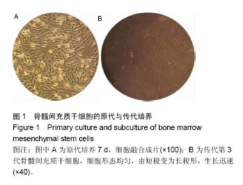
2.1 兔骨髓间充质干细胞的生长情况及形态特征 原代培养 4 d后,换液弃去未贴壁的细胞,细胞呈短梭形集落样生长;7 d后,集落连成一片,融合度达到80%-90%,进行传代(图1A);第3代细胞形态均匀,由短梭变为长梭形,生长迅速(图1B)。"
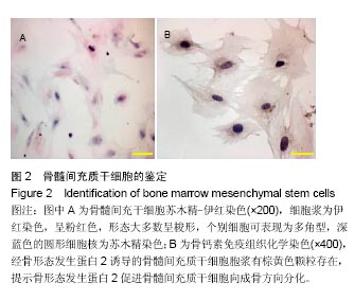
2.2 骨髓间充质干细胞的鉴定 苏木精-伊红染色:镜下见第3代骨髓间充质干细胞形态不同,细胞浆为伊红染色,呈粉红色,形态大多数呈梭形,个别细胞可表现为多角型,深蓝色的圆形细胞核为苏木精染色,并可见个别细胞核正一分为二的分裂期,细胞排列紧密(图2A)。 骨钙素免疫组织化学染色:经骨形态发生蛋白2诱导的骨髓间充质干细胞胞浆有棕黄色颗粒存在,提示骨形态发生蛋白2促进骨髓间充质干细胞向成骨方向分化,符合骨髓间充质干细胞细胞特性(图2B)。"
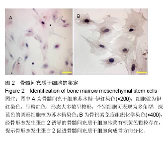

2.3 扫描电镜观察纳米羟基磷灰石粉末及纳米羟基磷灰石/壳聚糖复合支架材料 电镜下可见纳米羟基磷灰石粉末结构清晰,平均长度30-70 nm,直径10-20 nm,棒状(图3A);可注射性纳米羟基磷灰石/壳聚糖复合支架大体观为白色胶状(图3B),其在电镜下表面凸凹不平,大小不等的圆状颗粒排列紧密(图3C),固化后可见支架表面有大小不等的散在孔隙,表面粗糙(图3D)。"
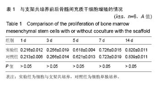
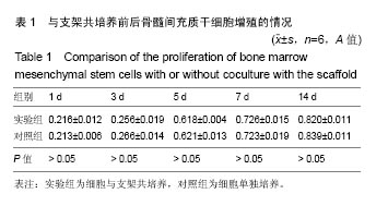
2.4 CCK-8检测与支架复合培养前后细胞的增殖 随着培养时间的延长,实验组和对照组A值不断增长,在14 d时达到最大值,两组各时间点A值比较差异均无显著性意义(P > 0.05),见表1。 "
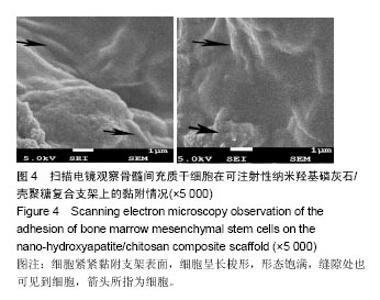
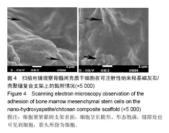
2.5 扫描电镜观察复合支架上细胞的黏附 骨髓间充质干细胞与可注射性纳米羟基磷灰石/壳聚糖复合支架共同培养7 d后,细胞紧紧黏附支架表面,细胞呈长梭形,形态饱满,缝隙处也可见到细胞(图4)。"
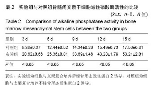
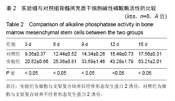
2.6 骨形态发生蛋白2诱导前后复合支架上细胞的碱性磷酸酶活性 实验组碱性磷酸酶活性均明显高于对照组(P < 0.05),见表2,说明复合于支架上的骨髓间充质干细胞在骨形态发生蛋白2诱导作用下成骨表达碱性磷酸酶增强。"

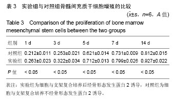
2.7 骨形态发生蛋白2诱导前后支架上细胞的增殖 实验组各个时间点A值均大于对照组(P < 0.05),见表3。"

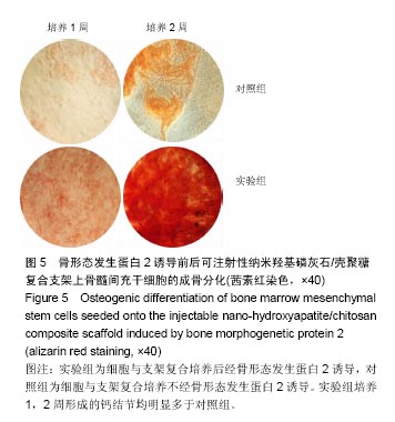
2.8 骨形态发生蛋白2诱导前后支架上细胞的茜素红染色 实验组培养1,2周形成的钙结节均明显多于对照组(图5)。"
| [1]Jain S,Krishna Meka SR,Chatterjee K.Curcumin eluting nanofibers augment osteogenesis toward phytochemical based bone tissue engineering.Biomedi Mater.2016;11(5):055007.[2]Yu X,Tang X,Gohil SV,et al.Biomaterials for Bone Regenerative Engineering.Adv Healthc Mater.2015;4(9):1268-1285.[3]Wang ZX,Chen C,Zhou Q,et al.The Treatment Efficacy of Bone Tissue Engineering Strategy for Repairing Segmental Bone Defects Under Osteoporotic Conditions.Tissue Eng Part A.2015;21(17-18):2346.[4]Barabaschi GD,Manoharan V,Li Q,et al.Engineering Pre-vascularized Scaffolds for Bone Regeneration.Adv Exp Med Biol.2015;881:79-94.[5]杨俊丽,韩霞,孙明启,等.兔骨髓间充质干细胞的生物学特征及原代培养[J].中国组织工程研究,2015,19(50):8043-8047.[6]Gupta P,Adhikary M,Joseph CM,et al.Biomimetic,Osteoconductive Non-mulberry Silk Fiber Reinforced Tricomposite Scaffolds for Bone Tissue Engineering.ACS Appl Mater Interfaces.2016;8(45):30797.[7]Camareroespinosa S,Rothenrutishauser B,Weder C,et al.Directed cell growth in multi-zonal scaffolds for cartilage tissue engineering. Biomaterials.2016;74:42.[8]Bhattacharya I,Ghayor C,Weber FE.The Use of Adipose Tissue-Derived Progenitors in Bone Tissue Engineering - a Review.Transfus Med Hemother.2016;43(5):336.[9]Yan Y,Zhang X,Li C,et al.Preparation and characterization of chitosan-silver/hydroxyapatite composite coatings onTiO 2, nanotube for biomedical applications.Appl Surf Sci.2015;332(4):62-69.[10]李珺,李晓桐,赵明.无机纳米材料及其在生物医学方面的应用研究[J].医疗卫生装备,2015,36(7):97-101.[11]Wang X,Li G,Guo J,et al.Hybrid composites of mesenchymal stem cell sheets, hydroxyapatite, and platelet-rich fibrin granules for bone regeneration in a rabbit calvarial critical-size defect model.Exp Ther Med.2017;13(5):1891-1899.[12]Shabbir A,Cox A,Rodriguez-Menocal L,et al.Mesenchymal Stem Cell Exosomes Induce Proliferation and Migration of Normal and Chronic Wound Fibroblasts, and Enhance Angiogenesis In Vitro.Stem Cells Dev.2015;24(14):1635.[13]de Oliveira Gonçalves JB,Buchaim DV,de Souza Bueno CR,et al.Effects of low-level laser therapy on autogenous bone graft stabilized with a new heterologous fibrin sealant.J Photochem Photobiol B. 2016;162:663-668.[14]De Windt TS,Vonk LA,Slaper‐Cortenbach IC,et al.Allogeneic Mesenchymal Stem Cells Stimulate Cartilage Regeneration and are Safe for Single‐stage Cartilage repair in Humans Upon Mixture with recycled Autologous Chondrons.Stem Cells.2017;35(1):256.[15]Jadalannagari S,Converse G,Mcfall C,et al.Decellularized Wharton's Jelly from human umbilical cord as a novel 3D scaffolding material for tissue engineering applications.PloS One.2017;12(2):e0172098.[16]Yang W, Zheng H, Wang Y, et al. Nesprin-1 has key roles in the process of mesenchymal stem cell differentiation into cardiomyocyte-like cells in vivo and in vitro. Molecular Medicine Reports, 2015, 11(1):133-142.[17]Russmueller G,Moser D,Spassova E,et al.Tricalcium phosphate-based biocomposites for mandibular bone regeneration—A histological study in sheep.J Craniomaxillofac Surg.2015;43(5):696-704.[18]姚彪.骨缺损修复材料α-CSH/BCP体外降解性能及细胞相容性的实验研究[D].南京中医药大学,2015.[19]Geller DS,Singh MY,Zhang W,et al.Development of a Model System to Evaluate Local Recurrence in Osteosarcoma and Assessment of the Effects of Bone Morphogenetic Protein-2.Clin Cancer Res. 2015; 21(13):3003.[20]Anusuya GS,Kandasamy M,Jacob Raja SA,et al.Bone morphogenetic proteins: Signaling periodontal bone regeneration and repair.J Pharm Bioallied Sci.2016;8(Suppl 1):S39-S41.[21]Dettori JR,Chapman JR,Devine JG,et al.The Risk of Cancer With the Use of Recombinant Human Bone Morphogenetic Protein in Spine Fusion.Spine(Phila Pa 1976).2016;41(16):1317.[22]Kuzaka B,Janiak M,W?odarski KH,et al.Expression of bone morphogenetic protein-2 and -7 in urinary bladder cancer predicts time to tumor recurrence.Arch Med Sci.2015;11(2):378-384.[23]Lappin DF,Abuserriah M,Hunter KD.Effects of recombinant human bone morphogenetic protein 7 (rhBMP-7) on the behaviour of oral squamous cell carcinoma: a preliminary in vitro study.Br J Oral Maxillofac Surg.2015;53(2):158-163.[24]Cong Y,Li CJ,Zhao JN,et al.Associations of polymorphisms in the bone morphogenetic protein-2 gene with risk and prognosis of osteosarcoma in a Chinese population.Tumor Biol. 2015;36(3): 2059-2064.[25]耿欣宇,刘冰熔.骨形态发生蛋白与肿瘤发生发展的研究进展[J].实用临床医药杂志,2015,19(23):237-239.[26]Bez M,Sheyn D,Tawackoli W,et al.In situ bone tissue engineering via ultrasound-mediated gene delivery to endogenous progenitor cells in mini-pigs.Sci Transl Med.2017;9(390):eaal3128.[27]Wang QF,Huang Y,He GC,et al.Osteoblast differentiation of rabbit adipose-derived stem cells by polyethylenimine-mediated BMP-2 gene transfection in vitro.Genet Mol Res.2017;16(1):doi: 10.4238/gmr16015358. [28]Salazar VS,Gamer LW,Rosen V.BMP signalling in skeletal development, disease and repair.Nat Rev Endocrinol.2016;12(4):203.[29]Salifu AA,Lekakou C,Labeed F.Multilayer cellular stacks of gelatin-hydroxyapatite fiber scaffolds for bone tissue engineering.J Biomed Mater Res A.2017;105(3):779-789.[30]Deepthi S,Venkatesan J,Kim SK,et al.An overview of chitin or chitosan/nano ceramic composite scaffolds for bone tissue engineering.Int J Biol Macromol.2016;93:1338-1353. |
| [1] | Zhang Tongtong, Wang Zhonghua, Wen Jie, Song Yuxin, Liu Lin. Application of three-dimensional printing model in surgical resection and reconstruction of cervical tumor [J]. Chinese Journal of Tissue Engineering Research, 2021, 25(9): 1335-1339. |
| [2] | Li Cai, Zhao Ting, Tan Ge, Zheng Yulin, Zhang Ruonan, Wu Yan, Tang Junming. Platelet-derived growth factor-BB promotes proliferation, differentiation and migration of skeletal muscle myoblast [J]. Chinese Journal of Tissue Engineering Research, 2021, 25(7): 1050-1055. |
| [3] | Liu Cong, Liu Su. Molecular mechanism of miR-17-5p regulation of hypoxia inducible factor-1α mediated adipocyte differentiation and angiogenesis [J]. Chinese Journal of Tissue Engineering Research, 2021, 25(7): 1069-1074. |
| [4] | Zeng Yanhua, Hao Yanlei. In vitro culture and purification of Schwann cells: a systematic review [J]. Chinese Journal of Tissue Engineering Research, 2021, 25(7): 1135-1141. |
| [5] | Ma Zetao, Zeng Hui, Wang Deli, Weng Jian, Feng Song. MicroRNA-138-5p regulates chondrocyte proliferation and autophagy [J]. Chinese Journal of Tissue Engineering Research, 2021, 25(5): 674-678. |
| [6] | Xu Dongzi, Zhang Ting, Ouyang Zhaolian. The global competitive situation of cardiac tissue engineering based on patent analysis [J]. Chinese Journal of Tissue Engineering Research, 2021, 25(5): 807-812. |
| [7] | Wang Yujiao, Liu Dan, Sun Song, Sun Yong. Biphasic calcium phosphate loaded with advanced platelet rich fibrin can promote the activity of rabbit bone marrow mesenchymal stem cells [J]. Chinese Journal of Tissue Engineering Research, 2021, 25(4): 504-509. |
| [8] | Zhou Jihui, Yao Meng, Wang Yansong, Li Xinzhi, Zhou You, Huang Wei, Chen Wenyao. Influence of novel nanoscaffolds on biological behaviors of neural stem cells and the related gene expression [J]. Chinese Journal of Tissue Engineering Research, 2021, 25(4): 532-536. |
| [9] | Wu Zijian, Hu Zhaoduan, Xie Youqiong, Wang Feng, Li Jia, Li Bocun, Cai Guowei, Peng Rui. Three-dimensional printing technology and bone tissue engineering research: literature metrology and visual analysis of research hotspots [J]. Chinese Journal of Tissue Engineering Research, 2021, 25(4): 564-569. |
| [10] | Li Li, Ma Li. Immobilization of lactase on magnetic chitosan microspheres and its effect on enzymatic properties [J]. Chinese Journal of Tissue Engineering Research, 2021, 25(4): 576-581. |
| [11] | Chang Wenliao, Zhao Jie, Sun Xiaoliang, Wang Kun, Wu Guofeng, Zhou Jian, Li Shuxiang, Sun Han. Material selection, theoretical design and biomimetic function of artificial periosteum [J]. Chinese Journal of Tissue Engineering Research, 2021, 25(4): 600-606. |
| [12] | Liu Fei, Cui Yutao, Liu He. Advantages and problems of local antibiotic delivery system in the treatment of osteomyelitis [J]. Chinese Journal of Tissue Engineering Research, 2021, 25(4): 614-620. |
| [13] | Li Xiaozhuang, Duan Hao, Wang Weizhou, Tang Zhihong, Wang Yanghao, He Fei. Application of bone tissue engineering materials in the treatment of bone defect diseases in vivo [J]. Chinese Journal of Tissue Engineering Research, 2021, 25(4): 626-631. |
| [14] | Zhang Zhenkun, Li Zhe, Li Ya, Wang Yingying, Wang Yaping, Zhou Xinkui, Ma Shanshan, Guan Fangxia. Application of alginate based hydrogels/dressings in wound healing: sustained, dynamic and sequential release [J]. Chinese Journal of Tissue Engineering Research, 2021, 25(4): 638-643. |
| [15] | Chen Jiana, Qiu Yanling, Nie Minhai, Liu Xuqian. Tissue engineering scaffolds in repairing oral and maxillofacial soft tissue defects [J]. Chinese Journal of Tissue Engineering Research, 2021, 25(4): 644-650. |
| Viewed | ||||||
|
Full text |
|
|||||
|
Abstract |
|
|||||