Chinese Journal of Tissue Engineering Research ›› 2018, Vol. 22 ›› Issue (2): 222-227.doi: 10.3969/j.issn.2095-4344.0010
Previous Articles Next Articles
Mitochondria-mediated synthesis of gold-silver alloy nanoparticles
- Institute of Applied Mechanics and Biomedical Engineering, Taiyuan University of Technology, Taiyuan 030024, Shanxi Province, China
-
Received:2017-08-22Online:2018-01-18Published:2018-01-18 -
Contact:Chen Wei-yi, M.D., Professor, Doctoral supervisor, Institute of Applied Mechanics and Biomedical Engineering, Taiyuan University of Technology, Taiyuan 030024, Shanxi Province, China -
About author:Meng Biao, Master, Institute of Applied Mechanics and Biomedical Engineering, Taiyuan University of Technology, Taiyuan 030024, Shanxi Province, China -
Supported by:the National Natural Science Foundation of China, No. 81602506; the Innovative Program of Shanxi High Educations, No. 2016143
CLC Number:
Cite this article
Meng Biao, Chen Wei-yi, Zhang Yi-xia, Wen Xu-dong, Li Kai .
share this article
Add to citation manager EndNote|Reference Manager|ProCite|BibTeX|RefWorks
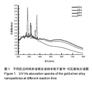
2.1 金银合金纳米粒子的表征结果 2.1.1 紫外可见吸收光谱 图1是制得的不同反应时间的金银合金纳米粒子的紫外-可见吸收光谱,在550 nm处出现的一处明显吸收峰即是金银合金纳米颗粒的特征峰。因为只有一处吸收峰,不同于会同时出现金和银两处吸收峰的金银的简单混合和核壳结构的金银纳米粒子,表明合成的是合金结构的纳米粒子,也与文献报道相一致。同时,由图1还可看出反应到12 h时还没有出现明显特征峰。反应到16 h时开始出现特征峰。在大约24 h时特征吸收峰保持不再改变,还原反应结束。"
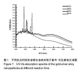
2.1.2 扫描电镜和能谱分析 通过扫描电镜与能谱图来进一步观察所制备金银合金纳米颗粒的结构特性,如图2所示,所制备的金银合金合金纳米粒子,平均粒径20- 30 nm,基本为球形分布,粒度比较均一,说明合成的纳米粒子是合金结构。能谱图说明所获得金银合金纳米颗粒的元素组成为金银两种元素。"

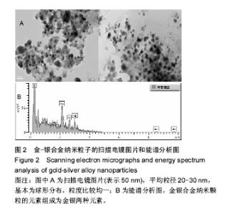
2.1.3 X射线衍射图谱 在X射线衍射图谱中,2θ在77.5°、86°、38°、46°、64.8°附近的5个特征衍射峰,经分析依次对的银纳米颗粒的(311)和(222)的X射线衍射特征峰与金纳米颗粒(111)、(200)、(220)的X射线衍射特征峰。(200)相应的峰值平面比其他平面更为强烈,表明(200)对应平面是主要的取向,说明纳米金银合金是高结晶度的。用◆标记的衍射峰与反应过程中线粒体中产生的有机生物活性分子相关,如图3所示。"


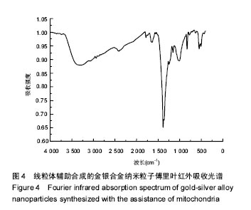
2.1.4 傅里叶红外光谱分析 图4为所制得的金银合金纳米粒子的傅里叶红外光谱图谱,-OH或N-H的伸缩振动产生在3 400 cm-1处强度较大的特征吸收峰。-CH3的伸缩振动引起2 900 cm-1处出现2个强度较弱和面积较窄的特征吸收峰。同时在1 600 cm-1的强特征吸收带,可能是由于酰胺键中羰基的拉伸振动造成的。在1 500 cm-1的特征峰,可能是由于多肽中二级酰胺基团的 N-H键的伸缩振动所产生的。从金银纳米粒子的傅里叶红外光谱图分析来看,线粒体中的蛋白质不仅介导了金银纳米粒子的合成,还包裹在纳米粒子表面,成为纳米粒子的表面稳定剂。"
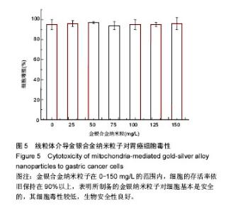
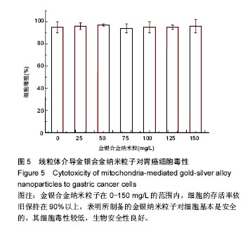
2.2 金银合金纳米粒子细胞毒性实验结果 纳米材料的生物安全性是其应用的前提条件,实验采用MTT法测试所制备金银合金纳米粒子对癌细胞的细胞毒性。如图5,金银合金纳米粒子在0-150 mg/L的范围内,细胞的存活率依旧保持在90%以上,表明所制备的金银纳米粒子对细胞基本是安全的,其细胞毒性较低,生物安全性良好。因为纳米粒子表面被生物分子包裹,是所得金银纳米粒子不与细胞直接接触,因而使其细胞毒性降低。"

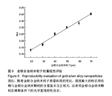
2.3 金银合金纳米粒子的体外稳定性评价 由于金银纳米粒子的生物学应用一般都在很低的浓度范围内,要求在稀释条件下不会影响纳米材料的光学稳定性。保持将超纯水不断滴入金银合金纳米颗粒的溶液中,然后测试其光学的特性变化。在图6中易观察到,随着金银合金纳米粒子质量浓度的变化,强度最大的特征吸收峰与金银合金纳米颗粒的含量基本呈正相关。这表明所得的金银合金纳米颗粒在稀释条件下的光学重现特性良好。"
| [1]Huang J,Wang W,Lin L,et al.A general strategy for the biosynthesis of gold nanoparticles by traditional Chinese medicines and their potential application as catalysts.Chem Asian J. 2009;4(7):1050-1054.[2]Ray PC,Darbha GK,Ray A,et al.A gold-nanoparticle-based fluorescence resonance energy transfer probe for multiplexed hybridization detection: accurate identification of bio-agents DNA. Nanotechnology.2007;18:1-6.[3]Schwartzberg AM,Grant CD,Wolcott A,et al.Unique gold nanoparticle aggregates as a highly active surface-enhanced Raman scattering substrate.J Phys Chem B. 2004;108(50): 19191-19197.[4]Pissuwan D,Niidome T,Cortie MB.The forthcoming applications of gold nanoparticles in drug and gene delivery systems.J Controlled Release.2011;149(1):65.[5]Boisselier E,Astruc D.Gold nanoparticles in nanomedicine: preparations, imaging, diagnostics, therapies and toxicity. Chem Soc Rev.2009;38(6):1759-1782. [6]Li Z,Huang P,Zhang X,et al.RGD-Conjugated Dendrimer-Modified Gold Nanorods for in Vivo Tumor Targeting and Photothermal Therapy.Mol Pharm.2009;7(1):94-104. [7]Chen DH,Chen CJ.Formation and characterization of Au–Ag bimetallic nanoparticles in water-in-oil microemulsions.J Mater Chem.2002;12(5):1557-1562.[8]Papavassiliou GC.Surfaceplasmons in small Au-Ag alloy particles.J Phys Metal Phys.1976;6(4):L103.[9]Link S,Wang ZL,El-Sayed MA.Alloy formation of gold-silver nanoparticles and the dependence of the plasmon absorption on their composition.J Phys Chem B.1999;103(18): 3529-3533.[10]Shankar SS,Ahmad A,Pasricha R,et al.Bioreduction of chloroaurate ions by geranium leaves and its endophytic fungus yields gold nanoparticles of different shapes.J Mater Chem.2003;13(7):1822-1826.[11]Shankar S,Ahmad A,Sastry M.Geranium leaf assisted biosynthesis of silver nanoparticles. Biotechnol Prog. 2003; 19(6):1627-1631.[12]Narayanan KB,Sakthivel N.Phytosynthesis of gold nanoparticles using leaf extract of Coleus amboinicusLour.Mater Charact.2010;61:1232-1238.[13]Faramarzi MA,Forootanfar H.Biosynthesis and characterization of gold nanoparticles produced by laccase from Paraconiothyriumvariabile.Colloids Surf B Biointerfaces. 2011;87(1):23-27.[14]Philip D.Honey mediated green synthesis of gold nanoparticles.Spectrochim Acta A Mol Biomol Spectrosc. 2009;73(4):650-653. [15]Nune SK,Chanda N,Shukla R,et al.Green nanotechnology from tea: phytochemicals in tea as building blocks for production of biocompatible gold nanoparticles.J Mater Chem.2009;19(19):2912-2920.[16]Katz E,Willner I,Wang J.Electroanalytical and bioelectroanalytical systems based on metal and semiconductor nanoparticles.Electroanalysis. 2004;16(1-2): 19-44.[17]Tisch U,Haick H.Arrays of chemisensitive monolayer-capped metallic nanoparticles for diagnostic breath testing.Rev Chem Eng.2011;26(5-6):171-179.[18]Ahn H,Chandekar A,Kang B,et al.Electrical conductivity and vapor-sensing properties of ω-(3-thienyl) alkanethiol-protected gold nanoparticle films.Chem Mater. 2004;16(17):3274-3278.[19]Wang AQ,Chang CM,Mou CY.Evolution of catalytic activity of Au-Ag bimetallic nanoparticles on mesoporous support for CO oxidation.J Phys Chem B.2005;109(40):18860-18867.[20]Mallin MP,Murphy CJ.Solution-phase synthesis of sub-10 nm Au-Ag alloy nanoparticles.Nano Lett.2002;2(11):1235-1237.[21]Endo T,Yoshimura T,Esumi K.Synthesis and catalytic activity of gold silver binary nanoparticles stabilized by PAMAM dendrimer.J Colloid Interface Sci.2005;286(2):602-609.[22]Boisselier E,Astruc D.Gold nanoparticles in nanomedicine: preparations, imaging, diagnostics,therapies and toxicity. Chem Soc Rev.2009;38(6):1759-1782.[23]Bond GC. Gold: a relatively new catalyst. Catal Today. 2002;72(1-2): 5-9.[24]Li Z,Huang P,Zhang X,et al.RGD-conjugated dendrimer- modified gold nanorods for in vivo tumor targeting and photothermal therapy.Mol Pharm.2010;7(1):94-104.[25]Cu D,Zhang H,Wang K,et al.Gold Nanoparticles Enhance Efficiency of In Vitro Gene Transcription-Translation System.Nano Biomed Eng.2011;3(2):120-125.[26]Gole A, Murphy CJ.Seed-mediated synthesis of gold nanorods: role of the size and nature of the seed.Chem Mater.2004;16(19):3633-3640. [27]Meltzer S,Resch R,Koel BE,et al.Fabrication of nanostructures by hydroxylamine seeding of gold nanoparticle templates.Langmuir.2001;17(5):1713-1718. [28]Westcott SL,Oldenburg SJ,Lee TR, et al.Formation and adsorption of clusters of gold nanoparticles onto functionalized silica nanoparticle surfaces. Langmuir. 1998;14(19):5396-5401. [29]Ahmad A,Senapati S,Khan MI,et al.Extracellular biosynthesis of monodisperse gold nanoparticles by a novel extremophilic actinomycete, Thermomonospora sp. Langmuir.2003;19(8):3550-3553. [30]Mukherjee P,Senapati S,Mandal D,et al.Extracellular synthesis of gold nanoparticles b the fungus Fusarium oxysporum.Chembiochem.2002;3(5):461-463. [31]Xie J,Lee JY,Wang DIC,et al.High-yield synthesis of complex gold nanostructures in a fungal system. J Phys Chem C. 2007;111(45):16858-16865. |
| [1] | Zhang Tongtong, Wang Zhonghua, Wen Jie, Song Yuxin, Liu Lin. Application of three-dimensional printing model in surgical resection and reconstruction of cervical tumor [J]. Chinese Journal of Tissue Engineering Research, 2021, 25(9): 1335-1339. |
| [2] | Zeng Yanhua, Hao Yanlei. In vitro culture and purification of Schwann cells: a systematic review [J]. Chinese Journal of Tissue Engineering Research, 2021, 25(7): 1135-1141. |
| [3] | Xu Dongzi, Zhang Ting, Ouyang Zhaolian. The global competitive situation of cardiac tissue engineering based on patent analysis [J]. Chinese Journal of Tissue Engineering Research, 2021, 25(5): 807-812. |
| [4] | Wu Zijian, Hu Zhaoduan, Xie Youqiong, Wang Feng, Li Jia, Li Bocun, Cai Guowei, Peng Rui. Three-dimensional printing technology and bone tissue engineering research: literature metrology and visual analysis of research hotspots [J]. Chinese Journal of Tissue Engineering Research, 2021, 25(4): 564-569. |
| [5] | Chang Wenliao, Zhao Jie, Sun Xiaoliang, Wang Kun, Wu Guofeng, Zhou Jian, Li Shuxiang, Sun Han. Material selection, theoretical design and biomimetic function of artificial periosteum [J]. Chinese Journal of Tissue Engineering Research, 2021, 25(4): 600-606. |
| [6] | Liu Fei, Cui Yutao, Liu He. Advantages and problems of local antibiotic delivery system in the treatment of osteomyelitis [J]. Chinese Journal of Tissue Engineering Research, 2021, 25(4): 614-620. |
| [7] | Li Xiaozhuang, Duan Hao, Wang Weizhou, Tang Zhihong, Wang Yanghao, He Fei. Application of bone tissue engineering materials in the treatment of bone defect diseases in vivo [J]. Chinese Journal of Tissue Engineering Research, 2021, 25(4): 626-631. |
| [8] | Zhang Zhenkun, Li Zhe, Li Ya, Wang Yingying, Wang Yaping, Zhou Xinkui, Ma Shanshan, Guan Fangxia. Application of alginate based hydrogels/dressings in wound healing: sustained, dynamic and sequential release [J]. Chinese Journal of Tissue Engineering Research, 2021, 25(4): 638-643. |
| [9] | Chen Jiana, Qiu Yanling, Nie Minhai, Liu Xuqian. Tissue engineering scaffolds in repairing oral and maxillofacial soft tissue defects [J]. Chinese Journal of Tissue Engineering Research, 2021, 25(4): 644-650. |
| [10] | Xing Hao, Zhang Yonghong, Wang Dong. Advantages and disadvantages of repairing large-segment bone defect [J]. Chinese Journal of Tissue Engineering Research, 2021, 25(3): 426-430. |
| [11] | Bai Shengchao, Gao Yang, Wang Bo, Li Junping, Wang Ruiyuan. Dynamic changes of mitochondrial function of the skeletal muscle after acupuncture intervention in rats with heavy load exercise-induced injury [J]. Chinese Journal of Tissue Engineering Research, 2021, 25(23): 3648-3653. |
| [12] | Chen Siqi, Xian Debin, Xu Rongsheng, Qin Zhongjie, Zhang Lei, Xia Delin. Effects of bone marrow mesenchymal stem cells and human umbilical vein endothelial cells combined with hydroxyapatite-tricalcium phosphate scaffolds on early angiogenesis in skull defect repair in rats [J]. Chinese Journal of Tissue Engineering Research, 2021, 25(22): 3458-3465. |
| [13] | Wang Hao, Chen Mingxue, Li Junkang, Luo Xujiang, Peng Liqing, Li Huo, Huang Bo, Tian Guangzhao, Liu Shuyun, Sui Xiang, Huang Jingxiang, Guo Quanyi, Lu Xiaobo. Decellularized porcine skin matrix for tissue-engineered meniscus scaffold [J]. Chinese Journal of Tissue Engineering Research, 2021, 25(22): 3473-3478. |
| [14] | Mo Jianling, He Shaoru, Feng Bowen, Jian Minqiao, Zhang Xiaohui, Liu Caisheng, Liang Yijing, Liu Yumei, Chen Liang, Zhou Haiyu, Liu Yanhui. Forming prevascularized cell sheets and the expression of angiogenesis-related factors [J]. Chinese Journal of Tissue Engineering Research, 2021, 25(22): 3479-3486. |
| [15] | Liu Chang, Li Datong, Liu Yuan, Kong Lingbo, Guo Rui, Yang Lixue, Hao Dingjun, He Baorong. Poor efficacy after vertebral augmentation surgery of acute symptomatic thoracolumbar osteoporotic compression fracture: relationship with bone cement, bone mineral density, and adjacent fractures [J]. Chinese Journal of Tissue Engineering Research, 2021, 25(22): 3510-3516. |
| Viewed | ||||||
|
Full text |
|
|||||
|
Abstract |
|
|||||