Chinese Journal of Tissue Engineering Research ›› 2026, Vol. 30 ›› Issue (31): 8068-8076.doi: 10.12307/2026.883
Previous Articles Next Articles
miR-122-5p-transfected exosomes from human adipose-derived mesenchymal stem cells for treatment of intrauterine adhesions in rats
Qin Ying1, 2, Chen Jibing3, Liang Weiping3, Jing Wenyao1, Zhu Xiangyun1, He Junkui1, Pang Lihong1, Li Hongli3
- 1Department of Prenatal Diagnosis and Genetic Diagnosis, First Affiliated Hospital of Guangxi Medical University, Nanning 530021, Guangxi Zhuang Autonomous Region, China; 2Department of Obstetrics, Guigang City People's Hospital, Guigang 537100, Guangxi Zhuang Autonomous Region, China; 3Center for Translational Medical Research in Integrative Chinese and Western Medicine, Ruikang Hospital Affiliated to Guangxi University of Chinese Medicine, Nanning 530012, Guangxi Zhuang Autonomous Region, China
-
Received:2026-01-14Accepted:2026-02-13Online:2026-11-08Published:2026-05-22 -
Contact:Li Hongli, PhD, Assistant researcher, Center for Translational Medical Research in Integrative Chinese and Western Medicine, Ruikang Hospital Affiliated to Guangxi University of Chinese Medicine, Nanning 530012, Guangxi Zhuang Autonomous Region, China; Co-corresponding author: Pang Lihong, PhD, Chief physician, Professor, Department of Prenatal Diagnosis and Genetic Diagnosis, First Affiliated Hospital of Guangxi Medical University, Nanning 530021, Guangxi Zhuang Autonomous Region, China -
About author:Qin Ying, MD candidate, Associate chief physician, Department of Prenatal Diagnosis and Genetic Diagnosis, First Affiliated Hospital of Guangxi Medical University, Nanning 530021, Guangxi Zhuang Autonomous Region, China; Department of Obstetrics, Guigang City People's Hospital, Guigang 537100, Guangxi Zhuang Autonomous Region, China -
Supported by:National Natural Science Foundation of China, No. 82260306, 81960281 (to PLH); Construction of Clinical Intervention Protocols Guangxi Key R & D Program, No. AB24010080 (to PLH); Doctoral Research Startup Fund of Guangxi University of Chinese Medicine, No. 2021BS032 (to LHL)

CLC Number:
Cite this article
Qin Ying, Chen Jibing, Liang Weiping, Jing Wenyao, Zhu Xiangyun, He Junkui, Pang Lihong, Li Hongli. miR-122-5p-transfected exosomes from human adipose-derived mesenchymal stem cells for treatment of intrauterine adhesions in rats[J]. Chinese Journal of Tissue Engineering Research, 2026, 30(31): 8068-8076.
share this article
Add to citation manager EndNote|Reference Manager|ProCite|BibTeX|RefWorks
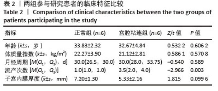
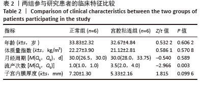
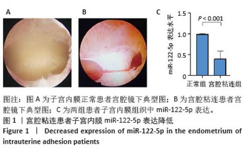
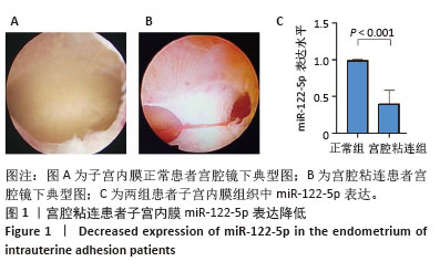
2.1 miR-122-5p在宫腔粘连患者子宫内膜中的表达 共纳入12例患者,其中6例中重度宫腔粘连患者作为宫腔粘连组,6例子宫内膜正常者作为正常组。如表2所示,两组年龄、体质量指数、月经周期和子宫内膜厚度均无统计学差异(P > 0.05)。然而,宫腔粘连组的流产次数显著多于正常组(P < 0.05)。宫腔镜检查结果显示正常组宫腔呈倒三角形,宫底平坦,四壁内膜平滑,无异常赘生物(图1A);宫腔粘连组宫腔形态改变,子宫前后壁可见片状致密物累及宫腔1/2(图1B)。qPCR检测结果进一步表明:宫腔粘连组子宫内膜组织中miR-122-5p的相对表达水平显著低于正常组(图1C)。 "
"
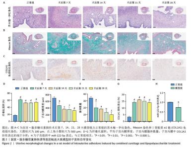
2.2 大鼠宫腔粘连模型构建 采用双重损伤方法建立大鼠宫腔粘连模型,模拟临床不孕患者子宫内膜损伤的病理特征。如图2A,B所示,苏木精-伊红染色、Masson染色显示,与正常组相比,模型组出现典型纤维化病变,子宫内膜纤维化面积在术后第7天达到高峰(61.51%),虽于第14天略有回落(53.10%),但仍然显著高于正常组,并持续至术后第28天,仍维持在较高水平,未见明显恢复(图2D);同时,子宫内膜厚度和腺体数量自术后第7天起显著减少,随着观察时间的延长呈逐渐下降趋势(图2E,F)。此外,免疫组织化学染色显示,模型组Ⅰ型胶原α1链的阳性表达在术后第7天显著增强,并持续高表达至第28天(图2C,G)。上述结果表明宫腔粘连模型构建成功且在一定时间内保持稳定。基于上述结果,选取纤维化程度最高的术后第7天作为干预时间点。qPCR检测显示,模型组子宫内膜中miR-122-5p的表达水平显著低于正常组(图2H)。因此,后续研究于造模后第7天进行外泌体治疗,以评估其对子宫内膜纤维化的干预效果及作用机制。 "
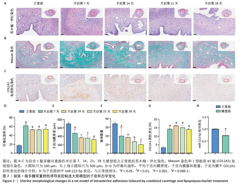
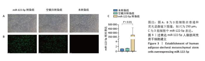
2.3 过表达miR-122-5p脂肪间充质干细胞建立 通过慢病毒感染脂肪间充质干细胞,获得稳定表达miR-122-5p的脂肪间充质干细胞和空载脂肪间充质干细胞。为了观察慢病毒感染效率,感染48 h后使用含嘌呤霉素的培养基对3组细胞进行筛选,荧光显微镜下观察,当感染效率在80%左右,未转染慢病毒组细胞基本全部死亡,无绿色荧光表达,miR-122-5p转染组和空载体转染组细胞生长良好且荧光表达丰度高(图3A,B)。qPCR检查3组细胞中miR-122-5p的表达水平,如图3C所示,与未转染慢病毒组和空载体转染组相比,miR-122-5p转染组脂肪间充质干细胞中miR-122-5p表达显著升高,表明稳定过表达miR-122-5p脂肪间充质干细胞系构建成功。 "
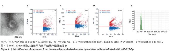
2.4 miR-122-5p转染人脂肪间充质干细胞外泌体的分离与鉴定 外泌体在透射电镜下呈扁圆形、球形或杯状的形态,见图4A。BCA测定外泌体蛋白质量浓度为10.64 mg/mL,流式细胞检测显示,特异性表面标志物CD9、CD63和CD81均呈阳性(图4B-D),纳米颗粒跟踪分析显示外泌体粒径为56 nm(图4E)。"
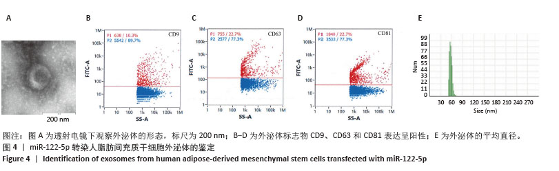
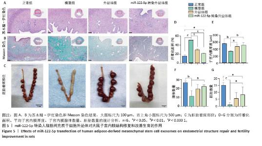
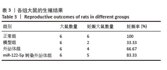
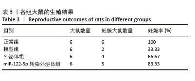
2.5 miR-122-5p转染人脂肪间充质干细胞外泌体对宫腔粘连大鼠治疗效果分析 苏木精-伊红染色和Masson染色显示,与正常组相比,模型组子宫内膜厚度明显降低[(555.83±14.20) μm vs. (341.44± 10.49) μm,P < 0.01)];与模型组相比,外泌体组和miR-122-5p转染外泌体组子宫内膜厚度均明显增加[(341.44±10.49) μm vs. (474.24± 50.37) μm,P < 0.05;(341.44±10.49) μm vs. (510.22±73.76) μm,P < 0.01](图5A,E);正常组腺体结构完整,腺体数量一致,与正常组相比,模型组大鼠子宫腺体数量减少(26.50±2.00 vs. 11.27±6.13,P < 0.01);与模型组相比,外泌体组和miR-122-5p转染外泌体组腺体数量均有所增加(11.27±6.13 vs. 19.50±4.92,P > 0.05;11.27±6.13 vs. 22.33±1.59,P < 0.05](图5A,F);与正常组相比,模型组大鼠子宫内膜纤维化面积明显增加[(15.72±2.71)% vs. (50.11±0.91)%,P < 0.000 1];与模型组相比,外泌体组和miR-122-5p转染外泌体组子宫内膜纤维化水平明显改善[(50.11±0.91)% vs. (29.02±2.38)%,P < 0.000 1;(50.11±0.91)% vs. (21.34±1.24)%,P < 0.000 1],且miR-122-5p转染外泌体组的治疗效果更加显著(图5B,D)。 各组大鼠胚胎着床部位见图5C。与正常组相比,模型组的妊娠率明显降低(100% vs. 33.3%),外泌体组和miR-122-5p转染外泌体组的妊娠率均高于模型组(66.67%,83.33% vs. 33.3%),见表3;与正常组相比,模型组胚胎数量显著下降(15.17±1.83 vs. 1.00±1.67,P < 0.000 1);与模型组相比,外泌体组和miR-122-5p转染外泌体组胚胎数量均有所升高(1.00±1.67 vs. 5.50±4.42,P > 0.05;1.00±1.67 vs. 8.33±5.54,P < 0.05),miR-122-5p转染外泌体组的改善效果更为显著(图5G)。 "
"
| [1] YU S, ZHANG X, LI W, et al. Thermosensitive hydrogel as a sustained release carrier for mesenchymal stem cell-derived extracellular vesicles in the treatment of intrauterine adhesion. J Nanobiotechnology. 2024;22(1):570. [2] LEE WL, LIU CH, CHENG M, et al. Focus on the Primary Prevention of Intrauterine Adhesions: Current Concept and Vision. Int J Mol Sci. 2021;22(10):5175. [3] PENG X, WANG T, DAI B, et al. Gene Therapy for Inflammatory Cascade in Intrauterine Injury with Engineered Extracellular Vesicles Hybrid Snail Mucus-enhanced Adhesive Hydrogels. Adv Sci (Weinh). 2025;12(1):e2410769. [4] XIN L, WEI C, TONG X, et al. In situ delivery of apoptotic bodies derived from mesenchymal stem cells via a hyaluronic acid hydrogel: A therapy for intrauterine adhesions. Bioact Mater. 2021;12:107-119. [5] CAO Y, WU J, HUANG J, et al. Human embryonic stem cell-derived immunity-and-matrix-regulatory cells promote endometrial repair and fertility restoration in IUA rats. Stem Cell Res Ther. 2025;16(1):204. [6] CHEN P, YE C, HUANG Y, et al. Glutaminolysis regulates endometrial fibrosis in intrauterine adhesion via modulating mitochondrial function. Biol Res. 2024; 57(1):13. [7] DEANS R, ABBOTT J. Review of intrauterine adhesions. J Minim Invasive Gynecol. 2010;17(5):555-569. [8] ZHENG X, HUANG R, YIN L, et al. Injectable antioxidant hyaluronan/chitosan hydrogel as a platelet-rich plasma and stem cell carrier to promote endometrial regeneration and fertility restoration. Acta Biomater. 2025;195:201-215. [9] LIU Y, JIA D, LI L, et al. Advances in Nanomedicine and Biomaterials for Endometrial Regeneration: A Comprehensive Review. Int J Nanomedicine. 2024;19:8285-8308. [10] XIONG Z, HU Y, JIANG M, et al. Hypoxic bone marrow mesenchymal stem cell exosomes promote angiogenesis and enhance endometrial injury repair through the miR-424-5p-mediated DLL4/Notch signaling pathway. PeerJ. 2024;12:e16953. [11] YU D, WONG YM, CHEONG Y, et al. Asherman syndrome--one century later. Fertil Steril. 2008;89(4):759-779. [12] GAO M, YU Z, YAO D, et al. Mesenchymal stem cells therapy: A promising method for the treatment of uterine scars and premature ovarian failure. Tissue Cell. 2022;74:101676. [13] GÓRNICKI T, JÓZKOWIAK M, DATA K, et al. Wharton’s jelly mesenchymal stem cells (WJ-MSCs), a “Holy Grail” in tissue bioengineering and reconstructive medicine. Biomed Pharmacother. 2025;192:118570. [14] KOISHYBAYEVA D, AMIRASHOV A, BALMAGAMBETOVA S, et al. Mesenchymal stem cell therapy for diabetes: An umbrella review. Tissue Cell. 2025;96:103043. [15] YI X, LIU F, GAO K, et al. Reconstructable Uterus-Derived Materials for Uterus Recovery toward Efficient Live Births. Adv Mater. 2022;34(8):e2106510. [16] CAO Y, SUN H, ZHU H, et al. Allogeneic cell therapy using umbilical cord MSCs on collagen scaffolds for patients with recurrent uterine adhesion: a phase I clinical trial. Stem Cell Res Ther. 2018;9(1):192. [17] YU X, SHI L, ZHANG Y, et al. A Comparative Study of Mesenchymal Stem Cells from Various Sources in Endometrial Repair: the Significant Advantages of Decidual Mesenchymal Stem Cells. Stem Cell Rev Rep. 2025;21(8):2667-2670. [18] LI Q, LIAO Z, HU X, et al. Phase 1 dose-escalation trial of sub-endometrial injection of human embryonic stem cells-derived immunity-and-matrix-regulatory cells to promote endometrial angiogenesis in refractory intrauterine adhesion. Mol Ther. 2026;34(1):423-442. [19] LIANG Y, SHUAI Q, ZHANG X, et al. Incorporation of Decidual Stromal Cells Derived Exosomes in Sodium Alginate Hydrogel as an Innovative Therapeutic Strategy for Advancing Endometrial Regeneration and Reinstating Fertility. Adv Healthc Mater. 2024;13(13):e2303674. [20] TAN F, LI X, WANG Z, et al. Clinical applications of stem cell-derived exosomes. Signal Transduct Target Ther. 2024;9(1):17. [21] LI B, CAO Y, SUN M, et al. Expression, regulation, and function of exosome-derived miRNAs in cancer progression and therapy. FASEB J. 2021;35(10):e21916. [22] KALLURI R, LEBLEU VS. The biology, function, and biomedical applications of exosomes. Science. 2020;367(6478):eaau6977. [23] KARIM RONY RMI, TOMPKINS JD. Cardiac repair and regeneration: cell therapy, in vivo reprogramming, and the promise of extracellular vesicles. Exp Mol Med. 2025;57(10):2182-2200. [24] SONG J, LIU J, CUI C, et al. Mesenchymal stromal cells ameliorate diabetes-induced muscle atrophy through exosomes by enhancing AMPK/ULK1-mediated autophagy. J Cachexia Sarcopenia Muscle. 2023;14(2):915-929. [25] NAZDIKBIN YAMCHI N, AHMADIAN S, MOBARAK H, et al. Amniotic fluid-derived exosomes attenuated fibrotic changes in POI rats through modulation of the TGF-β/Smads signaling pathway. J Ovarian Res. 2023;16(1):118. [26] PARK HS, CETIN E, SIBLINI H, et al. Therapeutic Potential of Mesenchymal Stem Cell-Derived Extracellular Vesicles to Treat PCOS. Int J Mol Sci. 2023;24(13):11151. [27] ZHANG H, FANG Y, GAO Y, et al. Brown adipose tissue-derived exosomes delay fertility decline in aging mice. Front Endocrinol (Lausanne). 2023;14:1180104. [28] LIAO Z, LIU C, WANG L, et al. Therapeutic Role of Mesenchymal Stem Cell-Derived Extracellular Vesicles in Female Reproductive Diseases. Front Endocrinol (Lausanne). 2021;12:665645. [29] LIU HD, WANG SW. Role of noncoding RNA in the pathophysiology and treatment of intrauterine adhesion. Front Genet. 2022;13:948628. [30] SUN D, JIANG Z, CHEN Y, et al. MiR-455-5p upregulation in umbilical cord mesenchymal stem cells attenuates endometrial injury and promotes repair of damaged endometrium via Janus kinase/signal transducer and activator of transcription 3 signaling. Bioengineered. 2021;12(2):12891-12904. [31] CHEN JM, HUANG QY, CHEN WH, et al. Transcriptomics of tissue exosomes to investigate miR-195-5p’s amelioration of endometrial fibrosis via the YAP-Smad7 pathway: an animal study. J Transl Med. 2024;22(1):1050. [32] LI X, DUAN H, WANG S, et al. Umbilical cord mesenchymal stem cell-derived exosomes reverse endometrial fibrosis by the miR-145-5p/ZEB2 axis in intrauterine adhesions. Reprod Biomed Online. 2023;46(2):234-243. [33] SHAO X, QIN J, WAN C, et al. ADSC Exosomes Mediate lncRNA-MIAT Alleviation of Endometrial Fibrosis by Regulating miR-150-5p. Front Genet. 2021;12:679643. [34] CHEN S, MA Y, QIU X, et al. MicroRNA-122-5p alleviates endometrial fibrosis via inhibiting the TGF-β/SMAD pathway in Asherman’s syndrome. Reprod Biomed Online. 2023;47(5):103253. [35] ZHANG H, XING J, SUN M, et al. Engineered exosomes for targeted microRNA delivery to reverse liver fibrosis. Biomaterials. 2026;324:123510. [36] IQBAL Z, REHMAN K, MAHMOOD A, et al. Exosome for mRNA delivery: strategies and therapeutic applications. J Nanobiotechnology. 2024;22(1):395. [37] AAGL ADVANCING MINIMALLY INVASIVE GYNECOLOGY WORLDWIDE. AAGL practice report: practice guidelines for management of intrauterine synechiae. J Minim Invasive Gynecol. 2010;17(1):1-7. [38] 杨玉伟,李万里,陈继冰,等.间充质干细胞包裹人胰岛减轻即刻经血液介导的炎症反应的体外研究 [J].器官移植,2023,14(4):562-569. [39] LI J, PAN Y, YANG J, et al. Tumor necrosis factor-α-primed mesenchymal stem cell-derived exosomes promote M2 macrophage polarization via Galectin-1 and modify intrauterine adhesion on a novel murine model. Front Immunol. 2022;13:945234. [40] CHEN TQ, WEI XJ, LIU HY, et al. Telocyte-Derived Exosomes Provide an Important Source of Wnts That Inhibits Fibrosis and Supports Regeneration and Repair of Endometrium. Cell Transplant. 2023;32:9636897231212746. [41] SUN W, XU W, XIAO M, et al. Enhancing exosome stability and delivery with natural polymers to prevent intrauterine adhesions and promote endometrial regeneration: a review. J Nanobiotechnology. 2025;23(1):529. [42] LIN Y, LI Y, CHEN P, et al. Exosome-Based Regimen Rescues Endometrial Fibrosis in Intrauterine Adhesions Via Targeting Clinical Fibrosis Biomarkers. Stem Cells Transl Med. 2023;12(3):154-168. [43] QIN Z, YU Q, LONG Y. Unveiling the Pathological Landscape of Intrauterine Adhesion: Mechanistic Insights and Exosome-Biomaterial Therapeutic Innovations. Int J Nanomedicine. 2025;20:9667-9694. [44] ZHOU Z, WANG H, ZHANG X, et al. Defective autophagy contributes to endometrial epithelial-mesenchymal transition in intrauterine adhesions. Autophagy. 2022;18(10):2427-2442. [45] ZHAI X, PU D, WANG R, et al. Gas6/AXL pathway: immunological landscape and therapeutic potential. Front Oncol. 2023;13:1121130. [46] ZHOU L, DONG L, LI H, et al. Mesenchymal stem cell-derived exosomes ameliorate TGF-β1-induced endometrial fibrosis by altering their miRNA profile. Am J Transl Res. 2023;15(5):3203-3216. [47] ZHANG Z, HU J. DKK1 loss promotes endometrial fibrosis via autophagy and exosome-mediated macrophage-to-myofibroblast transition. J Transl Med. 2024;22(1):617. [48] LIANG Y, MENG J, YU Z, et al. Ru single-atom nanozymes targeting ROS-ferroptosis pathways for enhanced endometrial regeneration in intrauterine adhesion therapy. Biomaterials. 2025;315:122923. [49] SONG M, MA L, ZHU Y, et al. Umbilical cord mesenchymal stem cell-derived exosomes inhibits fibrosis in human endometrial stromal cells via miR-140-3p/FOXP1/Smad axis. Sci Rep. 2024;14(1):8321. [50] YUAN D, GUO T, QIAN H, et al. Exosomal miR-543 derived from umbilical cord mesenchymal stem cells ameliorates endometrial fibrosis in intrauterine adhesion via downregulating N-cadherin. Placenta. 2023;131:75-81. [51] WANG S, LIU T, NAN N, et al. Exosomes from Human Umbilical Cord Mesenchymal Stem Cells Facilitates Injured Endometrial Restoring in Early Repair Period through miR-202-3p Mediating Formation of ECM. Stem Cell Rev Rep. 2023;19(6): 1954-1964. [52] XIAO B, ZHU Y, LIU M, et al. miR-340-3p-modified bone marrow mesenchymal stem cell-derived exosomes inhibit ferroptosis through METTL3-mediated m6A modification of HMOX1 to promote recovery of injured rat uterus. Stem Cell Res Ther. 2024;15(1):224. [53] LU W, GUO Y, LIU H, et al. The Inhibition of Fibrosis and Inflammation in Obstructive Kidney Injury via the miR-122-5p/SOX2 Axis Using USC-Exos. Biomater Res. 2024;28:0013. [54] CABALLERO-VALDERRAMA MR, BEVILACQUA E, ECHEVARRÍA M, et al. Early Myocardial Strain Reduction and miR-122-5p Elevation Associated with Interstitial Fibrosis in Anthracycline-Induced Cardiotoxicity. Biomedicines. 2024;13(1):45. [55] LI B, LI Y, LI S, et al. Circ_MTM1 knockdown inhibits the progression of HBV-related liver fibrosis via regulating IL7R expression through targeting miR-122-5p. Am J Transl Res. 2022;14(4):2199-2211. [56] YAO S, ZHOU Z, WANG L, et al. Targeting endometrial inflammation in intrauterine adhesion ameliorates endometrial fibrosis by priming MSCs to secrete C1INH. iScience. 2023;26(7):107201. [57] XI J, PAN Y, JIN C, et al. Evaluation of different rat models intrauterine adhesion models and improvement of the technique for their establishment. Exp Anim. 2023;72(2):274-284. |
| [1] | Tao Daiju, Su Haiyu, Wang Yuqi, Shen Zhiqiang, He Bo . Construction and identification of stable PC12 cell lines with high/low expression of miR-122-5p [J]. Chinese Journal of Tissue Engineering Research, 2026, 30(7): 1790-1799. |
| [2] | Wang Baiyan, Yang Shu, Wang Yiming, Wu Mengqing, Xiao Yu, Guo Zixuan, Zhang Boyi, Feng Shuying. Exosome-delivered CRISPR/Cas system enables gene editing in target cells [J]. Chinese Journal of Tissue Engineering Research, 2026, 30(7): 1839-1849. |
| [3] | Wang Yanfei, Jin Lianhai, Li Qingya, Fu Yuanfei, Tan Huangsheng, Deng Pengwei, Gao Kun. Synovial fluid exosome-mediated crosstalk between synoviocytes and chondrocytes in development and progression of knee osteoarthritis [J]. Chinese Journal of Tissue Engineering Research, 2026, 30(34): 9032-9040. |
| [4] | Zheng Ying, Li Mengyao, Zheng Fanfan, He Zhao, Zhang Ning, Zou Jialun, Li Youlei, Gao Feng. Mechanism of action of extracellular vesicles loaded with biomaterials in repairing spinal cord injury [J]. Chinese Journal of Tissue Engineering Research, 2026, 30(32): 8544-8554. |
| [5] | Zhang Chuanlin, Wu Yuhuai, Zhou Haixiang, Liu Jianping, Xu Qian, Zhang Lili, Li Cuie. An innovative strategy for engineered exosomal long non-coding RNAs in treatment of osteoporosis [J]. Chinese Journal of Tissue Engineering Research, 2026, 30(31): 8264-8271. |
| [6] | Jiao Ziyi, Enhejirigala, Shi Guopeng, Yan Dayang, Wang Wei, Wang Xing, Liu Fuling, Wang Haiyan, Li Xiaohe. Bone tissue repair with animal/human-derived exosomes: visualization analysis of development trends [J]. Chinese Journal of Tissue Engineering Research, 2026, 30(31): 8293-8301. |
| [7] | Wang Jing, Shi Haijie, Zhang Mengmeng, Zhang Jinyong, Wang Miaomiao, Tang Lin. Interleukin-4 modulates macrophage polarization and osteogenic differentiation of adipose-derived mesenchymal stem cells [J]. Chinese Journal of Tissue Engineering Research, 2026, 30(31): 8077-8083. |
| [8] | Liu Jinrui, Yang Xianguang, Liu Haolong, Li Li, Ruan Zhaohui, Li Yanlin. Exosomes from different sources and osteoarthritis: engineering technology, targeting, and drug carrier function [J]. Chinese Journal of Tissue Engineering Research, 2026, 30(31): 8219-8229. |
| [9] | He Jinwen, Li Jin. Safety, stability, and action mechanism of mesenchymal stem cell-derived exosomes in treatment of viral infectious diseases [J]. Chinese Journal of Tissue Engineering Research, 2026, 30(31): 8230-8236. |
| [10] | Tian Yu, Guo Ying, Yiershatijiang Aniwaer, Aihematijiang Refuhaiti, Maierdana Maimaitireyimu. Molecular mechanisms by which Fusobacterium nucleatum regulates colonic polyp formation in mice [J]. Chinese Journal of Tissue Engineering Research, 2026, 30(29): 7603-7611. |
| [11] | Li Tianbo, Yu Zeyang, Qin Xinyuan, Wang Jiangning, Gao Lei. Exosomes derived from human umbilical cord mesenchymal stem cells in treatment of diabetic foot ulcers [J]. Chinese Journal of Tissue Engineering Research, 2026, 30(19): 4897-4901. |
| [12] | Jian Xichao, Shao Jingjie, Tang Shihan, Qi Fang, Deng Chengliang. Exosomes promote diabetic wound healing: a visual analysis of research hotspots and evolutionary trends [J]. Chinese Journal of Tissue Engineering Research, 2026, 30(19): 5072-5081. |
| [13] | Wang Xinyue, Li Hongli, Guo Chunhui, Chen Jibing, Yu Hua. Changes in the expression of six microRNAs in ovarian tissue from animal models of premature ovarian failure and in peripheral blood of patients with premature ovarian failure [J]. Chinese Journal of Tissue Engineering Research, 2026, 30(18): 4675-4684. |
| [14] | Kong Xiaojuan, Tan Zhenyu, Lei Lei. Repair of rat endometrial injury by using miR-424-5p modified exosome/Poloxam 407 hydrogel [J]. Chinese Journal of Tissue Engineering Research, 2026, 30(14): 3626-3635. |
| [15] | Lu Biqiong, Wei Zhongjian. Skeletal muscle-derived exosome-mediated regulation of bone formation and role of exercise intervention [J]. Chinese Journal of Tissue Engineering Research, 2026, 30(13): 3379-3391. |
| Viewed | ||||||
|
Full text |
|
|||||
|
Abstract |
|
|||||