Chinese Journal of Tissue Engineering Research ›› 2026, Vol. 30 ›› Issue (31): 8230-8236.doi: 10.12307/2026.829
Previous Articles Next Articles
Safety, stability, and action mechanism of mesenchymal stem cell-derived exosomes in treatment of viral infectious diseases
He Jinwen1, Li Jin1, 2
- 1Key Laboratory of Infectious Disease and Biosafety, Guizhou Provincial Department of Education, Zunyi Medical University, Zunyi 563006, Guizhou Province, China; 2School of Preclinical Medicine of Zunyi Medical University, Zunyi 563006, Guizhou Province, China
-
Received:2025-09-25Accepted:2026-01-06Online:2026-11-08Published:2026-05-25 -
Contact:Li Jin, Associate professor, Master’s supervisor, Key Laboratory of Infectious Disease and Biosafety, Guizhou Provincial Department of Education, Zunyi Medical University, Zunyi 563006, Guizhou Province, China; School of Preclinical Medicine of Zunyi Medical University, Zunyi 563006, Guizhou Province, China -
About author:He Jinwen, MS candidate, Key Laboratory of Infectious Disease and Biosafety, Guizhou Provincial Department of Education, Zunyi Medical University, Zunyi 563006, Guizhou Province, China -
Supported by:National Natural Science Foundation of China (Regional Project), No. 82360859 (to LJ); Guizhou Provincial Science and Technology Program Project, No. ZK[2024]287 (to LJ)

CLC Number:
Cite this article
He Jinwen, Li Jin. Safety, stability, and action mechanism of mesenchymal stem cell-derived exosomes in treatment of viral infectious diseases[J]. Chinese Journal of Tissue Engineering Research, 2026, 30(31): 8230-8236.
share this article
Add to citation manager EndNote|Reference Manager|ProCite|BibTeX|RefWorks
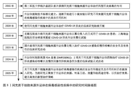
2.1 间充质干细胞来源外泌体在病毒感染性疾病中的研究时间脉络图 见图3。"
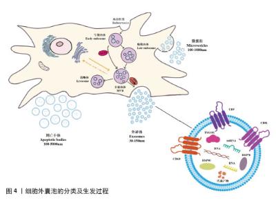
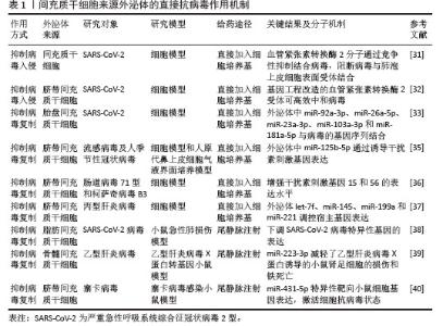
2.2 外泌体的生物发生与分子组成 2.2.1 外泌体的形成与释放 细胞外囊泡是一类具有重要生理学功能的纳米级膜性结构,从细胞生物学角度来看,细胞外囊泡是由健康细胞、病态细胞以及病毒感染细胞自然分泌的亚细胞颗粒,表面具有与母细胞相似的脂质双分子层结构[13],能递送化疗药物等小分子化合物、蛋白质、小干扰RNA以及miRNA等多种生物活性分子。凭借独特的转运机制,细胞外囊泡能潜在规避溶酶体的降解,高效递送生物大分子[14]。根据大小和生物发生的不同,细胞外囊泡主要分为以下3类:外泌体、微囊泡和凋亡小体[15]。其中,外泌体和微囊泡作为细胞外囊泡的关键效应分子,它们的形成与释放极具特点,在多种生理过程中发挥重要作用[16]。 微囊泡是细胞受特定的信号刺激或活化后,细胞质膜直接向外出芽、脱落形成的细胞外囊泡,它的形成过程主要是受信号刺激使得质膜磷脂双分子层发生不对称的结构改变,导致特定的膜转运蛋白被激活,进一步触发膜上脂质的重新分布,细胞骨架蛋白发生解聚,最终质膜局部向外凸起形成芽体,芽体经过膜裂变将包含细胞质成分的微囊泡释放到细胞外,参与细胞间通讯及多种病理生理过程[17]。 相比之下,外泌体的释放过程则有着更为复杂的细胞内调控机制,需要包括Rab GTP 酶、内体分选复合物、膜联蛋白、脂筏蛋白以及四跨膜蛋白(如CD63、CD8和CD9)在内的多种关键蛋白协同完成[15],具体过程可以概括为细胞膜内陷将细胞外物质通过内吞作用形成早期内体,这些早期内体在质子泵的作用下逐渐酸化成熟转变为晚期内体,随后晚期内体的内膜向内出芽,在内体分选复合物、相关脂蛋白以及四跨膜蛋白的作用下,将特定蛋白、核酸等物质分选包裹形成腔内囊泡,这些腔内囊泡共同构成的多泡体在Rab GTP酶等蛋白的调控下借助细胞骨架和微管系统转运至细胞膜附近,在特定的信号刺激下与细胞质膜发生融合,最终以胞吐的方式将腔内囊泡释放到细胞外,这些被释放的囊泡即为外泌体[18]。细胞外囊泡的分类及释放过程,见图4。 "
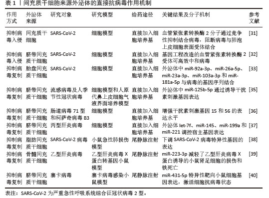
2.2.2 间充质干细胞来源外泌体的分子组成 间充质干细胞来源外泌体具有与母细胞相似的双分子层膜结构,表面富含CD9、CD81和CD63等四跨膜蛋白和整合素,这些蛋白有助于间充质干细胞来源外泌体与靶细胞之间的黏附、侵入以及膜融合过程,使它们能够精准定位特定的细胞与组织器官[19]。间充质干细胞来源外泌体内含的热休克蛋白70和热休克蛋白90,可以与抗原结合,参与抗原呈递过程,调控细胞应激反应[20]。此外,间充质干细胞来源外泌体携带的白细胞介素10、转化生长因子β等抗炎因子,还能帮助调节机体免疫平衡,抑制由病毒感染引起的过度炎症反应,增强宿主细胞的整体免疫能力[8,21]。 间充质干细胞来源外泌体的膜结构中还含有丰富的脂质成分,不仅能够维持自身结构的完整性,而且脂质在膜上的不对称分布还能进一步增强与靶细胞之间的相互作用效率[16,22],这一特性对于提高抗病毒药物的靶向递送具有重要意义。间充质干细胞来源外泌体携带的脂质代谢酶还可以调节受体细胞内的脂质稳态从而保护细胞免受伤害[23]。此外,间充质干细胞来源外泌体中的一些活性代谢物质,如乙酰-DL-缬氨酸,还可以通过恢复小鼠肠道菌群稳态,有效改善小鼠爆发性肝炎引起的肝损伤[24]。由此可以看出这些脂质成分在细胞保护和活性物质递送方面存在巨大潜力。 间充质干细胞可以从骨髓、脂肪、胎盘和脐带等多种组织中分离获取[25],并且不同来源间充质干细胞分泌的外泌体所携带的DNA、RNA、脂质、代谢物以及功能蛋白都存在一定的差异[18],这不仅反映了间充质干细胞来源外泌体组成的复杂性,还提示存在潜在的多功能性,为开展间充质干细胞来源外泌体的抗病毒研究提供了可能。 2.3 间充质干细胞来源外泌体的直接抗病毒作用 病毒是一类严格依赖宿主细胞才能复制传播的微小病原体,它们必须利用宿主细胞的生物合成系统才能完成整个生命周期[26-27]。病毒的复制过程复杂且高度特异性,通常包括识别与入侵、脱壳与基因释放、复制与合成、组装与释放这几个关键步骤[28]。近年来,研究者证实了外泌体能够通过递送病毒组分或宿主调控因子,直接参与调节病毒的生命周期,从而改变病毒的复制效率和致病过程[29]。下文将从直接抑制病毒入侵和复制的角度阐释间充质干细胞来源外泌体发挥作用的具体机制。 2.3.1 抑制病毒入侵 Ⅱ型肺泡上皮细胞是严重急性呼吸系统综合征冠状病毒2型(Severe acute respiratory syndrome coronavirus 2,SARS-Cov-2)感染的主要靶细胞,细胞表面的血管紧张素转换酶2受体在病毒入侵过程中发挥关键作用,介导了病毒对宿主细胞的感染[30]。间充质干细胞来源外泌体能够通过与病毒竞争宿主细胞表面受体的方式,有效抑制病毒入侵宿主细胞。MANZOOR等[31]对间充质干细胞来源细胞外囊泡在感染性疾病中的治疗作用一文中总结分析,发现间充质干细胞来源细胞外囊泡上的血管紧张素转换酶2受体可以通过竞争性抑制SARS-COV-2与肺泡细胞结合,从而有效阻断病毒与宿主肺泡上皮细胞表面受体的结合,显著降低病毒感染和入侵的风险。在进一步研究中发现,借助基因工程技术可增强这种抗病毒效应。在一项外泌体工程化研究中,利用慢病毒载体对间充质干细胞进行遗传修饰后,使细胞能够持续分泌高亲和力工程化血管紧张素转换酶2蛋白,这些经过改造的血管紧张素转换酶2分子在病毒接触宿主细胞之前就能高效中和病毒,并且它与SARS-CoV-2刺突蛋白的结合亲和力优于宿主细胞受体,能有效抑制病毒刺突蛋白与宿主细胞天然血管紧张素转换酶2受体的结合,阻止病毒入侵宿主细胞[32]。 2.3.2 抑制病毒复制 间充质干细胞来源外泌体所携带的多种miRNA可通过直接和间接双重机制有效抑病毒的复制,这些miRNA一方面能够直接靶向病毒基因组,干扰病毒复制,在PARK等[33]报道的胎盘间充质干细胞来源外泌体中存在5种miRNA能够结合SARS-CoV-2病毒的3′非翻译保守区域,抑制病毒RNA复制;另一方面还能调控宿主细胞中对病毒生命周期至关重要的细胞因子,在病毒复制中发挥双重调控作用[34]。研究证实,脐带间充质干细胞来源外泌体携带的特异性miRNA能显著抑制包括流感病毒、人类季节性冠状病毒、肠道病毒71型、柯萨奇病毒B3、丙型肝炎病毒和SARS-Cov-2在内的多种病毒的复制及其子代的产生,具有广谱抗病毒潜力。 间充质干细胞来源外泌体的直接抗病毒作用机制见表1[31-33,35-40]。 "
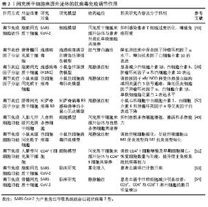
2.4 间充质干细胞来源外泌体的抗病毒免疫调节作用 2.4.1 调节免疫细胞活性 间充质干细胞来源外泌体在抵抗病毒感染过程中还发挥重要的免疫调节作用,可通过多种机制调节宿主T细胞、B细胞和自然杀伤细胞等免疫细胞的活性,从而增强机体的抗病毒免疫能力。现有研究证实间充质干细胞来源外泌体可以抑制T细胞的增殖和活化,减少炎症因子的产生[41]。此外,间充质干细胞来源外泌体还能调控模式识别受体、调节B细胞活性、促进抗体产生以及引导巨噬细胞向抗炎表型转化,进一步实现对T细胞活性的调节,展现出强大的免疫调节能力[42]。在一项利用脂肪间充质干细胞来源外泌体治疗COVID-19感染患者的临床研究中发现,间充质干细胞来源外泌体显著降低了患者T细胞的过度活化,使患者过度炎症反应明显减轻并增强了患者免疫耐受能力[43],从而实现抗击病毒感染的目的。 2.4.2 调节炎症因子分泌 间充质干细胞来源外泌体具有很强的抗炎作用,能减少炎症细胞聚集和炎症因子释放,有效缓解由病毒感染引起的严重炎症反应,从而保护组织免受过度损伤[44]。在流感病毒感染的猪急性肺损伤模型中,间充质干细胞来源外泌体能明显降低动物体内促炎因子(如肿瘤坏死因子α)水平,增加抗炎因子白细胞介素10分泌,显著抑制病毒诱导的肺上皮细胞凋亡[45]。此外,间充质干细胞来源外泌体还能通过影响一些关键的炎症信号通路来调节炎症因子的分泌。如间充质干细胞来源外泌体通过调控核因子κB/NOD样受体热蛋白结构域相关蛋白3炎症信号通路,小鼠肺组织及支气管肺泡灌洗液中的肿瘤坏死因子α、白细胞介素1β和单核细胞趋化蛋白1等促炎因子表达水平降低,从而有效减轻由巨细胞病毒感染引发的小鼠肺部炎症和纤维化问题[8]。 2.4.3 调节免疫细胞功能 间充质干细胞来源外泌体还能调节免疫细胞代谢,改变免疫细胞的存活和增殖能力[46],以此发挥抗病毒作用。自然杀伤细胞是先天免疫系统的关键组成部分,在维持免疫监视、预防癌症以及抵御包括反转录病毒在内的各类病毒方面发挥着重要作用[47]。自然杀伤细胞表面受体在识别病毒感染细胞后被激活,通过释放细胞毒性颗粒、分泌细胞因子以及实施抗体依赖性细胞毒性作用等抗病毒效应,来清除被病毒感染的细胞[48]。研究发现,间充质干细胞来源外泌体能够一定程度抑制自然杀伤细胞的增殖、激活和杀伤能力,减少自然杀伤细胞在病变部位的转运,从而减轻炎症反应[49]。间充质干细胞来源外泌体还可以将巨噬细胞从M1表型转移到M2表型,进一步抑制巨噬细胞的促炎状态。在由脂多糖诱导的小鼠急性肺损伤模型中,骨髓间充质干细胞来源外泌体抑制小鼠肺泡巨噬细胞的M1型极化,促进M2型极化,有效改善了小鼠肺部炎症反应及肺组织病理损伤[50]。此外,间充质干细胞来源外泌体还能通过调控免疫细胞亚群比例,增强宿主免疫防御能力并优化免疫细胞正常发育与功能发挥。以CD4+ T细胞为例,间充质干细胞来源外泌体可精准调控细胞增殖进程及早期凋亡现象,有效恢复CD4+ T细胞在数量与功能上的平衡[51],从而全方位提升宿主免疫系统抵御病毒感染的能力。 间充质干细胞来源外泌体的抗病毒免疫调节作用,见表2[38,43,49,51-54]。 "
| [1] ALSHIBAN NM, ALEYIYDI MS, NASSAR MS, et al. Epidemiologic and clinical updates on viral infections in Saudi Arabia. Saudi Pharm J. 2024; 32(7):102126. [2] POIRIER EZ, VIGNUZZI M. Virus population dynamics during infection. Curr Opin Virol. 2017; 23:82-87. [3] HIRANO J, YOSHIO S, SAKAI Y, et al. Hepatitis C virus modulates signal peptide peptidase to alter host protein processing. Proc Natl Acad Sci U S A. 2021;118(22):e2026184118. [4] PEPIN KM, RILEY S, GRENFELL BT. Effects of influenza antivirals on individual and population immunity over many epidemic waves. Epidemiol Infect. 2013;141(2):366-376. [5] KNIPE DM, RAJA P, LEE J. Viral gene products actively promote latent infection by epigenetic silencing mechanisms. Curr Opin Virol. 2017; 23:68-74. [6] SARGSYAN K, MAZMANIAN K, LIM C. A strategy for evaluating potential antiviral resistance to small molecule drugs and application to SARS-CoV-2. Sci Rep. 2023;13(1):502. [7] SI YL, ZHAO YL, HAO HJ, et al. MSCs: Biological characteristics, clinical applications and their outstanding concerns. Ageing Res Rev. 2011; 10(1):93-103. [8] CHEN F, CHEN Z, WU HT, et al. Mesenchymal Stem Cell-Derived Exosomes Attenuate Murine Cytomegalovirus-Infected Pneumonia via NF-κB/NLRP3 Signaling Pathway. Viruses. 2024;16(4):619. [9] ROCHA JLM, DE OLIVEIRA WCF, NORONHA NC, et al. Mesenchymal Stromal Cells in Viral Infections: Implications for COVID-19. Stem Cell Rev Rep. 2021;17(1):71-93. [10] HASTUTI S, IDROES R, IMRAN I, et al. hUMSC vs. hUMSC-Exosome: Which One Is Better for Epilepsy? Pharmaceuticals (Basel). 2022; 15(10):1247. [11] LI N, ROCHETTE L, WU Y, et al. New Insights into the Role of Exosomes in the Heart After Myocardial Infarction. J Cardiovasc Transl Res. 2019;12(1):18-27. [12] SONG Y, LIANG F, TIAN W, et al. Optimizing therapeutic outcomes: preconditioning strategies for MSC-derived extracellular vesicles. Front Pharmacol. 2025;16:1509418. [13] DE ABREU RC, FERNANDES H, DA COSTA MARTINS PA, et al. Native and bioengineered extracellular vesicles for cardiovascular therapeutics. Nat Rev Cardiol. 2020;17(11):685-697. [14] VADER P, MOL EA, PASTERKAMP G, et al. Extracellular vesicles for drug delivery. Adv Drug Deliv Rev. 2016;106(Pt A):148-156. [15] KOU M, HUANG L, YANG J, et al. Mesenchymal stem cell-derived extracellular vesicles for immunomodulation and regeneration: a next generation therapeutic tool? Cell Death Dis. 2022;13(7):580. [16] TENCHOV R, SASSO JM, WANG X, et al. Exosomes─Nature’s Lipid Nanoparticles, a Rising Star in Drug Delivery and Diagnostics. ACS Nano. 2022;16(11):17802-17846. [17] VAN NIEL G, D’ANGELO G, RAPOSO G. Shedding light on the cell biology of extracellular vesicles. Nat Rev Mol Cell Biol. 2018;19(4):213-228. [18] KALLURI R, LEBLEU VS. The biology, function, and biomedical applications of exosomes. Science. 2020;367(6478):eaau6977. [19] HE J, REN W, WANG W, et al. Exosomal targeting and its potential clinical application. Drug Deliv Transl Res. 2022;12(10):2385-2402. [20] CHEN YF, LUH F, HO YS, et al. Exosomes: a review of biologic function, diagnostic and targeted therapy applications, and clinical trials. J Biomed Sci. 2024;31(1):67. [21] 蒲倩,修光辉,孙洁,等.间充质干细胞外泌体在脓毒症多器官功能障碍中作用的研究进展[J].中华危重病急救医学,2021,33(6):757-760. [22] SKOTLAND T, HESSVIK NP, SANDVIG K, et al. Exosomal lipid composition and the role of ether lipids and phosphoinositides in exosome biology. J Lipid Res. 2019;60(1):9-18. [23] YAGHOUBI Y, MOVASSAGHPOUR A, ZAMANI M, et al. Human umbilical cord mesenchymal stem cells derived-exosomes in diseases treatment. Life Sci. 2019;233:116733. [24] YANG F, NI B, LIANG X, et al. Mesenchymal stromal cell-derived extracellular vesicles as nanotherapeutics for concanavalin a-induced hepatitis: modulating the gut‒liver axis. Stem Cell Res Ther. 2025;16(1):4. [25] MUTHU S, BAPAT A, JAIN R, et al. Exosomal therapy-a new frontier in regenerative medicine. Stem Cell Investig. 2021;8:7. [26] TRAINOR BM, SHCHERBIK N. Short and Sweet: Viral 5`-UTR as a Canonical and Non-Canonical Translation Initiation Switch. J Cell Immunol. 2021;3(5):296-304. [27] KHAN D, FOX PL. Host-like RNA Elements Regulate Virus Translation. Viruses. 2024;16(3):468. [28] JONES JE, LE SAGE V, LAKDAWALA SS. Viral and host heterogeneity and their effects on the viral life cycle. Nat Rev Microbiol. 2021;19(4):272-282. [29] SHI Y, DU L, LV D, et al. Emerging role and therapeutic application of exosome in hepatitis virus infection and associated diseases. J Gastroenterol. 2021;56(4):336-349. [30] LI Y, ZHOU W, YANG L, et al. Physiological and pathological regulation of ACE2, the SARS-CoV-2 receptor. Pharmacol Res. 2020;157:104833. [31] MANZOOR T, SALEEM A, FAROOQ N, et al. Extracellular vesicles derived from mesenchymal stem cells - a novel therapeutic tool in infectious diseases. Inflamm Regen. 2023;43(1):17. [32] HASHEMI ZS, KHALILI S, BAROUGH MS, et al. Characterization of an engineered ACE2 protein for its improved biological features and its transduction into MSCs: A novel approach to combat COVID-19 infection. Int J Biol Macromol. 2024;277(Pt 3):134066. [33] PARK JH, CHOI Y, LIM CW, et al. Potential Therapeutic Effect of Micrornas in Extracellular Vesicles from Mesenchymal Stem Cells against SARS-CoV-2. Cells. 2021;10(9):2393. [34] MAO L, CHEN Y, GU J, et al. Roles and mechanisms of exosomal microRNAs in viral infections. Arch Virol. 2023;168(4):121. [35] OH SJ, LEE EN, PARK JH, et al. Anti-Viral Activities of Umbilical Cord Mesenchymal Stem Cell-Derived Small Extracellular Vesicles Against Human Respiratory Viruses. Front Cell Infect Microbiol. 2022;12:850744. [36] MENG Y, LI C, LIANG Y, et al. Umbilical Cord Mesenchymal-Stem-Cell-Derived Exosomes Exhibit Anti-Oxidant and Antiviral Effects as Cell-Free Therapies. Viruses. 2023;15(10):2094. [37] QIAN X, XU C, FANG S, et al. Exosomal MicroRNAs Derived From Umbilical Mesenchymal Stem Cells Inhibit Hepatitis C Virus Infection. Stem Cells Transl Med. 2016;5(9):1190-1203. [38] LEE JH, JEON H, LÖTVALL J, et al. Therapeutic potential of mesenchymal stem cell-derived extracellular vesicles in SARS-CoV-2 and H1N1 influenza-induced acute lung injury. J Extracell Vesicles. 2024;13(9):e12495. [39] CHEN Y, YANG X, FENG M, et al. Exosomal miR-223-3p from bone marrow mesenchymal stem cells targets HDAC2 to downregulate STAT3 phosphorylation to alleviate HBx-induced ferroptosis in podocytes. Front Pharmacol. 2024;15:1327149. [40] YUAN M, TIAN X, MA W, et al. miRNA-431-5p enriched in EVs derived from IFN-β stimulated MSCs potently inhibited ZIKV through CD95 downregulation. Stem Cell Res Ther. 2024; 15(1):435. [41] HARRELL CR, DJONOV V, VOLAREVIC A, et al. Molecular Mechanisms Responsible for the Therapeutic Potential of Mesenchymal Stem Cell-Derived Exosomes in the Treatment of Lung Fibrosis. Int J Mol Sci. 2024;25(8):4378. [42] ALSHAHRANI MY, JASIM SA, ALTALBAWY FMA, et al. A comprehensive insight into the immunomodulatory role of MSCs-derived exosomes (MSC-Exos) through modulating pattern-recognition receptors (PRRs). Cell Biochem Funct. 2024;42(4):e4029. [43] MOTALLEBNEZHAD M, HAZRATI A, ESMAEILI GOUVARCHIN GHALEH H, et al. Exosomes from Adipose Tissue-derived Mesenchymal Stem Cells Induce Regulatory T Cells in COVID-19 Patients. Iran J Allergy Asthma Immunol. 2023;22(3):233-244. [44] HARRELL CR, JOVICIC N, DJONOV V, et al. Mesenchymal Stem Cell-Derived Exosomes and Other Extracellular Vesicles as New Remedies in the Therapy of Inflammatory Diseases. Cells. 2019;8(12):1605. [45] KHATRI M, RICHARDSON LA, MEULIA T. Mesenchymal stem cell-derived extracellular vesicles attenuate influenza virus-induced acute lung injury in a pig model. Stem Cell Res Ther. 2018;9(1):17. [46] SHEN Z, HUANG W, LIU J, et al. Effects of Mesenchymal Stem Cell-Derived Exosomes on Autoimmune Diseases. Front Immunol. 2021; 12:749192. [47] SCHIMMER S, SRIDHAR V, SATAN Z, et al. Iron improves the antiviral activity of NK cells. Front Immunol. 2025;15:1526197. [48] LITTWITZ-SALOMON E, DITTMER U, SUTTER K. Insufficient natural killer cell responses against retroviruses: how to improve NK cell killing of retrovirus-infected cells. Retrovirology. 2016;13(1):77. [49] FAN Y, HERR F, VERNOCHET A, et al. Human Fetal Liver Mesenchymal Stem Cell-Derived Exosomes Impair Natural Killer Cell Function. Stem Cells Dev. 2019;28(1):44-55. [50] DENG H, WU L, LIU M, et al. Bone Marrow Mesenchymal Stem Cell-Derived Exosomes Attenuate LPS-Induced ARDS by Modulating Macrophage Polarization Through Inhibiting Glycolysis in Macrophages. Shock. 2020;54(6): 828-843. [51] MA D, WU Z, ZHAO X, et al. Immunomodulatory effects of umbilical mesenchymal stem cell-derived exosomes on CD4+ T cells in patients with primary Sjögren’s syndrome. Inflammopharmacology. 2023;31(4):1823-1838. [52] GU X, LI Y, CHEN K, et al. Exosomes derived from umbilical cord mesenchymal stem cells alleviate viral myocarditis through activating AMPK/mTOR-mediated autophagy flux pathway. J Cell Mol Med. 2020;24(13):7515-7530. [53] ZHU YG, SHI MM, MONSEL A, et al. Nebulized exosomes derived from allogenic adipose tissue mesenchymal stromal cells in patients with severe COVID-19: a pilot study. Stem Cell Res Ther. 2022;13(1):220. [54] SENGUPTA V, SENGUPTA S, LAZO A, et al. Exosomes Derived from Bone Marrow Mesenchymal Stem Cells as Treatment for Severe COVID-19. Stem Cells Dev. 2020;29(12): 747-754. [55] BOUSALI M, PAPATHEODORIDIS G, PARASKEVIS D, et al. Hepatitis B Virus DNA Integration, Chronic Infections and Hepatocellular Carcinoma. Microorganisms. 2021;9(8):1787. [56] RAMCHANDAR N, DING Y, FARNAES L, et al. Diagnosis of cytomegalovirus infection from clinical whole genome sequencing. Sci Rep. 2020;10(1):11020. [57] TAJIK S, FARAHANI AV, ARDEKANI OS, et al. Zika virus tropism and pathogenesis: understanding clinical impacts and transmission dynamics. Virol J. 2024;21(1):271. [58] SHAH S, MANSOUR HM, AGUILAR TM, et al. Mesenchymal Stem Cell-Derived Exosomes as a Neuroregeneration Treatment for Alzheimer’s Disease. Biomedicines. 2024;12(9):2113. [59] ZHOU T, HE C, LAI P, et al. miR-204-containing exosomes ameliorate GVHD-associated dry eye disease. Sci Adv. 2022;8(2):eabj9617. [60] Efficacy and safety of wharton’s jelly-derived mesenchymal stem cell exosomes in the treatment of diabetic foot ulcers: a randomized controlled trial. (2025-02-02)[2024-04-06]. https://clinicaltrials.gov/study/NCT06812637. [61] The protocol of evaluation of safety and efficiency of method of exosome inhalation in SARS-CoV-2 associated two-sided pneumonia. (2020-07-27)[2020-07-20]. https://clinicaltrials.gov/study/NCT04491240. [62] LU M, LOU A, GAO J, et al. Quercetin-primed MSC exosomes synergistically attenuate osteoarthritis progression. J Orthop Surg Res. 2025;20(1):373. [63] WANG B, JIA H, ZHANG B, et al. Pre-incubation with hucMSC-exosomes prevents cisplatin-induced nephrotoxicity by activating autophagy. Stem Cell Res Ther. 2017;8(1):75. |
| [1] | Wu Yanting, Li Yu, Liao Jinfeng. Magnesium oxide nanoparticles regulate osteogenesis- and angiogenesis-related gene expressions to promote bone defect healing [J]. Chinese Journal of Tissue Engineering Research, 2026, 30(8): 1885-1895. |
| [2] | Jiang Xinghai, Song Yulin, Li Dejin, Shao Jianmin, Xu Junzhi, Liu Huakai, Wu Yingguo, Shen Yuehui, Feng Sicheng. Vascular endothelial growth factor 165 genes transfected into bone marrow mesenchymal stem cells to construct a vascularized amphiphilic peptide gel module [J]. Chinese Journal of Tissue Engineering Research, 2026, 30(8): 1903-1911. |
| [3] | Hu Xiongke, Liu Shaohua, Tan Qian, Liu Kun, Zhu Guanghui. Shikonin intervention with bone marrow mesenchymal stem cells improves microstructure of femur in aged mice [J]. Chinese Journal of Tissue Engineering Research, 2026, 30(7): 1609-1615. |
| [4] | Cai Ziming, Yu Qinghe, Ma Pengfei, Zhang Xin, Zhou Longqian, Zhang Chongyang, Lin Wenping. Heme oxygenase-1 alleviates lipopolysaccharide-induced inflammatory response in nucleus pulposus mesenchymal stem cells [J]. Chinese Journal of Tissue Engineering Research, 2026, 30(7): 1624-1631. |
| [5] | Yuan Xiaoshuang, Yang Xu, Yang Bo, Chen Xiaoxu, Tian Ting, Wang Feiqing, Li Yanju, Liu Yang, Yang Wenxiu. Effect of conditioned medium of diffuse large B-cell lymphoma cells on proliferation and apoptosis of human bone marrow mesenchymal stem cells [J]. Chinese Journal of Tissue Engineering Research, 2026, 30(7): 1632-1640. |
| [6] | Li Zhenyu, Zhang Siming, Bai Jiaxiang, Zhu Chen. Osthole improves osteogenic differentiation function of bone marrow mesenchymal stem cells under high-glucose conditions [J]. Chinese Journal of Tissue Engineering Research, 2026, 30(7): 1641-1648. |
| [7] | Han Nianrong, Huang Yifei, Akram · Osman, Liu Yanlu, Hu Wei . Programmed cell death receptor-1 suppresses osteogenic differentiation of rat bone marrow mesenchymal stem cells in a high-glucose microenvironment [J]. Chinese Journal of Tissue Engineering Research, 2026, 30(7): 1649-1657. |
| [8] | Jin Dongsheng, Zhao Zhanghong, Zhu Ziyin, Zhang Sen, Sun Zuyan, Deng Jiang. Effects of icariin-loaded microsphere-three-dimensional scaffold on osteogenic differentiation of rabbit bone marrow mesenchymal stem cells [J]. Chinese Journal of Tissue Engineering Research, 2026, 30(7): 1658-1668. |
| [9] | Zou Yulian, Chen Chaopei, Huang Haixia, Lan Yuyan, Liu Min, Huang Ting. Resveratrol promotes osteogenic differentiation of bone marrow mesenchymal stem cells in an inflammatory microenvironment [J]. Chinese Journal of Tissue Engineering Research, 2026, 30(7): 1669-1678. |
| [10] | Wang Qiuhua, Du Ziwei, Wang Wenshuang, Zhao Dongmei, Zhang Xiaoqing. Differences in metabolism, proliferation, differentiation of adipose-derived mesenchymal stem cells, and differentiation into vascular smooth muscle cells between male and female rats [J]. Chinese Journal of Tissue Engineering Research, 2026, 30(7): 1687-1698. |
| [11] | He Jiale, Huang Xi, Dong Hongfei, Chen Lang, Zhong Fangyu, Li Xianhui. Acellular dermal matrix combined with adipose-derived stem cell exosomes promotes burn wound healing [J]. Chinese Journal of Tissue Engineering Research, 2026, 30(7): 1699-1710. |
| [12] | Xia Linfeng, Wang Lu, Long Qianfa, Tang Rongwu, Luo Haodong, Tang Yi, Zhong Jun, Liu Yang. Human umbilical cord mesenchymal stem cell-derived exosomes alleviate blood-brain barrier damage in mice with septic encephalopathy [J]. Chinese Journal of Tissue Engineering Research, 2026, 30(7): 1711-1719. |
| [13] | Ye Qianqian, Pan Hang, Tian Chuan, Zhu Xiangqing, Ye Li, Zhao Xiaojuan, Shu Liping, Pan Xinghua. Effects of highly active umbilical cord mesenchymal stem cells on structure and function of thymus in elderly tree shrews [J]. Chinese Journal of Tissue Engineering Research, 2026, 30(7): 1720-1729. |
| [14] | Cui Lianxu, Li Haomin, Xu Junrong, Tan Baodong, Lu Dahong, Peng Siwei, Wang Jinhui. Effect of umbilical cord mesenchymal stem cell conditioned medium on tissue repair after traumatic craniocerebral injury in miniature pigs [J]. Chinese Journal of Tissue Engineering Research, 2026, 30(7): 1730-1735. |
| [15] | Wang Baiyan, Yang Shu, Wang Yiming, Wu Mengqing, Xiao Yu, Guo Zixuan, Zhang Boyi, Feng Shuying. Exosome-delivered CRISPR/Cas system enables gene editing in target cells [J]. Chinese Journal of Tissue Engineering Research, 2026, 30(7): 1839-1849. |
| Viewed | ||||||
|
Full text |
|
|||||
|
Abstract |
|
|||||