Chinese Journal of Tissue Engineering Research ›› 2026, Vol. 30 ›› Issue (31): 8219-8229.doi: 10.12307/2026.783
Previous Articles Next Articles
Exosomes from different sources and osteoarthritis: engineering technology, targeting, and drug carrier function
Liu Jinrui, Yang Xianguang, Liu Haolong, Li Li, Ruan Zhaohui, Li Yanlin
- Department of Sports Medicine, First Affiliated Hospital of Kunming Medical University, Kunming 650032, Yunnan Province, China
-
Received:2025-09-22Accepted:2026-02-10Online:2026-11-08Published:2026-05-25 -
Contact:Li Yanlin, MD, Chief physician, Professor, Department of Sports Medicine, First Affiliated Hospital of Kunming Medical University, Kunming 650032, Yunnan Province, China -
About author:Liu Jinrui, MS, Department of Sports Medicine, First Affiliated Hospital of Kunming Medical University, Kunming 650032, Yunnan Province, China -
Supported by:Kunming Medical University Joint Special Project - Key Project, No. 202401AY070001-007 (to LYL); Yunnan Provincial Academician (Expert) Workstation Project, No. 202405AF140051 (to LYL); Joint Special Project - General Project of Kunming Medical University, No. 202401AY070001-242 (to YXG); Graduate Innovation Fund of Kunming Medical University, No. 2024S250 (to LJR)

CLC Number:
Cite this article
Liu Jinrui, Yang Xianguang, Liu Haolong, Li Li, Ruan Zhaohui, Li Yanlin. Exosomes from different sources and osteoarthritis: engineering technology, targeting, and drug carrier function[J]. Chinese Journal of Tissue Engineering Research, 2026, 30(31): 8219-8229.
share this article
Add to citation manager EndNote|Reference Manager|ProCite|BibTeX|RefWorks
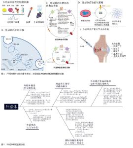
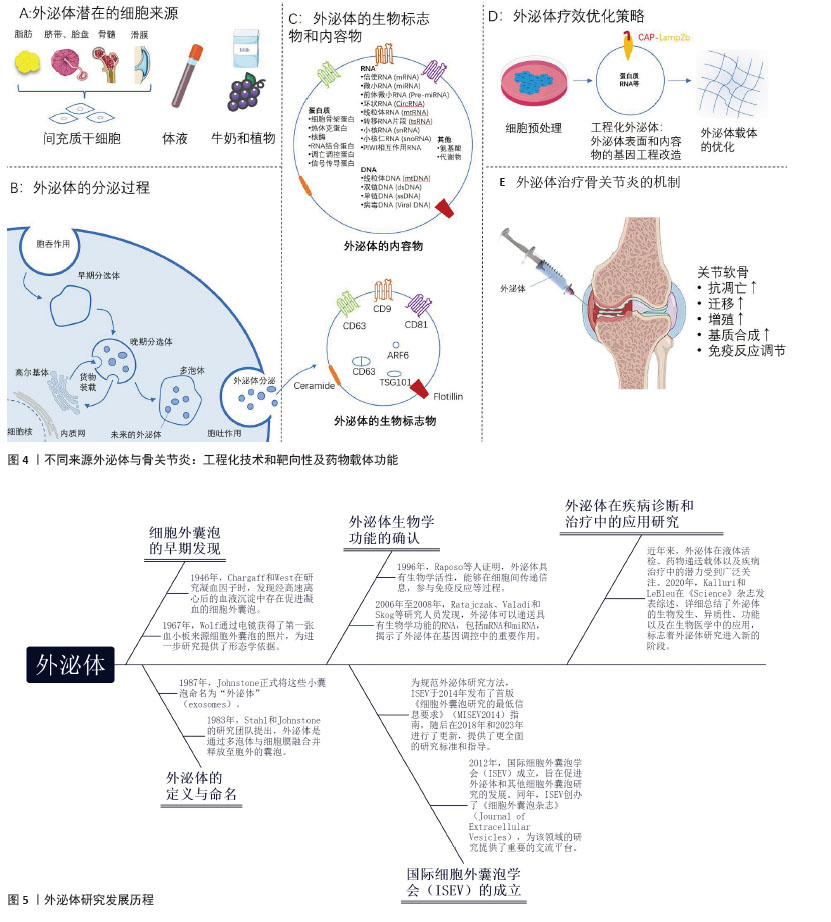
2.1 外泌体的生物学基础 外泌体是细胞释放到外部环境中的直径30-150 nm的膜性小囊泡,由细胞内的多泡体产生。它们存在于血液、尿液、唾液等多种体液中,具有携带RNA、DNA、蛋白质等分子并在细胞间传递信息的能力[10-12]。 外泌体的生物合成过程复杂,涉及内涵体分选复合物依赖和非依赖途径。当多泡体与细胞膜融合时,内含的囊泡被释放为外泌体,随后通过与其他细胞相互作用,影响目标细胞的功能[8,13-14]。外泌体的主要成分包括脂质、蛋白质和核酸,这些成分在免疫调节、疾病发展、组织修复等方面具有重要作用。外泌体具有一些特异性的蛋白可作为生物标志物,如表面蛋白CD9、CD63和CD81及其内容物蛋白ALIX等[8-9,15-16]。 外泌体的提取方法包括超速离心、超滤、尺寸排除色谱和免疫亲和分离。为维持外泌体的生物活性,通常在4 ℃短期保存和-80 ℃长期保存。鉴定外泌体需要结合透射电子显微镜、纳米颗粒跟踪分析和蛋白质印迹技术,以确保研究数据的准确性和可靠性[9]。 在骨关节炎研究中,不同来源(如间充质干细胞、体液、牛奶和植物等)外泌体在功能组成、生物标志物表达、药物递送能力及免疫调节潜能方面存在差异。通过工程化修饰、靶向改造或负载功能分子等手段,可有效提升外泌体在组织修复、抗炎、免疫调控等方面的治疗效应(图4)。外泌体研究经历了从细胞外囊泡的早期观察到生物学功能确认,并逐渐扩展至疾病诊断和治疗的应用探索。随着国际细胞外囊泡学会(International Society for Extracellular Vesicles,ISEV)的成立及外泌体研究规范的提出,外泌体相关研究持续拓展,机制与应用研究不断丰富,临床转化潜力逐步显现(图5)。"
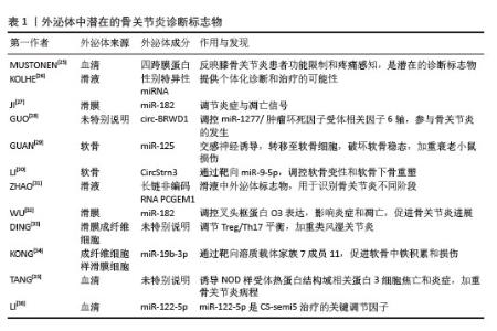
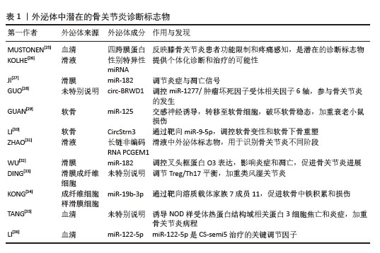
2.2 外泌体在骨关节炎早期诊断中的应用进展 骨关节炎在诊断明确时通常已经出现了显著的病理变化和临床症状,严重影响患者生活且目前暂无延缓骨关节炎发展的治疗方法。若能找到骨关节炎早期的诊断方法,则可早诊早治,减缓病程的发展,有效减少骨关节炎所带来的疾病负担。外泌体中包含的物质如信使RNA、微小RNA(miRNAs)、DNA和蛋白质,其含量通常受到原始细胞类型和状态的影响,这些物质可能成为早期诊断的靶标[17-18]。外泌体存在于大部分体液中,通过无创或微创的方式获取体液标本,从中提取外泌体进行液体活检,可为实时监测疾病的基因突变提供一种可行的方法。 外泌体的诊断潜力已在癌症诊断领域得到验证,目前已有多款试剂盒成功开发并用于临床。尿液外泌体ExoDx Prostate IntelliScore检测是一种非侵入性尿液测试,通过收集尿液中的外泌体,分析其中RNA的表达(PCA3、ERG、SPDEF),从而帮助早期诊断高等级前列腺癌,特别是针对前列腺特异性抗原水平在2-10 ng/mL之间的男性患者,可以减少不必要的组织病理活检[19-20]。ExoDx? Lung (ALK)是第一个基于外泌体RNA的液体活检试剂盒,旨在从血液样本中分离和分析外泌体RNA,标志着非侵入性诊断技术的重要进步。非小细胞肺癌是常见的肺癌类型,其中最常见的基因突变之一是表皮生长因子受体(epidermal growth factor receptor,EGFR)基因突变。CASTELLANOS-RIZALDOS等[21]研究表明,通过结合外泌体RNA和cfDNA可以更准确地检测EGFR T790M突变。KIM等[22]对54份血浆样本和13份胸腔积液中的EGFR突变情况进行比较,结果显示外泌体联合cfDNA能够更有效地检测和监测EGFR T790M突变。KRUG等[23]通过外泌体RNA结合循环肿瘤DNA检测方法提升了液体活检在非小细胞肺癌诊断中的应用潜力。以上研究为外泌体作为骨关节炎早期诊断标记物提供了参考方向。 对于早期骨关节炎,由于病理变化尚不明显,传统诊断方法难以有效检测。然而,通过富集关节滑液中的外泌体,分析外泌体表型和成分,可识别早期骨关节炎相关的生物标志物,有助于骨关节炎早诊早治。关节液和血液是两种较易获取的体液来源。外泌体由多种细胞和组织分泌,其中中性粒细胞和滑膜成纤维细胞分泌的外泌体已在类风湿关节炎和骨关节炎的关节滑液中检测到。SONG等[24]发现类风湿关节炎患者的滑液外泌体因高水平的核因子κB受体活化因子配体而更显著地促进破骨细胞生成,可能携带特异性“破骨细胞生成标志”。 外泌体在骨关节炎诊断与疾病进展研究中备受关注(表1)。MUSTONEN等[25]发现血清中细胞外囊泡的四跨膜蛋白谱可反映患者功能受限与疼痛感知,提示四跨膜蛋白作为潜在生物标志物的可能性。KOLHE等[26]报道多种miRNA在不同性别患者的滑液外泌体中存在显著差异表达,为骨关节炎的个体化诊断与治疗提供思路。JI等[27]和WU等[32]研究均聚焦滑膜特异性外泌体中的miR-182,发现其通过调控炎症和凋亡通路,加速骨关节炎进程。GUO等[28]、LI等[30]与GUAN等[29]则分别揭示了外泌体携带的环状RNA(circ-BRWD1、CircStrn3)和miRNA(miR-125等)如何通过特定分子轴或靶基因,影响软骨稳态、炎症反应及软骨下骨重塑,从而加剧骨关节炎病情。此外,ZHAO等[31]鉴定到滑液来源外泌体长链非编码RNA PCGEM1可作为识别骨关节炎不同分期的标志物,TANG等[35]研究表明血清外泌体能诱导NOD样受体热蛋白结构域相关蛋白3介导的细胞焦亡与炎症,进一步加重骨关节炎。LI等[36]利用生物信息学分析发现miR-122-5p在 CS-semi5治疗骨关节炎中起关键调控作用。由此可见,通过分析滑液、血浆等体液中外泌体的谱系差异,有望为骨关节炎及其他关节疾病的诊断和治疗提供重要理论基础。 "
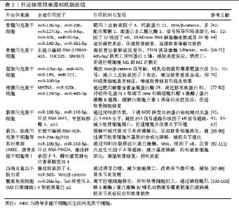
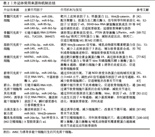
2.3 外泌体在骨关节炎治疗中的研究进展 在骨关节炎治疗中,间充质干细胞来源外泌体是当前研究的热点之一。间充质干细胞在许多临床试验中已展现出治疗潜力,间充质干细胞归巢至软骨缺损部位,通过软骨分化以修复软骨缺损,并通过旁分泌组织修复因子,起到免疫调节、组织修复和再生、抗炎作用[37-38]。然而,间充质干细胞治疗存在一些局限性,包括免疫反应与安全性、间充质干细胞老化、供体和受体的个体差异、潜在的伦理问题、致瘤性等[39-40]。 相比之下,外泌体作为一种细胞外囊泡,具有较低的免疫原性,不存在伦理问题。许多研究已证明,间充质干细胞来源外泌体在骨关节炎治疗中具有积极作用,主要包括促进软骨修复、减少炎症、调节免疫反应以及保护关节软骨和骨质免受进一步退化等[41-51]。间充质干细胞来源多样,包括骨髓、滑膜、脂肪、胎盘、脐带和羊水等。目前,尚未确定哪种间充质干细胞来源外泌体在骨关节炎治疗中的效果和安全性最优。除间充质干细胞外,其他细胞来源外泌体也体现出了骨关节炎治疗潜力。 2.3.1 骨髓间充质干细胞来源外泌体 来源于骨髓间充质干细胞的外泌体治疗骨关节炎具有显著效果。骨髓间充质干细胞来源外泌体可促进软骨修复、减轻炎症及疼痛[42,46,52-54]。例如,JAMMES等[55]研究表明,骨髓间充质干细胞来源外泌体能够显著改善马膝骨关节炎软骨细胞表型,增强软骨功能标志物的表达。CONTENTIN等[56]则通过优化外泌体纯化方法并进行促炎预处理,提高了骨髓间充质干细胞来源外泌体在马骨关节炎中的疗效。 在骨髓间充质干细胞来源外泌体中,miRNA的作用受到广泛关注。CHEN等[57]发现骨髓间充质干细胞来源外泌体中的miR-136-5p 能够抑制创伤性骨关节炎软骨细胞退变,HUANG等[58]指出miR-206通过减少上皮转录因子3的表达促进骨关节炎成骨细胞的增殖和分化。其他miRNA如miR-127-3p[59]、miR-140-3p[60]、miR-361-5p[61]、miR-9-5p[62]、miR-326[63]、miR-95-5p等在调节软骨细胞功能、缓解炎症、促进软骨修复方面发挥了重要作用[64]。此外,骨髓间充质干细胞来源外泌体中的miR-193b-3p和miR-29a也在调节软骨形成及细胞外基质重塑中发挥了关键作用[65-67]。 除了miRNA,长链非编码RNA在骨髓间充质干细胞来源外泌体中的作用也备受关注。骨髓间充质干细胞来源外泌体通过调控LYRM4-AS1/GRPR/miR-6515-5p信号通路[68]、TUC339的表达以及PINK1/Parkin信号通路[69-70],促进M1型巨噬细胞向M2型极化,减轻骨关节炎的炎症反应并促进软骨修复。此外,长链非编码RNA SNHG7通过调节miR-485-5p/FSP1轴,抑制炎症和铁死亡,进一步缓解骨关节炎症状[71]。 综上所述,骨髓间充质干细胞来源外泌体中的多种miRNA和长链非编码RNA通过调节细胞功能、抑制炎症及促进软骨修复,在骨关节炎治疗中发挥重要作用。骨髓间充质干细胞来源外泌体的主要挑战在于供者衰老和细胞增殖能力较低。随着年龄增长,骨髓中间充质干细胞的数量和质量下降,老年供者的骨髓间充质干细胞功能减弱,进而影响外泌体的治疗效果。此外,骨髓间充质干细胞的增殖能力较弱,限制了外泌体的大规模生产,且骨髓样本的获取具有一定侵入性,干细胞来源较为有限。因此,骨髓间充质干细胞来源外泌体在规模化应用中面临产量和一致性问题。 2.3.2 脂肪间充质干细胞来源外泌体 脂肪间充质干细胞来源外泌体在骨关节炎中同样展现出治疗潜力。脂肪间充质干细胞的常用来源有皮下脂肪和髌下脂肪垫,皮下脂肪可通过抽脂术获取,髌下脂肪垫可在膝关节置换术和关节镜手术中获取,这两种术中获取的脂肪组织可作为干细胞提取的良好来源。TOFI?O-VIAN等[51]研究发现脂肪间充质干细胞来源外泌体能够下调骨关节炎成骨细胞的衰老特征。脂肪间充质干细胞来源外泌体中的miRNA亦被广泛研究,LI等[72]研究表明,脂肪间充质干细胞来源外泌体通过转运miR-376c-3p并靶向Wnt/β-catenin信号轴来缓解骨关节炎。MENG等[73]发现脂肪间充质干细胞来源外泌体中的miR-429通过靶向FEZ2的自噬改善骨关节炎软骨损伤。ZHAO等[74]研究发现脂肪间充质干细胞来源外泌体通过上调miR-145和miR-221,促进软骨形成并抑制炎症。WU等[75]研究发现来自髌下脂肪垫间质干细胞的富含miR-100-5p的外泌体能够保护关节软骨,并通过抑制哺乳动物雷帕霉素靶蛋白改善步态异常。此外,过氧化还原酶6在人脂肪间充质干细胞微泡中发挥软骨保护作用。康俊峰等[76]发现脂肪间充质干细胞来源外泌体及膝痹宁Ⅱ均可以通过促进PINK1/Parkin信号通路表达,提高软骨细胞线粒体自噬水平,延缓软骨组织退变,且二者联用可以增强疗效。 脂肪间充质干细胞来源外泌体主要存在脂肪组织异质性和炎症状态问题。脂肪组织在肥胖个体中常伴有慢性低度炎症,这种炎症环境可能改变脂肪间充质干细胞及其外泌体的功能,影响在抗炎和再生治疗中的效果。此外,虽然脂肪组织来源广泛,获取相对容易,但从中分离出高纯度的干细胞仍具有挑战,比如需要避免脂肪前体细胞的混入。 2.3.3 滑膜间充质干细胞来源外泌体 滑膜是位于关节腔内的结缔组织,分泌滑液以润滑和营养关节。滑膜组织通常在膝关节置换手术和关节镜手术中获取,可用于提取滑膜间充质干细胞。LONG等[77]在体内和体外的研究均表明滑膜间充质干细胞来源外泌体通过MATN3可缓解膝骨关节炎。此外,KONG等[78-79]研究发现滑膜间充质干细胞来源外泌体中的miR-320c通过靶向ADAM19依赖的Wnt信号通路,促进软骨损伤修复。QIU等[80]发现人滑膜间充质干细胞来源外泌体介导的miR-129-5p通过靶向高迁移率族蛋白B1缓解白细胞介素1诱导的骨关节炎。QIU等[81]发现滑膜间充质干细胞来源外泌体 miR-485-3p通过靶向神经纤毛蛋白1介导的磷脂酰肌醇3激酶/蛋白激酶B通路缓解骨关节炎软骨损伤。王智超等[82]发现金天格胶囊治疗骨关节炎的药效作用明显,作用机制可能与促进滑膜间充质干细胞来源外泌体靶向关节软骨细胞转运miRNA进而调控Serpinb10、Ntn1、Il1b、Tgm2、Megf10 、Il11、Cd40、Slc15a3、Pou2f2等基因有关。 滑膜间充质干细胞来源外泌体的应用也受到滑膜组织来源的限制,滑膜组织通常需要通过手术获取,尽管比骨髓获取创伤性小,但仍然需要关节镜手术操作,尤其在健康供体中较难获取。此外,不同关节部位的滑膜组织异质性较大,可能导致滑膜间充质干细胞来源外泌体的功能存在差异性,影响治疗效果的稳定性。关节炎可能进一步改变滑膜组织质量,导致外泌体功能受影响。 2.3.4 脐带间充质干细胞来源外泌体 脐带间充质干细胞因获取过程相对简单且无伦理争议,在再生医学和组织工程中得到了广泛研究和应用。脐带间充质干细胞来源外泌体已被证实可通过促进软骨细胞增殖、减少凋亡、减轻炎症、调节巨噬细胞极化及软骨基质代谢相关蛋白的表达,减缓骨关节炎发展[41,43,83-85]。脐带间充质干细胞来源外泌体通过miR-100-5p/NOX4轴抑制活性氧产生和软骨细胞凋亡[86]。此外,研究发现,脐带间充质干细胞来源外泌体可使骨关节炎软骨细胞恢复活力,p53信号通路是其中的关键因素[44] 。人脐带间充质干细胞来源细胞外囊泡通过与甲基转移酶3相互作用降低巨噬细胞NOD样受体热蛋白结构域相关蛋白3的m6A水平,从而缓解小鼠膝骨关节炎[87]。 脐带间充质干细胞来源外泌体由于来源丰富、低免疫原性和广泛的治疗潜力,在再生医学中具有极大的应用前景。然而,要将脐带间充质干细胞来源外泌体广泛应用于临床,仍需要解决外泌体提取、纯化、异质性和规模化生产等方面的技术难题,同时加强对长期效能和安全性的研究。 2.3.5 其他来源外泌体 脐血、胎盘和胚胎来源间充质干细胞外泌体也显示出对骨关节炎的治疗作用。脐血间充质干细胞通过miR-29a-3p/FOS轴分泌的长链非编码RNA H19用于中枢性骨关节炎疼痛敏化[88]。在人胚胎间充质干细胞的研究中,外泌体被证明可以促进骨软骨细胞再生并通过平衡软骨细胞外基质的合成和降解来缓解骨关节炎[50,89]。此外,在大鼠骨关节炎模型中,人胎盘来源外泌体减轻了疼痛负担,恢复了软骨退化,并以剂量依赖的方式下调了促炎、分解代谢或凋亡蛋白的表达[90]。FOTOUHI等[91]证实来自Wharton’s Jelly的外泌体能显著减少滑膜细胞中关键炎症递质的表达,为骨关节炎治疗提供了潜在的抗炎疗效。 除了间充质干细胞外泌体,还有其他细胞和体液可作为外泌体的来源。诱导多能干细胞衍生的间充质干细胞(induced mesenchymal stem cells,iMSCs)结合了诱导多能干细胞的多能性和间充质干细胞的自我更新与多向分化能力,在再生医学、组织工程和细胞治疗等领域具有重要的应用前景。研究发现,与滑膜间充质干细胞来源外泌体相比,诱导多能干细胞衍生的间充质干细胞来源外泌体在骨关节炎治疗中表现出更显著的效果[92]。牙髓干细胞来源外泌体通过抑制瞬时感受器电位香草酸受体4介导的破骨细胞活化,减轻了小鼠膝关节炎[93] 。通过对miR-100-5p/哺乳动物雷帕霉素靶蛋白研究,人脱落乳牙干细胞外泌体在颞下颌关节软骨细胞中的抗炎作用也得到了证实[94]。LIN等[95]研究表明来自人乳牙干细胞的外泌体通过核因子κB途径显著抑制骨关节炎的炎症,促进软骨修复。ZHANG等[96]研究表明骨骼干细胞衍生外泌体通过促进细胞迁移、增加细胞外基质产生和减少炎症反应,有效促进了半月板撕裂的愈合和次发性骨关节炎的改善。 血液也包含大量的外泌体。以血清外泌体为载体,通过搭载激活转录因子4诱导自噬,可以抑制骨关节炎的发展[97]。富血小板血浆在临床上已用于多种疾病的治疗,而其来源外泌体在骨关节炎治疗中也显示出新的潜力。来自富血小板血浆的外泌体介导距下骨关节炎的软骨保护作用[98]。来源于富血小板血浆的外泌体通过Wnt/β-catenin信号通路促进软骨细胞增殖和抑制细胞凋亡[99]。 髓系免疫细胞来源外泌体也可参与骨关节炎的治疗。例如,M2巨噬细胞来源外泌体通过抑制磷脂酰肌醇3激酶/蛋白激酶B/哺乳动物雷帕霉素靶蛋白通路对膝骨关节炎大鼠发挥治疗作用[100]。此外,M2巨噬细胞来源外泌体中的miR-26b-5p通过靶向Toll样受体3和X型胶原蛋白α1调节巨噬细胞极化和软骨细胞肥大,从而缓解骨关节炎[101]。地高辛通过下调滑膜巨噬细胞M1样极化及其衍生的外泌体miR-146b-5p/Usp3&Sox5轴,改善关节炎症微环境[102]。SI等[103]揭示了M1型巨噬细胞来源外泌体中的miR-363通过靶向Ras-GTP酶激活蛋白2显著抑制软骨细胞的增殖和存活,并增强炎症反应。 关节中的组织和细胞分泌的外泌体亦可对骨关节炎产生影响,例如软骨祖细胞、骨细胞和滑膜成纤维细胞。LIU等[104]研究表明,敲低环状RNA PRKCH可以通过调节miR-502-5p/血小板反应蛋白解整合素金属肽酶5轴减轻白细胞介素1β处理的软骨细胞表型变化,而软骨细胞分泌的外泌体能够在细胞间转移环状RNA PRKCH。在小鼠软骨损伤模型中,MRL/MpJ“超级愈合”小鼠软骨源性祖细胞分泌的外泌体可以增强关节软骨修复[105]。XU等[106]研究表明,来自软骨干细胞/祖细胞的外泌体能显著促进亚急性骨关节炎大鼠膝关节软骨修复,这主要得益于外泌体中的周期蛋白依赖性激酶9,它对软骨细胞的生长和迁移起关键作用。骨细胞来源外泌体通过调节DLX2/Wnt通路,缓解骨关节炎,并介导软骨修复。滑膜成纤维细胞来源外泌体miR-214-3p抑制骨关节炎大鼠软骨组织的炎症和变性[107]。滑液外泌体来源miR-182-5p通过下调肿瘤坏死因子α诱导蛋白8和通过微管相关蛋白轻链 3信号促进自噬来缓解骨关节炎[27]。 此外, LIU等[108]研究表明过表达 miR-140-5p人尿源干细胞来源外泌体通过下调血管内皮生长因子A缓解大鼠膝骨关节炎。鹿茸也可作为一种干细胞来源,其外泌体可以缓解间充质干细胞衰老和骨关节炎[109]。来源于宿主和肠道微生物群的细胞外囊泡可作为靶向治疗骨质疏松和骨关节炎的有前途的纳米载体[110]。LIU等[111]证实大蒜衍生外泌体能通过抑制丝裂原活化蛋白激酶信号通路,减少软骨基质的降解和炎症,从而在体内外模型中有效缓解骨关节炎。 综上所述,外泌体通过多种机制发挥作用,包括促进软骨细胞增殖、减少凋亡、减轻炎症、调节巨噬细胞极化以及调控软骨基质代谢等。它们不仅直接作用于受损的软骨细胞,还通过调节免疫和炎症反应间接改善关节环境。尽管干细胞外泌体在骨关节炎治疗中展现出广阔的前景,但仍面临一些挑战。首先,需进一步深入研究外泌体的具体分子机制,以明确各类外泌体中关键成分的作用模式;其次,间充质干细胞及其外泌体的制备、纯化和质量控制方法亟需进一步优化,以确保在临床应用中的安全性和有效性;此外,尽管已有研究探讨了不同来源外泌体在骨关节炎治疗中的疗效和机制,但目前关于不同来源外泌体疗效的系统性比较研究尚不充分。因此,未来应开展更全面的研究,以系统评估不同来源外泌体的疗效和机制差异。由于外泌体的提取方法和使用剂量尚未统一,这可能导致临床效果的变异,因此标准化外泌体的提取、纯化过程及剂量使用,也是未来研究的方向。表2对不同来源外泌体进行归纳讨论。"
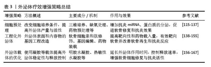
2.4 外泌体改善骨关节炎疗效的增强策略 天然外泌体在骨关节炎治疗中的效果和机制已得到广泛研究。然而,由于外泌体的治疗效果受到多种因素的影响,具不可控性,这是其临床应用的重要障碍之一。将药物递送到软骨细胞,仍然是骨关节炎分子治疗的一个重大挑战[112]。外泌体在治疗中需要满足高产量、靶向性、稳定性和有效性的要求。近年来,工程化外泌体因在生物医学领域的巨大潜力而备受关注。与天然外泌体相比,工程化外泌体通过表面修饰和内部治疗分子的修饰,具备更强的靶向性、更高的载药能力以及更优越的治疗效果[113]。对外泌体来源细胞的预处理可以提高外泌体的产量。优化外泌体的载体可有助于保持稳定性。随着生物材料科学的发展,凝胶作为外泌体的稳定载体,通过延长外泌体的作用时间、控制释放速率和增强靶向性,显著提高了外泌体的治疗效果[114]。以下就外泌体在骨关节炎治疗中疗效增强策略进行综述(表3)。 "
| [1] HUNTER DJ, BIERMA-ZEINSTRA S. Osteoarthritis. Lancet. 2019;393(10182):1745-1759. [2] LOESER RF, GOLDRING SR, SCANZELLO CR, et al. Osteoarthritis: a disease of the joint as an organ. Arthritis Rheum. 2012;64(6):1697-1707. [3] PERRUCCIO AV, YOUNG JJ, WILFONG JM, et al. Osteoarthritis year in review 2023: Epidemiology & therapy. Osteoarthritis Cartilage. 2024;32(2): 159-165. [4] GBD 2021 OSTEOARTHRITIS COLLABORATORS. Global, regional, and national burden of osteoarthritis, 1990-2020 and projections to 2050: a systematic analysis for the Global Burden of Disease Study 2021. Lancet Rheumatol. 2023; 5(9):e508-e522. [5] KATZ JN, ARANT KR, LOESER RF. Diagnosis and Treatment of Hip and Knee Osteoarthritis: A Review. JAMA. 2021;325(6):568. [6] GLYN-JONES S, PALMER AJ, AGRICOLA R, et al. Osteoarthritis. Lancet. 2015;386(9991):376-387. [7] NI Z, ZHOU S, LI S, et al. Exosomes: roles and therapeutic potential in osteoarthritis. Bone Res. 2020;8(1):25. [8] HE C, ZHENG S, LUO Y, et al. Exosome Theranostics: Biology and Translational Medicine. Theranostics. 2018;8(1):237-255. [9] ZHANG Y, BI J, HUANG J, et al. Exosome: A Review of Its Classification, Isolation Techniques, Storage, Diagnostic and Targeted Therapy Applications. Int J Nanomedicine. 2020;15:6917-6934. [10] KALLURI R, LEBLEU VS. The biology, function, and biomedical applications of exosomes. Science. 2020;367(6478):eaau6977. [11] VAN NIEL G, D’ANGELO G, RAPOSO G. Shedding light on the cell biology of extracellular vesicles. Nat Rev Mol Cell Biol. 2018;19(4):213-228. [12] KUMAR MA, BABA SK, SADIDA HQ, et al. Extracellular vesicles as tools and targets in therapy for diseases. Signal Transduct Target Ther. 2024;9(1):27. [13] WOLLERT T, HURLEY JH. Molecular mechanism of multivesicular body biogenesis by ESCRT complexes. Nature. 2010;464(7290):864-869. [14] MCKELVEY KJ, POWELL KL, ASHTON AW, et al. Exosomes: Mechanisms of Uptake. J Circ Biomark. 2015;4:7. [15] HAN QF, LI WJ, HU KS, et al. Exosome biogenesis: machinery, regulation, and therapeutic implications in cancer. Mol Cancer. 2022;21(1):207. [16] LIU C, XIA C, XIA C. Biology and function of exosomes in tumor immunotherapy. Biomed Pharmacother. 2023;169:115853. [17] CHEN A, CHEN Y, RONG X, et al. The application of exosomes in the early diagnosis and treatment of osteoarthritis. Front Pharmacol. 2023;14:1154135. [18] FAN WJ, LIU D, PAN LY, et al. Exosomes in osteoarthritis: Updated insights on pathogenesis, diagnosis, and treatment. Front Cell Dev Biol. 2022;10:949690. [19] MCKIERNAN J, DONOVAN MJ, O’NEILL V, et al. A Novel Urine Exosome Gene Expression Assay to Predict High-grade Prostate Cancer at Initial Biopsy. JAMA Oncol. 2016;2(7):882. [20] MCKIERNAN J, DONOVAN MJ, MARGOLIS E, et al. A Prospective Adaptive Utility Trial to Validate Performance of a Novel Urine Exosome Gene Expression Assay to Predict High-grade Prostate Cancer in Patients with Prostate-specific Antigen 2–10 ng/ml at Initial Biopsy. Eur Urol. 2018;74(6):731-738. [21] CASTELLANOS-RIZALDOS E, GRIMM DG, TADIGOTLA V, et al. Exosome-Based Detection of EGFR T790M in Plasma from Non-Small Cell Lung Cancer Patients. Clin Cancer Res. 2018; 24(12):2944-2950. [22] KIM Y, SHIN S, LEE KA. Exosome-based detection of EGFR T790M in plasma and pleural fluid of prospectively enrolled non-small cell lung cancer patients after first-line tyrosine kinase inhibitor therapy. Cancer Cell Int. 2021;21(1):50. [23] KRUG AK, ENDERLE D, KARLOVICH C, et al. Improved EGFR mutation detection using combined exosomal RNA and circulating tumor DNA in NSCLC patient plasma. Ann Oncol. 2018; 29(3):700-706. [24] SONG JE, KIM JS, SHIN JH, et al. Role of Synovial Exosomes in Osteoclast Differentiation in Inflammatory Arthritis. Cells. 2021;10(1):120. [25] MUSTONEN AM, PALVIAINEN M, SÄISÄNEN L, et al. Tetraspanin profiles of serum extracellular vesicles reflect functional limitations and pain perception in knee osteoarthritis. Arthritis Res Ther. 2024;26(1):33. [26] KOLHE R, HUNTER M, LIU S, et al. Gender-specific differential expression of exosomal miRNA in synovial fluid of patients with osteoarthritis. Sci Rep. 2017;7(1):2029. [27] JI Y, XIONG L, ZHANG G, et al. Synovial fluid exosome-derived miR-182-5p alleviates osteoarthritis by downregulating TNFAIP8 and promoting autophagy through LC3 signaling. Int Immunopharmacol. 2023;125(Pt A):111177. [28] GUO Z, WANG H, ZHAO F, et al. Exosomal circ-BRWD1 contributes to osteoarthritis development through the modulation of miR-1277/TRAF6 axis. Arthritis Res Ther. 2021;23(1):159. [29] GUAN Z, LIU Y, LUO L, et al. Sympathetic innervation induces exosomal miR-125 transfer from osteoarthritic chondrocytes, disrupting subchondral bone homeostasis and aggravating cartilage damage in aging mice. J Adv Res. 2025; 69:245-260. [30] LI B, DING T, CHEN H, et al. CircStrn3 targeting microRNA-9-5p is involved in the regulation of cartilage degeneration and subchondral bone remodelling in osteoarthritis. Bone Jt Res. 2023; 12(1):33-45. [31] ZHAO Y, XU J. Synovial fluid-derived exosomal lncRNA PCGEM1 as biomarker for the different stages of osteoarthritis. Int Orthop. 2018;42(12): 2865-2872. [32] WU S, LUO J, ZHANG X, et al. Synovia tissue-specific exosomes participate in the dual variation of the osteoarthritis microenvironment via miR-182. Exp Cell Res. 2024;436(2):113981. [33] DING Y, WANG L, WU H, et al. Exosomes derived from synovial fibroblasts under hypoxia aggravate rheumatoid arthritis by regulating Treg/Th17 balance. Exp Biol Med Maywood NJ. 2020;245(14):1177-1186. [34] KONG R, JI L, PANG Y, et al. Exosomes from osteoarthritic fibroblast-like synoviocytes promote cartilage ferroptosis and damage via delivering microRNA-19b-3p to target SLC7A11 in osteoarthritis. Front Immunol. 2023;14:1181156. [35] TANG Z, SHU L, CAO Z, et al. Osteoarthritis rat serum-derived extracellular vesicles aggravate osteoarthritis development by inducing NLRP3-mediated pyroptotic cell death and cellular inflammation. Hum Cell. 2024;37(6):1624-1637. [36] LI X, ZHOU Y, CHEN X, et al. Semi-synthetic chondroitin sulfate CS-semi5 upregulates miR-122-5p, conferring a therapeutic effect on osteoarthritis via the p38/MMP13 pathway. Acta Pharm Sin B. 2024;14(8):3528-3542. [37] GIORGINO R, ALBANO D, FUSCO S, et al. Knee Osteoarthritis: Epidemiology, Pathogenesis, and Mesenchymal Stem Cells: What Else Is New? An Update. Int J Mol Sci. 2023;24(7):6405. [38] ZHANG Z, ZHAO S, SUN Z, et al. Enhancement of the therapeutic efficacy of mesenchymal stem cell-derived exosomes in osteoarthritis. Cell Mol Biol Lett. 2023;28(1):75. [39] YANG X, MENG Y, HAN Z, et al. Mesenchymal stem cell therapy for liver disease: full of chances and challenges. Cell Biosci. 2020;10(1):123. [40] ZHANG Y, LIU K, MA X, et al. Therapeutic Effects of Puerarin Loaded Bone Marrow Mesenchymal Stem Cell‐derived Exosomes in a Rat Model of Osteoarthritis. Chem Biodivers. 2024;e202402095. [41] ZHANG S, CHUAH SJ, LAI RC, et al. MSC exosomes mediate cartilage repair by enhancing proliferation, attenuating apoptosis and modulating immune reactivity. Biomaterials. 2018; 156:16-27. [42] HE L, HE T, XING J, et al. Bone marrow mesenchymal stem cell-derived exosomes protect cartilage damage and relieve knee osteoarthritis pain in a rat model of osteoarthritis. Stem Cell Res Ther. 2020;11(1):276. [43] ZHANG S, TEO KYW, CHUAH SJ, et al. MSC exosomes alleviate temporomandibular joint osteoarthritis by attenuating inflammation and restoring matrix homeostasis. Biomaterials. 2019;200:35-47. [44] CAO H, CHEN M, CUI X, et al. Cell-Free Osteoarthritis Treatment with Sustained-Release of Chondrocyte-Targeting Exosomes from Umbilical Cord-Derived Mesenchymal Stem Cells to Rejuvenate Aging Chondrocytes. ACS Nano. 2023;17(14):13358-13376. [45] ZHANG J, RONG Y, LUO C, et al. Bone marrow mesenchymal stem cell-derived exosomes prevent osteoarthritis by regulating synovial macrophage polarization. Aging (Albany NY). 2020;12(24):25138-25152. [46] JIN Y, XU M, ZHU H, et al. Therapeutic effects of bone marrow mesenchymal stem cells-derived exosomes on osteoarthritis. J Cell Mol Med. 2021;25(19):9281-9294. [47] KWON DG, KIM MK, JEON YS, et al. State of the Art: The Immunomodulatory Role of MSCs for Osteoarthritis. Int J Mol Sci. 2022;23(3):1618. [48] BEI HP, HUNG PM, YEUNG HL, et al. Bone-a-Petite: Engineering Exosomes towards Bone, Osteochondral, and Cartilage Repair. Small. 2021; 17(50):e2101741. [49] COSENZA S, RUIZ M, TOUPET K, et al. Mesenchymal stem cells derived exosomes and microparticles protect cartilage and bone from degradation in osteoarthritis. Sci Rep. 2017;7(1):16214. [50] WANG Y, YU D, LIU Z, et al. Exosomes from embryonic mesenchymal stem cells alleviate osteoarthritis through balancing synthesis and degradation of cartilage extracellular matrix. Stem Cell Res Ther. 2017;8(1):189. [51] TOFIÑO-VIAN M, GUILLÉN MI, PÉREZ DEL CAZ MD, et al. Extracellular Vesicles from Adipose-Derived Mesenchymal Stem Cells Downregulate Senescence Features in Osteoarthritic Osteoblasts. Oxid Med Cell Longev. 2017;2017:7197598. [52] CHENG S, XU X, WANG R, et al. Chondroprotective effects of bone marrow mesenchymal stem cell-derived exosomes in osteoarthritis. J Bioenerg Biomembr. 2024;56(1):31-44. [53] ZHOU X, LIANG H, HU X, et al. BMSC-derived exosomes from congenital polydactyly tissue alleviate osteoarthritis by promoting chondrocyte proliferation. Cell Death Discov. 2020;6(1):142. [54] ZHOU L, YE H, LIU L, et al. Human Bone Mesenchymal Stem Cell-Derived Exosomes Inhibit IL-1β-Induced Inflammation in Osteoarthritis Chondrocytes. Cell J. 2021;23(4):485-494. [55] JAMMES M, CASSÉ F, VELOT E, et al. Pro-Inflammatory Cytokine Priming and Purification Method Modulate the Impact of Exosomes Derived from Equine Bone Marrow Mesenchymal Stromal Cells on Equine Articular Chondrocytes. Int J Mol Sci. 2023;24(18):14169. [56] CONTENTIN R, JAMMES M, BOURDON B, et al. Bone Marrow MSC Secretome Increases Equine Articular Chondrocyte Collagen Accumulation and Their Migratory Capacities. Int J Mol Sci. 2022;23(10):5795. [57] CHEN X, SHI Y, XUE P, et al. Mesenchymal stem cell-derived exosomal microRNA-136-5p inhibits chondrocyte degeneration in traumatic osteoarthritis by targeting ELF3. Arthritis Res Ther. 2020;22(1):256. [58] HUANG Y, ZHANG X, ZHAN J, et al. Bone marrow mesenchymal stem cell-derived exosomal miR-206 promotes osteoblast proliferation and differentiation in osteoarthritis by reducing Elf3. J Cell Mol Med. 2021;25(16):7734-7745. [59] DONG J, LI L, FANG X, et al. Exosome-Encapsulated microRNA-127-3p Released from Bone Marrow-Derived Mesenchymal Stem Cells Alleviates Osteoarthritis Through Regulating CDH11-Mediated Wnt/β-Catenin Pathway. J Pain Res. 2021;14:297-310. [60] HU Y, LIU HX, XU D, et al. The Anti-Inflammatory Effect of miR-140-3p in BMSCs-Exosomes on Osteoarthritis. Acta Chir Orthop Traumatol Cech. 2023;90(4):267-276. [61] TAO Y, ZHOU J, WANG Z, et al. Human bone mesenchymal stem cells-derived exosomal miRNA-361-5p alleviates osteoarthritis by downregulating DDX20 and inactivating the NF-κB signaling pathway. Bioorg Chem. 2021;113:104978. [62] JIN Z, REN J, QI S. Exosomal miR-9-5p secreted by bone marrow-derived mesenchymal stem cells alleviates osteoarthritis by inhibiting syndecan-1. Cell Tissue Res. 2020;381(1):99-114. [63] XU H, XU B. BMSC-Derived Exosomes Ameliorate Osteoarthritis by Inhibiting Pyroptosis of Cartilage via Delivering miR-326 Targeting HDAC3 and STAT1//NF-κB p65 to Chondrocytes. Mediators Inflamm. 2021;2021:9972805. [64] MAO G, HU S, ZHANG Z, et al. Exosomal miR-95-5p regulates chondrogenesis and cartilage degradation via histone deacetylase 2/8. J Cell Mol Med. 2018;22(11):5354-5366. [65] MENG F, LI Z, ZHANG Z, et al. MicroRNA-193b-3p regulates chondrogenesis and chondrocyte metabolism by targeting HDAC3. Theranostics. 2018;8(10):2862-2883. [66] YANG F, XIONG WQ, et al. Extracellular vesicles derived from mesenchymal stem cells mediate extracellular matrix remodeling in osteoarthritis through the transport of microRNA-29a. World J Stem Cells. 2024;16(2):191-206. [67] EL-DIN SS, ABOULHODA BE, HASSOUNA A, et al. The Role of Intra-Articular Delivery of BM-MSCs-Derived Exosomes in Improving Osteoarthritis: Implication of circYAP1/miRNA-21/TLR7 Axis. Discov Med. 2024;36(186):1420-1429. [68] WANG X, LI Z, CUI Y, et al. Exosomes Isolated From Bone Marrow Mesenchymal Stem Cells Exert a Protective Effect on Osteoarthritis via lncRNA LYRM4-AS1-GRPR-miR-6515-5p. Front Cell Dev Biol. 2021;9:644380. [69] SHEN X, QIN J, WEI Z, et al. Bone marrow mesenchymal stem cell exosome-derived lncRNA TUC339 influences the progression of osteoarthritis by regulating synovial macrophage polarization and chondrocyte apoptosis. Biomed Pharmacother. 2023;167:115488. [70] LI B, SHEN E, WU Z, et al. BMSC-Derived Exosomes Attenuate Rat Osteoarthritis by Regulating Macrophage Polarization through PINK1/Parkin Signaling Pathway. Cartilage. 2024: 19476035241245805. [71] WANG Y, HU K, LIAO C, et al. Exosomes-Shuttled lncRNA SNHG7 by Bone Marrow Mesenchymal Stem Cells Alleviates Osteoarthritis Through Targeting miR-485-5p/FSP1 Axis-Mediated Chondrocytes Ferroptosis and Inflammation. Tissue Eng Regen Med. 2024;21(8):1203-1216. [72] LI F, XU Z, XIE Z, et al. Adipose mesenchymal stem cells-derived exosomes alleviate osteoarthritis by transporting microRNA -376c-3p and targeting the WNT-beta-catenin signaling axis. Apoptosis. 2023;28(3-4):362-378. [73] MENG C, NA Y, HAN C, et al. Exosomal miR-429 derived from adipose-derived stem cells ameliorated chondral injury in osteoarthritis via autophagy by targeting FEZ2. Int Immunopharmacol. 2023;120:110315. [74] ZHAO C, CHEN JY, PENG WM, et al. Exosomes from adipose derived stem cells promote chondrogenesis and suppress inflammation by upregulating miR 145 and miR 221. Mol Med Rep. 2020;21(4):1881-1889. [75] WU J, KUANG L, CHEN C, et al. miR-100-5p-abundant exosomes derived from infrapatellar fat pad MSCs protect articular cartilage and ameliorate gait abnormalities via inhibition of mTOR in osteoarthritis. Biomaterials. 2019;206: 87-100. [76] 康俊峰,揭立士,伏厚宇,等.膝痹宁Ⅱ联合脂肪干细胞外泌体调控线粒体自噬改善膝OA的机制[J].中国实验方剂学杂志,2024,30(11): 111-119. [77] LONG L, ZOU G, CHENG Y, et al. MATN3 delivered by exosome from synovial mesenchymal stem cells relieves knee osteoarthritis: Evidence from in vitro and in vivo studies. J Orthop Translat. 2023;41: 20-32. [78] KONG R, GAO J, ZHANG J, et al. Synovial mesenchymal stem cell-derived exosomal miR-320c enhances chondrogenesis by targeting ADAM19. Future Med Chem. 2022;14(2):81-96. [79] KONG R, ZHANG J, JI L, et al. Synovial mesenchymal stem cell-derived exosomal microRNA-320c facilitates cartilage damage repair by targeting ADAM19-dependent Wnt signalling in osteoarthritis rats. Inflammopharmacology. 2023;31(2):915-926. [80] QIU M, LIU D, FU Q. MiR-129-5p shuttled by human synovial mesenchymal stem cell-derived exosomes relieves IL-1β induced osteoarthritis via targeting HMGB1. Life Sci. 2021;269:118987. [81] QIU M, XIE Y, TAN G, et al. Synovial mesenchymal stem cell-derived exosomal miR-485-3p relieves cartilage damage in osteoarthritis by targeting the NRP1-mediated PI3K/Akt pathway: Exosomal miR-485-3p relieves cartilage damage. Heliyon. 2024;10(2):e24042. [82] 王智超, 张雪, 张小飞,等.基于转录组测序探讨金天格胶囊调控滑膜间充质干细胞外泌体miRNA和关节软骨细胞mRNA治疗大鼠OA的作用机制[J].中国现代应用药学,2024, 41(11):1464-1475. [83] WANG S, JIANG W, LV S, et al. Human umbilical cord mesenchymal stem cells-derived exosomes exert anti-inflammatory effects on osteoarthritis chondrocytes. Aging (Albany NY). 2023;15(18):9544-9560. [84] LI P, LV S, JIANG W, et al. Exosomes derived from umbilical cord mesenchymal stem cells protect cartilage and regulate the polarization of macrophages in osteoarthritis. Ann Transl Med. 2022;10(18):976. [85] YANG H, ZHOU Y, YING B, et al. Effects of human umbilical cord mesenchymal stem cell-derived exosomes in the rat osteoarthritis models. Stem Cells Transl Med. 2024;13(8):803-811. [86] LI X, WANG Y, CAI Z, et al. Exosomes from human umbilical cord mesenchymal stem cells inhibit ROS production and cell apoptosis in human articular chondrocytes via the miR-100-5p/NOX4 axis. Cell Biol Int. 2021;45(10):2096-2106. [87] ZHOU H, SHEN X, YAN C, et al. Extracellular vesicles derived from human umbilical cord mesenchymal stem cells alleviate osteoarthritis of the knee in mice model by interacting with METTL3 to reduce m6A of NLRP3 in macrophage. Stem Cell Res Ther. 2022;13(1):322. [88] YANG Q, YAO Y, ZHAO D, et al. LncRNA H19 secreted by umbilical cord blood mesenchymal stem cells through microRNA-29a-3p/FOS axis for central sensitization of pain in advanced osteoarthritis. Am J Transl Res. 2021;13(3):1245-1256. [89] ZHANG S, CHU WC, LAI RC, et al. Exosomes derived from human embryonic mesenchymal stem cells promote osteochondral regeneration. Osteoarthritis Cartilage. 2016;24(12):2135-2140. [90] HUANG C, ZHAO Y, LIN S, et al. Characterization of human placenta-derived exosome (pExo) as a potential osteoarthritis disease modifying therapeutic. Arthritis Res Ther. 2023;25(1):229. [91] FOTOUHI A, HOSSEINI M, AGHEBATI-MALEKI A, et al. The Impact of Wharton’s Jelly-derived Exosomes on the Production of Inflammatory Mediators from HIG-82 Synoviocytes. Iran J Immunol. 2024;21(3):243-254. [92] ZHU Y, WANG Y, ZHAO B, et al. Comparison of exosomes secreted by induced pluripotent stem cell-derived mesenchymal stem cells and synovial membrane-derived mesenchymal stem cells for the treatment of osteoarthritis. Stem Cell Res Ther. 2017;8(1):64. [93] FU Y, CUI S, ZHOU Y, et al. Dental Pulp Stem Cell-Derived Exosomes Alleviate Mice Knee Osteoarthritis by Inhibiting TRPV4-Mediated Osteoclast Activation. Int J Mol Sci. 2023;24(5): 4926. [94] LUO P, JIANG C, JI P, et al. Exosomes of stem cells from human exfoliated deciduous teeth as an anti-inflammatory agent in temporomandibular joint chondrocytes via miR-100-5p/mTOR. Stem Cell Res Ther. 2019;10(1):216. [95] LIN CY, NARUPHONTJIRAKUL P, HUANG TY, et al. The Exosomes of Stem Cells from Human Exfoliated Deciduous Teeth Suppress Inflammation in Osteoarthritis. Int J Mol Sci. 2024;25(16):8560. [96] ZHANG FX, DOU Y, ZHANG B, et al. Skeletal Stem Cell-Derived Exosomes Promote Meniscal Tear Healing and Ameliorate Secondary Osteoarthritis. Am J Sports Med. 2024;52(10):2512-2523. [97] WANG Y, HE SH, LIANG X, et al. ATF4-modified serum exosomes derived from osteoarthritic mice inhibit osteoarthritis by inducing autophagy. IUBMB Life. 2021;73(1):146-158. [98] ZHANG Y, WANG X, CHEN J, et al. Exosomes derived from platelet-rich plasma administration in site mediate cartilage protection in subtalar osteoarthritis. J Nanobiotechnology. 2022; 20(1):56. [99] LIU X, WANG L, MA C, et al. Exosomes derived from platelet-rich plasma present a novel potential in alleviating knee osteoarthritis by promoting proliferation and inhibiting apoptosis of chondrocyte via Wnt/β-catenin signaling pathway. J Orthop Surg Res. 2019;14(1):470. [100] DA-WA ZX, JUN M, CHAO-ZHENG L, et al. Exosomes Derived from M2 Macrophages Exert a Therapeutic Effect via Inhibition of the PI3K/AKT/mTOR Pathway in Rats with Knee Osteoarthritic. Biomed Res Int. 2021;2021:7218067. [101] QIAN Y, CHU G, ZHANG L, et al. M2 macrophage-derived exosomal miR-26b-5p regulates macrophage polarization and chondrocyte hypertrophy by targeting TLR3 and COL10A1 to alleviate osteoarthritis. J Nanobiotechnology. 2024;22(1):72. [102] JIA H, DUAN L, YU P, et al. Digoxin ameliorates joint inflammatory microenvironment by downregulating synovial macrophage M1-like-polarization and its-derived exosomal miR-146b-5p/Usp3&Sox5 axis. Int Immunopharmacol. 2022;111:109135. [103] SI W, WEI H, CHEN W, et al. Exosomal microRNA-363 mediates the destructive effect of M1 macrophages on chondrocytes by repressing G3BP2. Exp Cell Res. 2024;442(2):114276. [104] LIU Z, CAO J, ZHANG L, et al. Knockdown of circ-PRKCH alleviates IL-1β-treated chondrocyte cell phenotypic changes through modulating miR-502-5p/ADAMTS5 axis. Autoimmunity. 2022;55(3):179-191. [105] WANG R, JIANG W, ZHANG L, et al. Intra-articular delivery of extracellular vesicles secreted by chondrogenic progenitor cells from MRL/MpJ superhealer mice enhances articular cartilage repair in a mouse injury model. Stem Cell Res Ther. 2020;11(1):93. [106] XU W, ZHANG Y, LI L, et al. Osteocyte-derived exosomes regulate the DLX2/wnt pathway to alleviate osteoarthritis by mediating cartilage repair. Autoimmunity. 2024;57(1):2364686. [107] LAI C, LIAO B, PENG S, et al. Synovial fibroblast-miR-214-3p-derived exosomes inhibit inflammation and degeneration of cartilage tissues of osteoarthritis rats. Mol Cell Biochem. 2023;478(3):637-649. [108] LIU Y, ZENG Y, SI HB, et al. Exosomes Derived From Human Urine-Derived Stem Cells Overexpressing miR-140-5p Alleviate Knee Osteoarthritis Through Downregulation of VEGFA in a Rat Model. Am J Sports Med. 2022;50(4):1088-1105. [109] LEI J, JIANG X, LI W, et al. Exosomes from antler stem cells alleviate mesenchymal stem cell senescence and osteoarthritis. Protein Cell. 2022;13(3):220-226. [110] CHEUNG KCP, JIAO M, XINGXUAN C, et al. Extracellular vesicles derived from host and gut microbiota as promising nanocarriers for targeted therapy in osteoporosis and osteoarthritis. Front Pharmacol. 2023;13:1051134. [111] LIU Y, NIE M, LI X, et al. Garlic-derived Exosomes Alleviate Osteoarthritis Through Inhibiting the MAPK Signaling Pathway. Appl Biochem Biotechnol. 2025;197(1):518-533. [112] DUAN L, XU X, XU L, et al. Exosome-Mediated Drug Delivery for Cell-Free Therapy of Osteoarthritis. Curr Med Chem. 2021;28(31):6458-6483. [113] LIANG Y, DUAN L, LU J, et al. Engineering exosomes for targeted drug delivery. Theranostics. 2021;11(7):3183-3195. [114] SADEGHI S, TEHRANI FR, TAHMASEBI S, et al. Exosome engineering in cell therapy and drug delivery. Inflammopharmacology. 2023;31(1): 145-169. [115] LEE SY, LEE JW. 3D Spheroid Cultures of Stem Cells and Exosome Applications for Cartilage Repair. Life (Basel). 2022;12(7):939. [116] DUAN L, LI X, XU X, et al. Large-scale Preparation of Synovial Fluid Mesenchymal Stem Cell-Derived Exosomes by 3D Bioreactor Culture. J Vis Exp. 2022;(185). doi: 10.3791/62221. [117] SAKAMOTO T, FUKU A, HORIE T, et al. A novel cell source for therapy of knee osteoarthritis using atelocollagen microsphere-adhered adipose-derived stem cells: Impact of synovial fluid exposure on cell activity. Regen Ther. 2024;27: 408-418. [118] ZHANG Y, QI G, YAN Y, et al. Exosomes derived from bone marrow mesenchymal stem cells pretreated with decellularized extracellular matrix enhance the alleviation of osteoarthritis through miR-3473b/phosphatase and tensin homolog axis. J Gene Med. 2023;25(8):e3510. [119] PFEIFER JPH, STIEVANI FC, FERNANDES CJDC, et al. Influence of inflammation on the expression of microRNA-140 in extracellular vesicles from 2D and 3D culture models of synovial-membrane-derived stem cells. Front Bioeng Biotechnol. 2024; 12:1416694. [120] HAN Y, REN J, BAI Y, et al. Exosomes from hypoxia-treated human adipose-derived mesenchymal stem cells enhance angiogenesis through VEGF/VEGF-R. Int J Biochem Cell Biol. 2019;109:59-68. [121] ZHAO J, SUN Y, SHENG X, et al. Hypoxia-treated adipose mesenchymal stem cell-derived exosomes attenuate lumbar facet joint osteoarthritis. Mol Med. 2023;29(1):120. [122] XIA P, WANG Q, SONG J, et al. Low-Intensity Pulsed Ultrasound Enhances the Efficacy of Bone Marrow-Derived MSCs in Osteoarthritis Cartilage Repair by Regulating Autophagy-Mediated Exosome Release. Cartilage. 2022;13(2):19476035221093060. [123] 徐扬,王谦,汪香秀,等.脉冲电磁场调控间充质干细胞外泌体抑制软骨细胞凋亡的研究[J].生物医学工程学杂志,2023,40(1):95-103. [124] YANG YH, WEN CS, KUO YL, et al. GuiLu-ErXian Glue extract promotes mesenchymal stem cells (MSC)-Induced chondrogenesis via exosomes release and delays aging in the MSC senescence process. J Ethnopharmacol. 2023;317:116784. [125] CAI J, XU J, YE Z, et al. Exosomes Derived From Kartogenin-Preconditioned Mesenchymal Stem Cells Promote Cartilage Formation and Collagen Maturation for Enthesis Regeneration in a Rat Model of Chronic Rotator Cuff Tear. Am J Sports Med. 2023;51(5):1267-1276. [126] LOU C, JIANG H, LIN Z, et al. MiR-146b-5p enriched bioinspired exosomes derived from fucoidan-directed induction mesenchymal stem cells protect chondrocytes in osteoarthritis by targeting TRAF6. J Nanobiotechnology. 2023;21(1):486. [127] QIU B, XU X, YI P, et al. Curcumin reinforces MSC-derived exosomes in attenuating osteoarthritis via modulating the miR-124/NF-kB and miR-143/ROCK1/TLR9 signalling pathways. J Cell Mol Med. 2020;24(18):10855-10865. [128] SHAO LT, LUO L, QIU JH, et al. PTH (1-34) enhances the therapeutic effect of bone marrow mesenchymal stem cell-derived exosomes by inhibiting proinflammatory cytokines expression on OA chondrocyte repair in vitro. Arthritis Res Ther. 2022;24(1):96. [129] DONG S, XU G, LI X, et al. Exosomes Derived from Quercetin-Treated Bone Marrow Derived Mesenchymal Stem Cells Inhibit the Progression of Osteoarthritis Through Delivering miR-124-3p to Chondrocytes. DNA Cell Biol. 2024;43(2):85-94. [130] WANG R, XU B, XU H. TGF-β1 promoted chondrocyte proliferation by regulating Sp1 through MSC-exosomes derived miR-135b. Cell Cycle. 2018;17(24):2756-2765. [131] WANG R, XU B. TGFβ1-modified MSC-derived exosome attenuates osteoarthritis by inhibiting PDGF-BB secretion and H-type vessel activity in the subchondral bone. Acta Histochem. 2022; 124(7):151933. [132] WANG R, XU B. TGF-β1-modified MSC-derived exosomal miR-135b attenuates cartilage injury via promoting M2 synovial macrophage polarization by targeting MAPK6. Cell Tissue Res. 2021;384(1):113-127. [133] MENG S, TANG C, DENG M, et al. Tropoelastin-Pretreated Exosomes from Adipose-Derived Stem Cells Improve the Synthesis of Cartilage Matrix and Alleviate Osteoarthritis. J Funct Biomater. 2023;14(4):203. [134] 高坤,陈大宇,张勇,等.牛膝醇提物调控滑膜成纤维细胞外泌体抑制软骨细胞外基质降解[J].中国组织工程研究,2021,25(23):3636-3640. [135] SANKARANARAYANAN J, LEE SC, KIM HK, et al. Cinnamaldehyde-Treated Bone Marrow Mesenchymal-Stem-Cell-Derived Exosomes via Aqueous Two-Phase System Attenuate IL-1β-Induced Inflammation and Catabolism via Modulation of Proinflammatory Signaling Pathways. Int J Mol Sci. 2024;25(13):7263. [136] WU J, WU J, XIANG W, et al. Engineering exosomes derived from TNF-α preconditioned IPFP-MSCs enhance both yield and therapeutic efficacy for osteoarthritis. J Nanobiotechnology. 2024;22(1):555. [137] 周俊,郭长青,王庆甫.脂多糖干预后的不同滑膜细胞来源炎性外泌体对软骨细胞的作用机制研究[J].安徽医科大学学报,2024,59(2):243-248. [138] LIANG Y, XU X, XU L, et al. Chondrocyte-specific genomic editing enabled by hybrid exosomes for osteoarthritis treatment. Theranostics. 2022; 12(11):4866-4878. [139] LIANG Y, XU X, LI X, et al. Chondrocyte-Targeted MicroRNA Delivery by Engineered Exosomes toward a Cell-Free Osteoarthritis Therapy. ACS Appl Mater Interfaces. 2020;12(33):36938-36947. [140] LIN Z, XU G, LU X,et al. Chondrocyte-targeted exosome-mediated delivery of Nrf2 alleviates cartilaginous endplate degeneration by modulating mitochondrial fission. J Nanobiotechnology. 2024;22(1):281. [141] CHEN M, LU Y, LIU Y, et al. Injectable Microgels with Hybrid Exosomes of Chondrocyte-Targeted FGF18 Gene-Editing and Self-Renewable Lubrication for Osteoarthritis Therapy. Adv Mater. 2024;36(16):e2312559. [142] ZHAO S, XIU G, WANG J, et al. Engineering exosomes derived from subcutaneous fat MSCs specially promote cartilage repair as miR-199a-3p delivery vehicles in Osteoarthritis. J Nanobiotechnology. 2023;21(1):341. [143] ZHANG H, YAN W, WANG J, et al. Surface functionalization of exosomes for chondrocyte-targeted siRNA delivery and cartilage regeneration. J Control Release. 2024;369:493-505. [144] WAN J, HE Z, PENG R, et al. Injectable photocrosslinking spherical hydrogel-encapsulated targeting peptide-modified engineered exosomes for osteoarthritis therapy. J Nanobiotechnology. 2023;21(1):284. [145] MONDAL J, PILLARISETTI S, JUNNUTHULA V, et al. Hybrid exosomes, exosome-like nanovesicles and engineered exosomes for therapeutic applications. J Control Release. 2023;353:1127-1149. [146] ZENG H, GUO S, REN X, et al. Current Strategies for Exosome Cargo Loading and Targeting Delivery. Cells. 2023;12(10):1416. [147] TIAN J, HAN Z, SONG D, et al. Engineered Exosome for Drug Delivery: Recent Development and Clinical Applications. Int J Nanomedicine. 2023;18:7923-7940. [148] ZHANG Y, LIU K, MA X, et al. Therapeutic Effects of Puerarin Loaded Bone Marrow Mesenchymal Stem Cell-Derived Exosomes in a Rat Model of Osteoarthritis. Chem Biodivers. 2025;22(3):e202402095. [149] TAO SC, YUAN T, ZHANG YL, et al. Exosomes derived from miR-140-5p-overexpressing human synovial mesenchymal stem cells enhance cartilage tissue regeneration and prevent osteoarthritis of the knee in a rat model. Theranostics. 2017;7(1):180-195. [150] LIN T, WU N, WANG L, et al. Inhibition of chondrocyte apoptosis in a rat model of osteoarthritis by exosomes derived from miR 140 5p overexpressing human dental pulp stem cells. Int J Mol Med. 2021;47(3):7. [151] 陈明伟, 余雯莉, 夏苏杭,等.携载microRNA-140外泌体/海藻酸钠/胶原水凝胶修复关节软骨损伤[J].中国组织工程研究, 2025,29(16):3326-3334. [152] WANG Z, YAN K, GE G, et al. Exosomes derived from miR-155-5p-overexpressing synovial mesenchymal stem cells prevent osteoarthritis via enhancing proliferation and migration, attenuating apoptosis, and modulating extracellular matrix secretion in chondrocytes. Cell Biol Toxicol. 2021; 37(1):85-96. [153] LIU Q, WU J, WANG H, et al. Human Infrapatellar Fat Pad Mesenchymal Stem Cell-derived Extracellular Vesicles Purified by Anion Exchange Chromatography Suppress Osteoarthritis Progression in a Mouse Model. Clin Orthop Relat Res. 2024;482(7):1246-1262. [154] ZHENG T, LI Y, ZHANG X, et al. Exosomes Derived From miR-212-5p Overexpressed Human Synovial Mesenchymal Stem Cells Suppress Chondrocyte Degeneration and Inflammation by Targeting ELF3. Front Bioeng Biotechnol. 2022;10:816209. [155] WANG H, ZHANG Y, ZHANG C, et al. Exosomes derived from miR-146a-overexpressing fibroblast-like synoviocytes in cartilage degradation and macrophage M1 polarization: a novel protective agent for osteoarthritis? Front Immunol. 2024; 15:1361606. [156] 吴志文,申恩谱,李贝贝,等.高表达软骨调素1骨髓间充质干细胞来源外泌体促进OA软骨细胞的增殖[J].中国组织工程研究,2023, 27(33):5277-5782. [157] PANG L, JIN H, LU Z, et al. Treatment with Mesenchymal Stem Cell-Derived Nanovesicle-Containing Gelatin Methacryloyl Hydrogels Alleviates Osteoarthritis by Modulating Chondrogenesis and Macrophage Polarization. Adv Healthc Mater. 2023;12(17):e2300315. [158] ZHANG FX, LIU P, DING W, et al. Injectable Mussel-Inspired highly adhesive hydrogel with exosomes for endogenous cell recruitment and cartilage defect regeneration. Biomaterials. 2021;278:121169. [159] MA T, XU G, GAO T, et al. Engineered Exosomes with ATF5-Modified mRNA Loaded in Injectable Thermogels Alleviate Osteoarthritis by Targeting the Mitochondrial Unfolded Protein Response. ACS Appl Mater Interfaces. 2024;16(17): 21383-21399. [160] ZHOU R, GUO J, JIN Z. Advancing osteoarthritis therapy with GMOCS hydrogel-loaded BMSCs-exos. J Nanobiotechnology. 2024;22(1):493. [161] SONG X, XIAO J, AI X, et al. An injectable thermosensitive hydrogel delivering M2 macrophage-derived exosomes alleviates osteoarthritis by promoting synovial lymphangiogenesis. Acta Biomater. 2024;189: 130-142. [162] CAO H, LI W, ZHANG H, et al. Bio-nanoparticles loaded with synovial-derived exosomes ameliorate osteoarthritis progression by modifying the oxidative microenvironment. J Nanobiotechnology. 2024;22(1):271. [163] KANG S, SHI X, CHEN Y, et al. Injectable decellularized Wharton’s jelly hydrogel containing CD56+ umbilical cord mesenchymal stem cell-derived exosomes for meniscus tear healing and cartilage protection. Mater Today Bio. 2024;29: 101258. [164] YANG L, LI W, ZHAO Y, et al. Magnetic Polysaccharide Mesenchymal Stem Cells Exosomes Delivery Microcarriers for Synergistic Therapy of Osteoarthritis. ACS Nano. 2024. doi: 10.1021/acsnano.4c01406. [165] LIU X, CHEN Y, ZHANG T. Mechanism study of BMSC-exosomes combined with hyaluronic acid gel in the treatment of posttraumatic osteoarthritis. Heliyon. 2024;10(14):e34192. [166] LI Z, LU H, FAN L, et al. Microneedle-Delivered PDA@Exo for Multifaceted Osteoarthritis Treatment via PI3K-Akt-mTOR Pathway. Adv Sci (Weinh). 2024;11(42):e2406942. [167] YANG L, LI W, ZHAO Y, et al. Stem cell recruitment polypeptide hydrogel microcarriers with exosome delivery for osteoarthritis treatment. J Nanobiotechnology. 2024;22(1):512. |
| [1] | Yu Cenqi, Liu Yang, Yu Jiangfeng, Kang Kang, Deng Yaoge, Xia Xiaowei, Zhang Yijian, Zhu Xuesong. Biomimetic Black Phosphorus Nanosystem Regulates Synovial Macrophage Polarization for Osteoarthritis Treatment [J]. Chinese Journal of Tissue Engineering Research, 2026, 30(在线): 1-13. |
| [2] | Fang Fengchao, Yang Wenwu, Wu Hongtao, Peng Peng, Liang Zhiye, Jiang Tao, Chen Weijian, Liu Wengang, Zhao Chuanxi. Association between mitochondrial dynamics imbalance and cartilage degeneration in osteoarthritis [J]. Chinese Journal of Tissue Engineering Research, 2026, 30(在线): 1-8. |
| [3] | Chen Qiuhan, Yang Long, Yuan Daizhu, Wu Zhanyu, Zou Zihao, Ye Chuan. Peri-knee osteotomy for treatment of knee osteoarthritis: optimization of treatment strategies [J]. Chinese Journal of Tissue Engineering Research, 2026, 30(9): 2303-2312. |
| [4] | Zhang Zizheng, Luo Wang, Liu Changlu. Application value of finite element analysis on unicompartmental knee arthroplasty for medial knee compartmental osteoarthritis [J]. Chinese Journal of Tissue Engineering Research, 2026, 30(9): 2313-2322. |
| [5] | Zhang Nan, Meng Qinghua, Bao Chunyu. Characteristics and clinical application of ankle joint finite element models [J]. Chinese Journal of Tissue Engineering Research, 2026, 30(9): 2343-2349. |
| [6] | Li Qingbin, Lin Jianhui, Huang Wenjie, Wang Mingshuang, Du Jiankai, Lao Yongqiang. Bone cement filling after enlarged curettage of giant cell tumor around the knee joint: a comparison of subchondral bone grafting and non-grafting [J]. Chinese Journal of Tissue Engineering Research, 2026, 30(8): 1896-1902. |
| [7] | Zhu Jing, Zhai Xiguo, Wu Qizhen, Wang Yupei, Lyu Ling, Hou Qinzheng. Development and application of human amniotic membrane in tissue engineering [J]. Chinese Journal of Tissue Engineering Research, 2026, 30(7): 1818-1827. |
| [8] | Wang Baiyan, Yang Shu, Wang Yiming, Wu Mengqing, Xiao Yu, Guo Zixuan, Zhang Boyi, Feng Shuying. Exosome-delivered CRISPR/Cas system enables gene editing in target cells [J]. Chinese Journal of Tissue Engineering Research, 2026, 30(7): 1839-1849. |
| [9] | Liu Xingyu, Li Lijie. Secretome of stem cells from human exfoliated deciduous teeth: a new hotspot in tissue engineering and stem cell therapy [J]. Chinese Journal of Tissue Engineering Research, 2026, 30(7): 1858-1868. |
| [10] | Ye Qianqian, Pan Hang, Tian Chuan, Zhu Xiangqing, Ye Li, Zhao Xiaojuan, Shu Liping, Pan Xinghua. Effects of highly active umbilical cord mesenchymal stem cells on structure and function of thymus in elderly tree shrews [J]. Chinese Journal of Tissue Engineering Research, 2026, 30(7): 1720-1729. |
| [11] | Li Hao, Tao Hongcheng, Zeng Ping, Liu Jinfu, Ding Qiang, Niu Chicheng, Huang Kai, Kang Hongyu. Mitogen-activated protein kinase signaling pathway regulates the development of osteoarthritis: guiding targeted therapy with traditional Chinese medicine [J]. Chinese Journal of Tissue Engineering Research, 2026, 30(6): 1476-1485. |
| [12] | Li Linzhen, Jiao Hongzhuo, Chen Weinan, Zhang Mingzhe, Wang Jianlong, Zhang Juntao. Effect of icariin-containing serum on lipopolysaccharide-induced inflammatory damage in human chondrocytes [J]. Chinese Journal of Tissue Engineering Research, 2026, 30(6): 1368-1374. |
| [13] | Chen Ju, Zheng Jinchang, Liang Zhen, Huang Chengshuo, Lin Hao, Zeng Li. Effect and mechanism of beta-caryophyllene in mice with osteoarthritis [J]. Chinese Journal of Tissue Engineering Research, 2026, 30(6): 1341-1347. |
| [14] | Lyu Guoqing, Aizimaitijiang·Rouzi, Xiong Daohai. Irisin inhibits ferroptosis in human articular chondrocytes: roles and mechanisms [J]. Chinese Journal of Tissue Engineering Research, 2026, 30(6): 1359-1367. |
| [15] | Zhang Qian, Huang Dongfeng. Weighted gene co-expression network analysis combined with machine learning to screen and validate biomarkers for osteoarthritis [J]. Chinese Journal of Tissue Engineering Research, 2026, 30(5): 1096-1105. |
| Viewed | ||||||
|
Full text |
|
|||||
|
Abstract |
|
|||||