Chinese Journal of Tissue Engineering Research ›› 2026, Vol. 30 ›› Issue (31): 8210-8218.doi: 10.12307/2026.437
Previous Articles Next Articles
Extracellular vesicles as gene therapy vectors: engineering strategies, mechanistic advantages, and clinical applications
Zhang Enqi1, Wu Hongwei1, Wu Chengliang1, Yang Tianqi1, Tang Weijie1, Wang Wenhui1, Yu Zeming1, Zhang Yanwen2, Wang Yuebing1, 3
- 1Nankai University School of Medicine, Tianjin 300071, China; 2College of Life Sciences, Nankai University, Tianjin 300071, China; 3First Central Hospital, Nankai University, Tianjin 300192, China
-
Received:2025-11-06Accepted:2026-01-29Online:2026-11-08Published:2026-05-25 -
Contact:Wang Yuebing, MD, Professor, Nankai University School of Medicine, Tianjin 300071, China; First Central Hospital, Nankai University, Tianjin 300192, China -
About author:Zhang Enqi, Nankai University School of Medicine, Tianjin 300071, China -
Supported by:National Natural Science Foundation of China, No. 32371426 (to WYB); Tianjin Natural Science Foundation, No. 24JCZDJC01380 (to WYB); Independent Research Project Fund of Institute of Transplant Medicine of Nankai University, No. NKTM2023005 (to WYB); 2024 Nankai University “National Undergraduate Innovation and Entrepreneurship Training Program”, No. 202410055093 (to WYB)

CLC Number:
Cite this article
Zhang Enqi, Wu Hongwei, Wu Chengliang, Yang Tianqi, Tang Weijie, Wang Wenhui, Yu Zeming, Zhang Yanwen, Wang Yuebing. Extracellular vesicles as gene therapy vectors: engineering strategies, mechanistic advantages, and clinical applications[J]. Chinese Journal of Tissue Engineering Research, 2026, 30(31): 8210-8218.
share this article
Add to citation manager EndNote|Reference Manager|ProCite|BibTeX|RefWorks
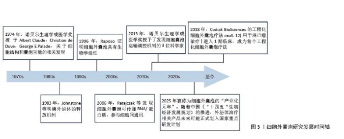
2.1 细胞外囊泡相关的研究路线 见图3。 "
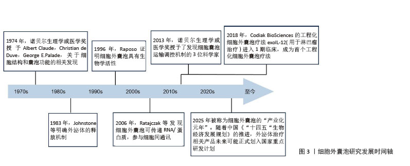
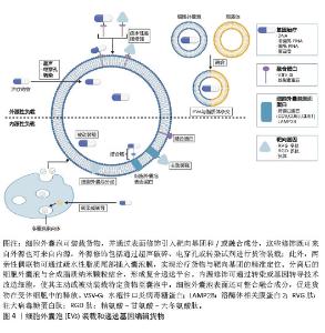
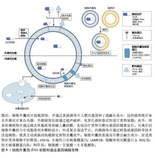
2.2 细胞外囊泡递送货物 细胞外囊泡可递送多类生物分子(包括编码RNA、非编码RNA、DNA及蛋白质),通过调控受体细胞的基因表达与功能实现治疗干预。细胞外囊泡具有优异的生物相容性、低细胞毒性、低免疫原性及可工程化修饰特性。在抵达靶部位前,细胞外囊泡的双层膜结构可保护核酸等生物大分子免受核酸酶降解。因此,细胞外囊泡可作为基因治疗的新型非病毒递送平台(图4)。"
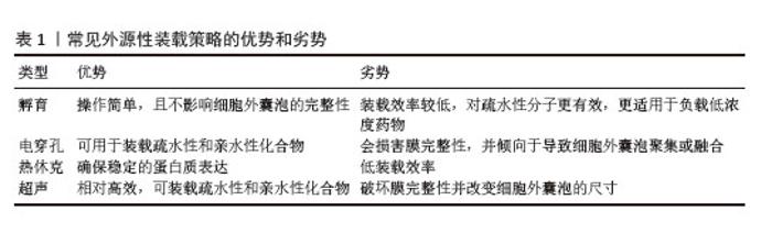
2.2.1 细胞外囊泡介导的RNA递送 治疗性RNA药物主要包括信使RNA、小干扰RNA、微小RNA、环状RNA及反义寡核苷酸等类型[3-4],尽管具有显著治疗潜力,这类分子仍面临多重递送挑战:体内易降解、递送效率低、脱靶效应及免疫原性风险。针对上述挑战,细胞外囊泡作为递送载体提供创新解决方案:天然磷脂双分子层结构可保护RNA免受核酸酶降解[5],且表面富集的整合素、四跨膜蛋白等配体可介导组织特异性靶向[6]。 微小RNA作为内源性非编码RNA,通过抑制靶信使RNA翻译或促进其降解调控基因表达[7]。特定微小RNA的表达失调可促进肿瘤侵袭、血管生成及转移等恶性表型[8-9],此类表达异常亦见于心血管疾病、神经退行性疾病等病理过程。因此,调控靶细胞内微小RNA水平已成为重要治疗策略[10]。基于细胞外囊泡的微小RNA递送系统可克服传统方法的靶向性不足、系统毒性高及体内不稳定等局限。相较于未修饰微小RNA,细胞外囊泡递送的微小RNA在体内稳定性显著提高,并可靶向富集于病变部位调控下游基因。人脐带间充质干细胞来源细胞外囊泡递送miR-451a可抑制类风湿关节炎滑膜成纤维细胞的增殖与侵袭,并改善胶原诱导性关节炎模型大鼠的关节炎症及影像学表现[11]。 小干扰RNA通过干扰机制特异性降解靶信使RNA,实现基因沉默[12],使它成为恶性肿瘤、遗传性疾病及病毒感染的治疗策略[13]。然而,传统载体(如脂质纳米颗粒)面临显著局限:高剂量小干扰RNA可能引发免疫激活、肝毒性及脱靶效应,导致临床试验终止。相较之下,细胞外囊泡递送系统具有显著优势:通过将小干扰RNA整合至pre-miR-451骨架,可提升细胞外囊泡的小干扰RNA装载效率。与脂质纳米颗粒相比,细胞外囊泡递送系统仅需1/10的小干扰RNA剂量即可实现同等基因沉默效果,显著降低细胞毒性风险[14]。 信使RNA疗法因不整合宿主基因组、快速表达功能性蛋白等优势,已成为基因治疗的重要策略[15-16]。例如,细胞外囊泡递送 Ldlr-mRNA至Ldlr 缺陷小鼠模型,可治疗家族性高胆固醇血症[17]。研究表明,细胞外囊泡递送的Ldlr-mRNA在体内稳定性显著提高,在肝细胞中高效表达功能性LDLR蛋白,改善疾病表型。细胞外囊泡的双层膜结构可有效保护信使RNA 免受核酸酶降解,显著提升给药稳定性。 2.2.2 细胞外囊泡介导的DNA递送 相较于RNA,基于细胞外囊泡的治疗性DNA(如质粒、反义寡核苷酸)递送系统具有独特价值:与RNA相比,细胞外囊泡介导的DNA递送在基因添加、抑制或编辑方面具有更高的稳定性优势。基于细胞外囊泡的治疗性DNA(如质粒、反义寡核苷酸)递送系统相较于质粒具有独特的稳定性结构。DNA的表达可由细胞类型特异性或细胞状态依赖性启动子调控,从而生成多种RNA及蛋白质。 质粒可在宿主细胞内自主复制、利用宿主转录翻译系统高效表达治疗性基因[18-19]。NGUYEN等[20]通过电穿孔法将绿色荧光蛋白编码质粒装载至细胞外囊泡,可有效转染THP-1巨噬细胞,结果显示绿色荧光蛋白在部分巨噬细胞亚群中稳定表达,该研究为胎盘细胞外囊泡的规模化生产提供依据,其内源性特征可能降低免疫原性风险[21]。 反义寡核苷酸与靶RNA 结合后,可激活核糖核酸酶H介导的降解作用,阻断致病基因表达[22]。此外,反义寡核苷酸可通过与靶RNA杂交形成DNA/RNA双链,产生空间位阻效应抑制基因功能[23]。为增强稳定性,反义寡核苷酸常进行化学修饰:如骨架硫代(磷酸二酯键转换为磷酸硫代酯键)或糖环修饰[24-25]。但化学修饰增加分子质量及亲脂性需依赖递送载体来克服生物屏障。细胞外囊泡递送的锁核酸修饰反义寡核苷酸(细胞外囊泡-锁核酸)较口服锁核酸有效降低靶基因表达,证明了载体的必要性[26]。 在免疫性疾病治疗中,细胞外囊泡-锁核酸递送系统同样具有应用价值:靶向miR-210的反义寡核苷酸(反义寡核苷酸-210)可抑制银屑病模型免疫失衡及炎症进展;细胞外囊泡递送反义寡核苷酸-210(细胞外囊泡-反义寡核苷酸-210)较游离反义寡核苷酸-210更能有效减轻银屑病症状,表明细胞外囊泡递送显著增强了反义寡核苷酸-210的治疗效果[27]。 CRISPR/Cas9基因编辑系统通过靶向DNA切割实现精准基因组修饰,在遗传病及肿瘤治疗领域具有重要价值,但临床应用受递送效率低、脱靶效应等安全性问题限制[28-29]。细胞外囊泡作为CRISPR/Cas9递送载体可突破上述局限:XU等[30]开发嵌合抗原受体修饰的细胞外囊泡(嵌合抗原受体-细胞外囊泡)可在体内外高效递送功能性CRISPR/Cas9,实现MYC癌基因靶向敲除。癌细胞来源细胞外囊泡具有天然肿瘤归巢能力,在SKOV3人源化卵巢癌模型中可特异性富集于病灶;利用此特性递送CRISPR/Cas9可抑制多聚ADP核糖聚合酶1表达,诱导癌细胞凋亡[31]。尽管目前细胞外囊泡-CRISPR疗法尚处临床转化早期阶段,但临床前研究已证实肿瘤基因靶向抑制潜力,为精准治疗提供新策略[32-33]。 2.2.3 细胞外囊泡介导的蛋白质递送 治疗性蛋白质包括基因组编辑酶、表观遗传修饰酶、转录因子、单克隆抗体、激素等,其有效的细胞内递送可以治疗多种疾病。已有研究中大部分使用治疗性蛋白质的用途包括缺陷蛋白质补偿、阻碍肿瘤进展、抑制凋亡蛋白、发挥酶作用、抗氧化等[34-35]。如MIZRAK等[36]通过负载自杀基因mRNA和尿嘧啶磷酸核糖转移酶的微囊泡来治疗神经鞘瘤小鼠,结果显示这些肿瘤在用前体药物5-氟胞嘧啶全身治疗后消退;WANG等[37]则通过含有Arrestin结构域蛋白1介导的微囊泡负载p53诱导小鼠多个组织DNA损伤依赖性凋亡。与此同时,提高蛋白质的细胞外囊泡装载效率、促进治疗蛋白质进入靶细胞后能够提高治疗性蛋白质的使用效率,INANO等[38]研究表明表明短的CD9-INF/TAT标签可有效改善细胞外囊泡介导的蛋白质递送。 2.3 细胞外囊泡的工程化装载策略 治疗性分子可以通过合成生物学策略装载至细胞外囊泡中,核心方法包括改造源细胞的内源性装载与修饰分离细胞外囊泡的外源性装载[39]。具体而言,内源性装载指通过基因工程改造供体细胞,而外源性装载则直接在分离的细胞外囊泡中导入治疗分子,常用电穿孔、超声处理或孵育实现[40]。此外,创新性策略如细胞外囊泡-脂质体杂交载体通过膜融合技术整合双方优势:脂质体提升大分子载能,同时细胞外囊泡膜蛋白维持结构稳定性,提高货物递送效率。 2.3.1 细胞外囊泡内源性工程化装载策略 转染是最常见的内源性装载方法,通过转染源细胞可以将货物装载到细胞外囊泡中。例如采用Lipofectamine3000试剂,可将反义寡核苷酸高效装载至红细胞衍生细胞外囊泡;磷酸硫代修饰的CpG-反义寡核苷酸复合物则通过清道夫受体介导的内吞作用,被神经干细胞高效内化,经内体逃逸后被动装载至细胞外囊泡。载有信号转导和转录活化因子3靶向CpG-反义寡核苷酸的细胞外囊泡在胶质瘤小鼠模型中显著抑制肿瘤生长[41],该策略在表达清道夫受体的细胞类型中具有普适潜力。 蛋白质融合是工程化细胞外囊泡的关键策略:利用四跨膜蛋白(CD9/CD63)的跨膜结构域构建融合蛋白,可实现治疗分子的定向装载。ES-HAGHI等[42]将CD9与RNA结合蛋白融合,使细胞外囊泡的miRNA载量提升,受体细胞递送效率提高。类似地,通过构建CD9与HuR融合蛋白,实现了对miR-155特异性识别与结合,该融合蛋白能有效将miR-155封装至细胞外囊泡中,并在体外和体内环境中实现对靶细胞的精准递送[43]。BUI等[44]基于CD63构建非病毒融合系统,通过表达人合胞素1,货物的递送效率显著提高。 2.3.2 细胞外囊泡外源性工程化策略 治疗分子可通过膜穿孔或物理化学方法直接装载至细胞外囊泡。相较于需细胞改造的内源性装载策略,外源性方法因操作便捷、普适性强,在临床转化中更具时效性优势[45]。常用技术包括:孵育、电穿孔、超声处理及热休克[46],见表1。 孵育法依赖浓度梯度实现治疗分子被动扩散入细胞外囊泡,操作便捷但封装效率低。相较之下,电穿孔封装效率更高,且对细胞外囊泡表面蛋白损伤小,成为常用技术[47],但易致核酸聚集,降低封装效率。脉冲超声可高效装载小RNA[41],规避核酸聚集问题,但会损伤细胞外囊泡膜完整性。此外,热休克转染方法也是一种常用的方法。ZHANG等[48]使用改良的氯化钙介导的热休克转染方法将miRNA富集到外泌体中,并证明通过外泌体递送到受体细胞中的 miRNA 模拟物或抑制剂能够引起受体细胞中miRNA的有效过表达或缺失(表1)。 "
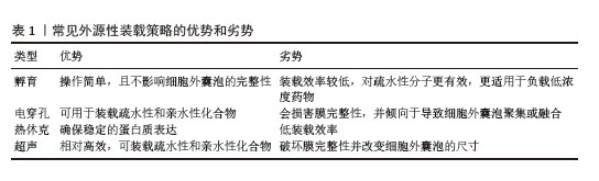
2.4 细胞外囊泡的靶向递送策略 实现精准靶向递送是细胞外囊泡临床转化的核心挑战。未经工程化修饰的天然细胞外囊泡在体内给药后有内在靶向特性,组织分布受表面脂质、蛋白质和糖类组成调控[49]。静脉给药后,外源性细胞外囊泡会迅速被单核吞噬细胞系统清除,导致递送效率显著降低[50]。因此,为实现细胞外囊泡对病灶区域的精准靶向递送,必须对细胞外囊泡表面进行工程化改造。 2.4.1 内在靶向 肿瘤来源细胞外囊泡展现出天然的肿瘤趋向性,能够实现针对同源肿瘤细胞的药物特异性递送[51-52]。例如,SKOV3卵巢癌细胞来源细胞外囊泡尾静脉注射后,在SKOV3卵巢癌异种移植小鼠模型中可选择性地富集于肿瘤组织,该研究进一步证实利用此类细胞外囊泡递送CRISPR-Cas9系统可有效沉默多聚ADP核糖聚合酶1基因表达,从而诱导癌细胞凋亡[53]。然而,这种内在靶向性存在显著的模型依赖性:QIAO等[54]研究表明,人纤维肉瘤细胞来源细胞外囊泡能够有效归巢至同源肿瘤组织,它的靶向效率显著优于Hela细胞来源细胞外囊泡;相比之下,SMYTH等[55]发现,小鼠乳腺癌细胞、人乳腺癌细胞和人前列腺癌细胞来源细胞外囊泡经静脉注射后,在响应肿瘤部位的蓄积量极低,这可能是由于血液清除速度快,这些肿瘤来源细胞外囊泡于各自模型中表现的内在靶向性的区别可能决定于细胞外囊泡本身天然的肿瘤趋向性的强弱程度以及细胞外囊泡注射入静脉后对于单核吞噬细胞系统抵抗能力的强弱。 克服单核吞噬细胞系统介导的快速清除,需要同步优化细胞外囊泡的体内循环时间与靶向能力。在延长循环时间方面:通过在细胞外囊泡表面过表达抗吞噬因子(如CD47),可抑制巨噬细胞的吞噬作用,显著延长血浆半衰期[56];聚乙二醇化修饰亦可减少细胞外囊泡的清除[57],通过延长循环时间增加细胞外囊泡在肿瘤组织的蓄积,但该修饰可能伴随潜在的免疫原性风险[58]。在增强主动靶向方面:经iRGD肽修饰的细胞外囊泡,能够特异性结合肿瘤血管内皮细胞高表达的αv整合素,从而在未分化甲状腺癌异种移植小鼠模型中显著提高细胞外囊泡在肿瘤部位的积累[59-60]。 值得注意的是,肿瘤来源细胞外囊泡可能具有促瘤效应,包括促进癌细胞增殖与存活[61]、抗肿瘤炎症以及增强肿瘤细胞的侵袭与转移能力[62-64]。因此,在考虑将肿瘤或永生化细胞系来源细胞外囊泡应用于治疗前,必须全面评估细胞外囊泡对不同细胞类型的生物学效应,并审慎权衡潜在风险与治疗获益。 2.4.2 工程化策略提高细胞外囊泡靶向性 对细胞外囊泡表面进行工程化修饰,可有效改善体内循环半衰期[65]、组织归巢能力及在靶标微环境中的蓄积效率[66-67]。其中,修饰细胞特异性靶向肽段是增强细胞外囊泡递送精准性的重要策略。例如,XU等[68]开发了一种双功能肽破骨细胞靶向肽-CP05,用于骨质疏松症的治疗。该肽包含两个功能域:CP05结构域能特异性识别并结合小鼠红细胞来源细胞外囊泡膜上的细胞外囊泡跨膜蛋白CD63[69],而破骨细胞靶向肽能够与破骨细胞膜表面高表达的抗酒石酸酸性磷酸酶相结合[70],通过破骨细胞靶向肽-CP05的桥接作用,可将红细胞来源细胞外囊泡精准导向破骨细胞。鉴于抑制破骨细胞的形成与活性是减少骨吸收进而逆转骨质疏松症进展的关键策略[71],研究者通过电穿孔法将抗miR-214(anti-miR)装载至红细胞来源细胞外囊泡中,形成载药复合体(红细胞来源细胞外囊泡/anti-miR-214),进一步利用破骨细胞靶向肽-CP05 对该复合体进行表面修饰,即可实现anti-miR-214向破骨细胞的高效靶向递送,从而发挥治疗作用。 类似地,抗体也可用于细胞外囊泡的功能化修饰,赋予细胞外囊泡对特定受体或表面蛋白的靶向能力。研究表明,经单链抗体片段scFv46.1修饰的细胞外囊泡在静脉注射后,能够通过转胞吞作用跨越小鼠血脑屏障,并在脑实质内显著富集,证实了该策略作为脑靶向递送平台的有效潜力[72]。 此外,既往研究中的工程化策略主要集中于利用蛋白质或肽作为靶向配体,而天然覆盖于细胞外囊泡表面的致密糖萼层在很大程度上尚未被充分开发利用[73]。然而,对细胞外囊泡表面糖萼进行工程化修饰同样是一件有效的靶向性提升策略。例如,通过对细胞外囊泡的母细胞进行基因工程改造,使其共表达融合至CD63蛋白大细胞外环的糖基化结构域、岩藻糖基转移酶 Ⅶ或Ⅸ。此策略成功使修饰后的细胞外囊泡表面分别呈现唾液酸 Lewis X或 Lewis X聚糖配体。这些工程化细胞外囊泡分别表现出对活化内皮细胞和树突状细胞的高度特异性靶向能力[74]。 除了对细胞外囊泡膜表面进行功能化修饰,物理改造也是一种重要的工程化策略。研究表明,通过外部物理干扰(如磁场、激光照射和超声波处理)可实现更精确的靶向效果[75]。例如,将超顺磁性氧化铁纳米颗粒修饰于中性粒细胞来源外泌体后,可在外部磁场引导下定向聚集于目标区域[76]。与磁场类似,激光照射也可用于增强靶向治疗效果:将细胞外囊泡与载药热敏脂质体融合形成的杂合纳米囊泡,不仅具备良好的靶向性,还能够在激光(如近红外光)照射下实现药物的可控释放[77-78]。然而,激光照射技术存在辐射损伤风险且成本较高。相比之下,超声波因较高的生物安全性引起了越来越多研究者的关注。例如,一项研究将二氢卟吩 e6与负载骨形态发生蛋白 7 mRNA的外泌体融合,并在其表面锚定 CP05-TK-mPEG(巯基酮-甲氧基聚乙二醇修饰的CP05)。在超声波作用下,二氢卟吩 e6产生的活性氧能够断裂巯基酮(TK)键,进而实现外泌体内包裹的骨形态发生蛋白 7 mRNA 的可控释放[79]。 此外,化学改造策略也增强细胞外囊泡的靶向治疗效果。研究者利用肿瘤微环境通常呈弱酸性的特点,设计了多种pH响应性递送系统。例如,一项研究采用表面修饰有i-基序(一种富含胞嘧啶的pH响应性DNA链)的细胞外囊泡递送阿霉素,使细胞外囊泡能够在酸性肿瘤微环境中选择性释放药物,从而增强阿霉素在肿瘤部位的蓄积,提高靶向治疗效率[80-81]。另一项研究将碳酸氢钠包裹进细胞外囊泡,囊泡被癌细胞内吞后,碳酸氢钠会迅速产生二氧化碳气泡,促进负载药物(如紫杉醇)的高效释放,进而增强抗肿瘤效果[82]。 2.5 细胞内递送与内体逃逸机制 细胞外囊泡介导的基因治疗效率,很大程度上取决于囊泡及其携带的治疗货物进入靶细胞的方式。细胞通过多种清除机制(如自噬、溶酶体降解及免疫应答)抵御外源物质入侵[83-85]。有效规避这些清除机制是提升细胞外囊泡基因递送效率的核心挑战。当前研究策略主要借鉴病毒载体和合成纳米颗粒的经验,聚焦于利用内体逃逸机制,避免治疗货物在溶酶体中被降解,从而保障治疗货物在靶细胞内的生物活性与功能。此外,通过对细胞外囊泡进行工程化改造,可进一步增强细胞外囊泡进入靶细胞并实现有效递送的能力。 2.5.1 内体逃逸机制 细胞外囊泡作为内源性纳米载体,具有独特的跨生物膜转运能力(尤其是内体逃逸机制),也是保障基因治疗递送效率的核心环节[86]。最新研究揭示,细胞外囊泡主要依赖四类分子机制介导内体逃逸(表2)[87-94],这些发现为新一代药物递送系统的开发提供了关键理论支撑。不同逃逸机制的相对效能受细胞外囊泡亚型、载货性质及靶细胞特性的显著调控。深入阐明这些复杂机制的时空动态特征,对设计下一代基于细胞外囊泡的药物递送平台具有决定性意义。 "
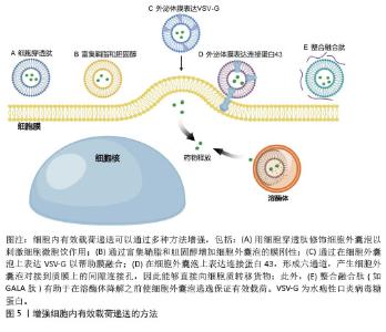
2.5.2 工程化策略提高有效载荷递送效率 为增强细胞外囊泡对细胞内有效载荷的递送效率,研究者开发了多种工程化改造策略,在外泌体膜表面进行生物修饰是重要手段(图5)。一种有效方法是在细胞外囊泡表面锚定细胞穿透肽,如富含精氨酸的细胞穿膜肽,通过刺激细胞微胞饮作用显著促进细胞外囊泡被细胞内化[95]。提高细胞外囊泡膜与细胞膜的融合效率是另一重要途径,其中通过富集鞘脂与胆固醇以增加膜刚性已被研究证实可有效提升融合效率[96]。此外,水泡性口炎病毒糖蛋白可模拟病毒膜融合机制,直接促进细胞外囊泡-细胞膜融合并增强有效载荷的细胞内递送[97]。细胞外囊泡与连接蛋白43的整合使细胞外囊泡能够停靠在嵌入受体细胞质膜中的间隙连接孔,因此为细胞外囊泡有效载荷的直接细胞质转移提供了另一种途径[98]。最后,工程化构建pH敏感的融合系统,如将细胞外囊泡与阳离子脂质及pH敏感的融合肽GALA复合,可增加细胞外囊泡-内体融合效率,从而促进货物的胞质释放[99]。 "
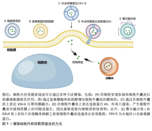
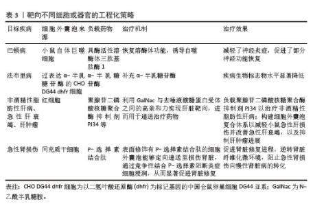
2.6 当前细胞外囊泡在不同细胞或器官中的靶向递送研究进展 细胞外囊泡因良好的靶向性、优异的生物相容性和稳定性等优势,已成为药物递送系统研究的热点。针对不同的器官或细胞类型,研究者通过多种工程化策略对其进行了广泛探索(表3)。 "
| [1] KIM HI, PARK J, ZHU Y, et al. Recent advances in extracellular vesicles for therapeutic cargo delivery. Exp Mol Med. 2024;56(4):836-849. [2] CECCHIN R, TROYER Z, WITWER K, et al. Extracellular vesicles: The next generation in gene therapy delivery. Mol Ther. 2023;31(5): 1225-1230. [3] SPARMANN A, VOGEL J. RNA-based medicine: from molecular mechanisms to therapy. EMBO J. 2023;42(21):e114760. [4] HASELTINE WA, HAZEL K, PATARCA R. RNA Structure: Past, Future, and Gene Therapy Applications. Int J Mol Sci. 2024;26(1):110. [5] SUN H, SU X, LI S, et al. Roles of glia-derived extracellular vesicles in central nervous system diseases: an update. Rev Neurosci. 2021;32(8):833-849. [6] WANG BZ, LUO LJ, VUNJAK-NOVAKOVIC G. RNA and Protein Delivery by Cell-Secreted and Bioengineered Extracellular Vesicles. Adv Healthc Mater. 2022;11(5):e2101557. [7] ROJAS-PIRELA M, ANDRADE-ALVIÁREZ D, MEDINA L, et al. MicroRNAs: master regulators in host-parasitic protist interactions. Open Biol. 2022;12(6):210395. [8] MENON A, ABD-AZIZ N, KHALID K, et al. miRNA: A Promising Therapeutic Target in Cancer. Int J Mol Sci. 2022;23(19):11502. [9] YANG H, LIU Y, CHEN L, et al. MiRNA-Based Therapies for Lung Cancer: Opportunities and Challenges? Biomolecules. 2023;13(6):877. [10] DIENER C, KELLER A, MEESE E. Emerging concepts of miRNA therapeutics: from cells to clinic. Trends Genet. 2022;38(6): 613-626. [11] MI L, GAO J, LI N, et al. Human umbilical cord mesenchymal stem cell-derived exosomes loaded miR-451a targets ATF2 to improve rheumatoid arthritis. Int Immunopharmacol. 2024;127:111365. [12] FRIEDRICH M, AIGNER A. Therapeutic siRNA: State-of-the-Art and Future Perspectives. BioDrugs. 2022;36(5):549-571. [13] HUANG X, GUO H, WANG L, et al. Biomimetic cell membrane-coated nanocarriers for targeted siRNA delivery in cancer therapy. Drug Discov Today. 2023;28(4):103514. [14] RESHKE R, TAYLOR JA, SAVARD A, et al. Reduction of the therapeutic dose of silencing RNA by packaging it in extracellular vesicles via a pre-microRNA backbone. Nat Biomed Eng. 2020;4(1):52-68. [15] SHEN G, LIU J, YANG H, et al. mRNA therapies: Pioneering a new era in rare genetic disease treatment. J Control Release. 2024;369: 696-721. [16] IQBAL Z, REHMAN K, MAHMOOD A, et al. Exosome for mRNA delivery: strategies and therapeutic applications. J Nanobiotechnology. 2024;22(1):395. [17] LI DF, LIU QS, YANG MF, et al. Nanomaterials for mRNA-based therapeutics: Challenges and opportunities. Bioeng Transl Med. 2023; 8(3):e10492. [18] SAYED N, ALLAWADHI P, KHURANA A, et al. Gene therapy: Comprehensive overview and therapeutic applications. Life Sci. 2022; 294:120375. [19] NEBOGATOVA J, HÄRK HH, PUSKAR A, et al. A Method for Using Cell-Penetrating Peptides for Loading Plasmid DNA into Secreted Extracellular Vesicles. Biomolecules. 2023;13(12):1751. [20] NGUYEN DB, TRAN HT, KAESTNER L, et al. The Relation Between Extracellular Vesicles Released From Red Blood Cells, Their Cargo, and the Clearance by Macrophages. Front Physiol. 2022;13:783260. [21] KANG M, HISEY C, TSAI B, et al. Placental Extracellular Vesicles Can Be Loaded with Plasmid DNA. Mol Pharm. 2023;20(4):1898-1913. [22] YAN W, LI Y, XIE S, et al. Chondrocyte-Targeted Delivery System of Sortase A-Engineered Extracellular Vesicles Silencing MMP13 for Osteoarthritis Therapy. Adv Healthc Mater. 2024;13(16):e2303510. [23] COLLOTTA D, BERTOCCHI I, CHIAPELLO E, et al. Antisense oligonucleotides: a novel Frontier in pharmacological strategy. Front Pharmacol. 2023;14:1304342. [24] GENNA V, IGLESIAS-FERNÁNDEZ J, REYES-FRAILE L, et al. Controlled sulfur-based engineering confers mouldability to phosphorothioate antisense oligonucleotides. Nucleic Acids Res. 2023;51(10):4713-4725. [25] YOSHIDA T, HAGIHARA T, UCHIDA Y, et al. Introduction of sugar-modified nucleotides into CpG-containing antisense oligonucleotides inhibits TLR9 activation. Sci Rep. 2024;14(1): 11540. [26] GROSSEN P, PORTMANN M, KOLLER E, et al. Evaluation of bovine milk extracellular vesicles for the delivery of locked nucleic acid antisense oligonucleotides. Eur J Pharm Biopharm. 2021;158:198-210. [27] ZHANG W, LIN J, SHI P, et al. Small Extracellular Vesicles Derived From MSCs Have Immunomodulatory Effects to Enhance Delivery of ASO-210 for Psoriasis Treatment. Front Cell Dev Biol. 2022;10:842813. [28] ZHU X, GAO M, YANG Y, et al. The CRISPR/Cas9 System Delivered by Extracellular Vesicles. Pharmaceutics. 2023;15(3):984. [29] DUBEY S, CHEN Z, JIANG YJ, et al. Small extracellular vesicles (sEVs)-based gene delivery platform for cell-specific CRISPR/Cas9 genome editing. Theranostics. 2024; 14(7):2777-2793. [30] XU Q, ZHANG Z, ZHAO L, et al. Tropism-facilitated delivery of CRISPR/Cas9 system with chimeric antigen receptor-extracellular vesicles against B-cell malignancies. J Control Release. 2020;326:455-467. [31] YANG S, IM SH, CHUNG JY, et al. An Antibody-CRISPR/Cas Conjugate Platform for Target-Specific Delivery and Gene Editing in Cancer. Adv Sci (Weinh). 2024;11(21):e2308763. [32] IQBAL Z, REHMAN K, XIA J, et al. Biomaterial-assisted targeted and controlled delivery of CRISPR/Cas9 for precise gene editing. Biomater Sci. 2023;11(11):3762-3783. [33] LIANG Y, IQBAL Z, WANG J, et al. Cell-derived extracellular vesicles for CRISPR/Cas9 delivery: engineering strategies for cargo packaging and loading. Biomater Sci. 2022;10(15):4095-4106. [34] HAN J, SUL JH, LEE J, et al. Engineered exosomes with a photoinducible protein delivery system enable CRISPR-Cas-based epigenome editing in Alzheimer’s disease. Sci Transl Med. 2024;16(759):eadi4830. [35] LE SAUX S, AUBERT-POUËSSEL A, MOHAMED KE, et al. Interest of extracellular vesicles in regards to lipid nanoparticle based systems for intracellular protein delivery. Adv Drug Deliv Rev. 2021;176:113837. [36] MIZRAK A, BOLUKBASI MF, OZDENER GB, et al. Genetically engineered microvesicles carrying suicide mRNA/protein inhibit schwannoma tumor growth. Mol Ther. 2013; 21(1):101-108. [37] WANG Q, YU J, KADUNGURE T, et al. ARMMs as a versatile platform for intracellular delivery of macromolecules. Nat Commun. 2018;9(1):960. [38] INANO S, KITANO T. A modified CD9 tag for efficient protein delivery via extracellular vesicles. PLoS One. 2024;19(10):e0310083. [39] YANG C, XUE Y, DUAN Y, et al. Extracellular vesicles and their engineering strategies, delivery systems, and biomedical applications. J Control Release. 2024;365:1089-1123. [40] DAD HA, GU TW, ZHU AQ, et al. Plant Exosome-like Nanovesicles: Emerging Therapeutics and Drug Delivery Nanoplatforms. Mol Ther. 2021;29(1):13-31. [41] DE ABREU RC, RAMOS CV, BECHER C, et al. Exogenous loading of miRNAs into small extracellular vesicles. J Extracell Vesicles. 2021;10(10):e12111. [42] ES-HAGHI M, NEUSTROEVA O, CHOWDHURY I, et al. Construction of Fusion Protein for Enhanced Small RNA Loading to Extracellular Vesicles. Genes (Basel). 2023;14(2):261. [43] LI Z, ZHOU X, WEI M, et al. In Vitro and in Vivo RNA Inhibition by CD9-HuR Functionalized Exosomes Encapsulated with miRNA or CRISPR/dCas9. Nano Lett. 2019;19(1):19-28. [44] BUI S, DANCOURT J, LAVIEU G. Virus-Free Method to Control and Enhance Extracellular Vesicle Cargo Loading and Delivery. ACS Appl Bio Mater. 2023;6(3):1081-1091. [45] TIAN J, HAN Z, SONG D, et al. Engineered Exosome for Drug Delivery: Recent Development and Clinical Applications. Int J Nanomedicine. 2023;18:7923-7940. [46] SADEGHI S, TEHRANI FR, TAHMASEBI S, et al. Exosome engineering in cell therapy and drug delivery. Inflammopharmacology. 2023;31(1):145-169. [47] MUKHOPADHYA A, TSIAPALIS D, MCNAMEE N, et al. Doxorubicin Loading into Milk and Mesenchymal Stem Cells’ Extracellular Vesicles as Drug Delivery Vehicles. Pharmaceutics. 2023;15(3):718. [48] ZHANG D, LEE H, ZHU Z, et al. Enrichment of selective miRNAs in exosomes and delivery of exosomal miRNAs in vitro and in vivo. Am J Physiol Lung Cell Mol Physiol. 2017;312(1):L110-L121. [49] ARMSTRONG JPK, STEVENS MM. Strategic design of extracellular vesicle drug delivery systems. Adv Drug Deliv Rev. 2018;130:12-16. [50] WEI Z, CHEN Z, ZHAO Y, et al. Mononuclear phagocyte system blockade using extracellular vesicles modified with CD47 on membrane surface for myocardial infarction reperfusion injury treatment. Biomaterials. 2021;275:121000. [51] SAARI H, LÁZARO-IBÁÑEZ E, VIITALA T, et al. Microvesicle- and exosome-mediated drug delivery enhances the cytotoxicity of Paclitaxel in autologous prostate cancer cells. J Control Release. 2015;220(Pt B):727-737. [52] ZHANG F, GUO J, ZHANG Z, et al. Application of engineered extracellular vesicles for targeted tumor therapy. J Biomed Sci. 2022; 29(1):14. [53] KIM SM, YANG Y, OH SJ, et al. Cancer-derived exosomes as a delivery platform of CRISPR/Cas9 confer cancer cell tropism-dependent targeting. J Control Release. 2017;266:8-16. [54] QIAO L, HU S, HUANG K, et al. Tumor cell-derived exosomes home to their cells of origin and can be used as Trojan horses to deliver cancer drugs. Theranostics. 2020;10(8): 3474-3487. [55] SMYTH T, KULLBERG M, MALIK N, et al. Biodistribution and delivery efficiency of unmodified tumor-derived exosomes. J Control Release. 2015;199:145-155. [56] OJHA T, PATHAK V, SHI Y, et al. Pharmacological and physical vessel modulation strategies to improve EPR-mediated drug targeting to tumors. Adv Drug Deliv Rev. 2017;119:44-60. [57] SHI S, LI T, WEN X, et al. Copper-64 Labeled PEGylated Exosomes for In Vivo Positron Emission Tomography and Enhanced Tumor Retention. Bioconjug Chem. 2019;30(10): 2675-2683. [58] D’AVANZO N, CELIA C, BARONE A, et al. Immunogenicity of Polyethylene Glycol Based Nanomedicines: Mechanisms, Clinical Implications and Systematic Approach. Advanced Therapeutics. 2020;3(3):1900170. [59] SUGAHARA KN, TEESALU T, KARMALI PP, et al. Tissue-penetrating delivery of compounds and nanoparticles into tumors. Cancer Cell. 2009;16(6):510-520. [60] TIAN Y, LI S, SONG J, et al. A doxorubicin delivery platform using engineered natural membrane vesicle exosomes for targeted tumor therapy. Biomaterials. 2014;35(7): 2383-2390. [61] BECKER A, THAKUR BK, WEISS JM, et al. Extracellular Vesicles in Cancer: Cell-to-Cell Mediators of Metastasis. Cancer Cell. 2016;30(6):836-848. [62] SMIRNOVA O, EFREMOV Y, KLYUCHEREV T, et al. Direct and cell-mediated EV-ECM interplay. Acta Biomater. 2024;186:63-84. [63] KALLURI R, MCANDREWS KM. The role of extracellular vesicles in cancer. Cell. 2023; 186(8):1610-1626. [64] PEREZ HURTADO EC, HENAO AGUDELO JS, FOGANHOLI DA SILVA RA, et al. The role of extracellular vesicles in cancer. Curr Top Membr. 2024;94:247-285. [65] LIANG X, NIU Z, GALLI V, et al. Extracellular vesicles engineered to bind albumin demonstrate extended circulation time and lymph node accumulation in mouse models. J Extracell Vesicles. 2022;11(7):e12248. [66] LIU X, CAO Z, WANG W, et al. Engineered Extracellular Vesicle-Delivered CRISPR/Cas9 for Radiotherapy Sensitization of Glioblastoma. ACS Nano. 2023;17(17):16432-16447. [67] VAN DER KOOG L, GANDEK TB, NAGELKERKE A. Liposomes and Extracellular Vesicles as Drug Delivery Systems: A Comparison of Composition, Pharmacokinetics, and Functionalization. Adv Healthc Mater. 2022; 11(5):e2100639. [68] XU L, XU X, LIANG Y, et al. Osteoclast-targeted delivery of anti-miRNA oligonucleotides by red blood cell extracellular vesicles. J Control Release. 2023;358:259-272. [69] GAO X, RAN N, DONG X, et al. Anchor peptide captures, targets, and loads exosomes of diverse origins for diagnostics and therapy. Sci Transl Med. 2018;10(444):eaat0195. [70] SHEU TJ, SCHWARZ EM, O’KEEFE RJ, et al. Use of a phage display technique to identify potential osteoblast binding sites within osteoclast lacunae. J Bone Miner Res. 2002; 17(5):915-922. [71] BI H, CHEN X, GAO S, et al. Key Triggers of Osteoclast-Related Diseases and Available Strategies for Targeted Therapies: A Review. Front Med (Lausanne). 2017;4:234. [72] YE Z, GASTFRIEND BD, UMLAUF BJ, et al. Antibody-Targeted Liposomes for Enhanced Targeting of the Blood-Brain Barrier. Pharm Res. 2022;39(7):1523-1534. [73] GERLACH JQ, GRIFFIN MD. Getting to know the extracellular vesicle glycome. Mol Biosyst. 2016;12(4):1071-1081. [74] ZHENG W, HE R, LIANG X, et al. Cell-specific targeting of extracellular vesicles through engineering the glycocalyx. J Extracell Vesicles. 2022;11(12):e12290. [75] ZHANG M, HU S, LIU L, et al. Engineered exosomes from different sources for cancer-targeted therapy. Signal Transduct Target Ther. 2023;8(1):124. [76] ZHANG J, JI C, ZHANG H, et al. Engineered neutrophil-derived exosome-like vesicles for targeted cancer therapy. Sci Adv. 2022;8(2): eabj8207. [77] CHENG L, ZHANG X, TANG J, et al. Gene-engineered exosomes-thermosensitive liposomes hybrid nanovesicles by the blockade of CD47 signal for combined photothermal therapy and cancer immunotherapy. Biomaterials. 2021;275:120964. [78] LI G, WANG J, XU M, et al. Engineered exosome for NIR-triggered drug delivery and superior synergistic chemo-phototherapy in a glioma model. Applied Materials Today. 2020;20:100723. [79] HUANG J, HE Y, YANG X, et al. I-motif-based nano-flares for sensing pH changes in live cells. Chem Commun (Camb). 2014;50(99): 15768-15771. [80] KIM JY, SONG J, JUNG H, et al. I-motif-coated exosomes as a pH-sensitive carrier for anticancer drugs. Applied Biological Chemistry. 2018;61(6):599-606. [81] NGUYEN CAO TG, KANG JH, KIM W, et al. Engineered extracellular vesicle-based sonotheranostics for dual stimuli-sensitive drug release and photoacoustic imaging-guided chemo-sonodynamic cancer therapy. Theranostics. 2022;12(3):1247-1266. [82] WILLIAMS RE, ADAMS HR, BLOHM M, et al. Management Strategies for CLN2 Disease. Pediatr Neurol. 2017;69:102-112. [83] DERETIC V. Autophagy in inflammation, infection, and immunometabolism. Immunity. 2021;54(3):437-453. [84] ZHANG Z, YUE P, LU T, et al. Role of lysosomes in physiological activities, diseases, and therapy. J Hematol Oncol. 2021;14(1):79. [85] HILL A, BEITELSHEES M, PFEIFER BA. Vaccine Delivery and Immune Response Basics. Methods Mol Biol. 2021;2183:1-8. [86] ZHAO J, ZHU W, MAO Y, et al. Unignored intracellular journey and biomedical applications of extracellular vesicles. Adv Drug Deliv Rev. 2024;212:115388. [87] O’BRIEN K, BREYNE K, UGHETTO S, et al. RNA delivery by extracellular vesicles in mammalian cells and its applications. Nat Rev Mol Cell Biol. 2020;21(10):585-606. [88] JIN Y, MA L, ZHANG W, et al. Extracellular signals regulate the biogenesis of extracellular vesicles. Biol Res. 2022;55(1):35. [89] BROCK DJ, KUSTIGIAN L, JIANG M, et al. Efficient cell delivery mediated by lipid-specific endosomal escape of supercharged branched peptides. Traffic. 2018;19(6):421-435. [90] SHETE HK, PRABHU RH, PATRAVALE VB. Endosomal escape: a bottleneck in intracellular delivery. J Nanosci Nanotechnol. 2014;14(1):460-474. [91] GANDEK TB, VAN DER KOOG L, NAGELKERKE A. A Comparison of Cellular Uptake Mechanisms, Delivery Efficacy, and Intracellular Fate between Liposomes and Extracellular Vesicles. Adv Healthc Mater. 2023;12(25):e2300319. [92] BEBELMAN MP, BUN P, HUVENEERS S, et al. Real-time imaging of multivesicular body-plasma membrane fusion to quantify exosome release from single cells. Nat Protoc. 2020;15(1):102-121. [93] PERRIN P, JANSSEN L, JANSSEN H, et al. Retrofusion of intralumenal MVB membranes parallels viral infection and coexists with exosome release. Curr Biol. 2021;31(17): 3884-3893.e4. [94] ILAHIBAKS NF, ARDISASMITA AI, XIE S, et al. TOP-EVs: Technology of Protein delivery through Extracellular Vesicles is a versatile platform for intracellular protein delivery. J Control Release. 2023;355:579-592. [95] NAKASE I, NOGUCHI K, FUJII I, et al. Vectorization of biomacromolecules into cells using extracellular vesicles with enhanced internalization induced by macropinocytosis. Sci Rep. 2016;6:34937. [96] PAROLINI I, FEDERICI C, RAGGI C, et al. Microenvironmental pH is a key factor for exosome traffic in tumor cells. J Biol Chem. 2009;284(49):34211-34222. [97] YANG Y, HONG Y, NAM GH, et al. Virus-Mimetic Fusogenic Exosomes for Direct Delivery of Integral Membrane Proteins to Target Cell Membranes. Adv Mater. 2017. doi: 10.1002/adma.201605604. [98] LU M, ZHAO X, XING H, et al. Cell-free synthesis of connexin 43-integrated exosome-mimetic nanoparticles for siRNA delivery. Acta Biomater. 2019;96:517-536. [99] NAKASE I, FUTAKI S. Combined treatment with a pH-sensitive fusogenic peptide and cationic lipids achieves enhanced cytosolic delivery of exosomes. Sci Rep. 2015;5:10112. [100] EL-HAGE N, HANEY MJ, ZHAO Y, et al. Extracellular Vesicles Released by Genetically Modified Macrophages Activate Autophagy and Produce Potent Neuroprotection in Mouse Model of Lysosomal Storage Disorder, Batten Disease. Cells. 2023;12(11):1497. [101] GERMAIN DP. Fabry disease. Orphanet J Rare Dis. 2010;5:30. [102] SERAS-FRANZOSO J, DÍAZ-RIASCOS ZV, CORCHERO JL, et al. Extracellular vesicles from recombinant cell factories improve the activity and efficacy of enzymes defective in lysosomal storage disorders. J Extracell Vesicles. 2021; 10(5):e12058. [103] SÁNCHEZ SV, OTAVALO GN, GAZEAU F, et al. Intranasal delivery of extracellular vesicles: A promising new approach for treating neurological and respiratory disorders. J Control Release. 2025;379:489-523. [104] HUANG X, YANG J, HU Y, et al. Protective Effects of GalNac-Modified Red Blood Cell-Derived Extracellular Vesicles Against Liver Diseases. Int J Nanomedicine. 2025;20: 8993-9017. [105] ZHANG X, ZHAO J, GE R, et al. Arg-Gly-Asp engineered mesenchymal stem cells as targeted nanotherapeutics against kidney fibrosis by modulating m6A. Acta Biomater. 2025;198:85-101. [106] ZHANG K, LI R, CHEN X, et al. Renal Endothelial Cell-Targeted Extracellular Vesicles Protect the Kidney from Ischemic Injury. Adv Sci (Weinh). 2023;10(3):e2204626. [107] COLAO IL, CORTELING R, BRACEWELL D, et al. Manufacturing Exosomes: A Promising Therapeutic Platform. Trends Mol Med. 2018; 24(3):242-256. |
| [1] | Zhou Sirui, Xu Yukun, Zhao Kewei. Ideas and methods of anti-melanogenesis of Angelica dahurica extracellular vesicles [J]. Chinese Journal of Tissue Engineering Research, 2026, 30(7): 1747-1754. |
| [2] | Huang Jiawen, Pan Zhiyi, Xue Wenjun, Lian Yuanpei, Xu Jianda. Plant-derived vesicles and malignant tumor therapy: cross-species communication and modulation of host cell responses [J]. Chinese Journal of Tissue Engineering Research, 2026, 30(7): 1828-1838. |
| [3] | Zhang Qian, Huang Dongfeng. Weighted gene co-expression network analysis combined with machine learning to screen and validate biomarkers for osteoarthritis [J]. Chinese Journal of Tissue Engineering Research, 2026, 30(5): 1096-1105. |
| [4] | Yang Xiao, Bai Yuehui, Zhao Tiantian, Wang Donghao, Zhao Chen, Yuan Shuo. Cartilage degeneration in temporomandibular joint osteoarthritis: mechanisms and regenerative challenges [J]. Chinese Journal of Tissue Engineering Research, 2026, 30(4): 926-935. |
| [5] | Xiao Yang, Gao Zibo, Hu Yuxiang, Kang Zhixin, Zhang Chaoxuan, Huang Chengyu, Liu Honglin, Chen Kai, Wang Hongshen, Li Yongjin. Molecular mechanism by which the imbalance of the functional network of tissue inhibitors of metalloproteinases drives intervertebral disc degeneration [J]. Chinese Journal of Tissue Engineering Research, 2026, 30(35): 9258-9268. |
| [6] | Liu Hongwei, Chang Lungji. Lentivirus-mediated gene therapy in a beta-thalassemia mouse model [J]. Chinese Journal of Tissue Engineering Research, 2026, 30(34): 8978-8985. |
| [7] | Liu Bangding, Tang Yongliang, Li Ni, Ren Bo. Quercetin-loaded hydrogel materials for treatment of infected bone defects [J]. Chinese Journal of Tissue Engineering Research, 2026, 30(32): 8427-8435. |
| [8] | Ding Hao, Gao Yuan, Li Bin, Yu Rui, Wang Jianru, Sun Yudie, Wang Xuanyang, Zhang Wenping, Zhu Mingjun. Potential and application prospects of combined treatment of acute myocardial infarction with hydrogel cardiac patches and traditional Chinese medicine [J]. Chinese Journal of Tissue Engineering Research, 2026, 30(32): 8479-8486. |
| [9] | Li Minghui, Qie Haoyu, Pan Min, Bi Ruijie, Lyu Xiaomeng, Zhang Haoya, Han Yifei. Hydrogel-based drug delivery systems for rheumatoid arthritis treatment [J]. Chinese Journal of Tissue Engineering Research, 2026, 30(32): 8496-8501. |
| [10] | Hu Kai, Chen Yulin, Yan Jing, He Yingying, Meng Yanhui, Li Runzhen, Tang Yaoping. Epimedium Sagittatum Maxim.-extracellular vesicles ameliorate vascular smooth muscle cell calcification: a proteomic analysis [J]. Chinese Journal of Tissue Engineering Research, 2026, 30(31): 8135-8145. |
| [11] | Wang Xuecheng, Zhao Liang, Wei Zhoubin. Transforming growth factor-beta regulates oxidative stress and participates in inflammatory process of pulmonary hypertension [J]. Chinese Journal of Tissue Engineering Research, 2026, 30(31): 8165-8173. |
| [12] | Zhang Chuanlin, Wu Yuhuai, Zhou Haixiang, Liu Jianping, Xu Qian, Zhang Lili, Li Cuie. An innovative strategy for engineered exosomal long non-coding RNAs in treatment of osteoporosis [J]. Chinese Journal of Tissue Engineering Research, 2026, 30(31): 8264-8271. |
| [13] | Wang Zhengye, Liu Wanlin, Zhao Zhenqun. Mechanism by which vascular endothelial growth factor A targets regulation of angiogenesis in the treatment of steroid-induced osteonecrosis of the femoral head [J]. Chinese Journal of Tissue Engineering Research, 2026, 30(3): 671-679. |
| [14] | Yang Hu, Zheng Yu, Jia Chengming, Wang Tong, Zhang Guangfei, Ji Yaoyao. Immune microenvironment regulates bone regeneration [J]. Chinese Journal of Tissue Engineering Research, 2026, 30(3): 701-710. |
| [15] | Wu Ningyuan, Wei Zhiyi, Feng Hao, Gao Ming. Frontiers and hot topics of nanobiomedicine in delaying the progression of osteoarthritis [J]. Chinese Journal of Tissue Engineering Research, 2026, 30(26): 6907-6915. |
| Viewed | ||||||
|
Full text |
|
|||||
|
Abstract |
|
|||||