Chinese Journal of Tissue Engineering Research ›› 2026, Vol. 30 ›› Issue (7): 1649-1657.doi: 10.12307/2026.086
Previous Articles Next Articles
Programmed cell death receptor-1 suppresses osteogenic differentiation of rat bone marrow mesenchymal stem cells in a high-glucose microenvironment
Han Nianrong, Huang Yifei, Akram · Osman, Liu Yanlu, Hu Wei
- Department of Spine II, Hospital of Traditional Chinese Medicine Affiliated to Xinjiang Medical University, Urumqi 830000, Xinjiang Uygur Autonomous Region, China
-
Received:2025-02-06Revised:2025-05-27Accepted:2025-06-20Online:2026-03-08Published:2025-08-18 -
Contact:Hu Wei, Chief physician, Department of Spine II, Hospital of Traditional Chinese Medicine Affiliated to Xinjiang Medical University, Urumqi 830000, Xinjiang Uygur Autonomous Region, China -
About author:Han Nianrong, Doctoral candidate, Attending physician, Department of Spine II, Hospital of Traditional Chinese Medicine Affiliated to Xinjiang Medical University, Urumqi 830000, Xinjiang Uygur Autonomous Region, China -
Supported by:Xinjiang Uygur Autonomous Region Natural Science Foundation - Outstanding Young Scientist Fund, No. 2022D01E29 (to HW)
CLC Number:
Cite this article
Han Nianrong, Huang Yifei, Akram · Osman, Liu Yanlu, Hu Wei . Programmed cell death receptor-1 suppresses osteogenic differentiation of rat bone marrow mesenchymal stem cells in a high-glucose microenvironment [J]. Chinese Journal of Tissue Engineering Research, 2026, 30(7): 1649-1657.
share this article
Add to citation manager EndNote|Reference Manager|ProCite|BibTeX|RefWorks
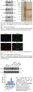
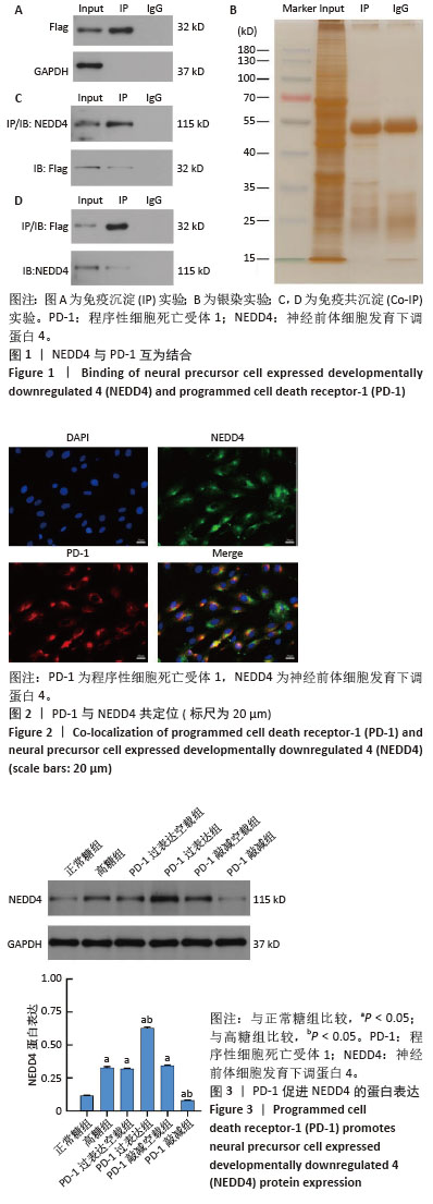
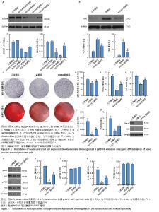
2.1 NEDD4是PD-1的交互蛋白 通过免疫沉淀下拉及质谱检测,发现蛋白NEDD4(图1A,B)。通过Co-IP实验,证实PD-1与NEDD4交互(图1C,D)。 2.2 PD-1与NEDD4共定位 免疫荧光染色发现,PD-1和NEDD4定位一致,集中在细胞膜及细胞浆位置(图2)。 2.3 PD-1促进NEDD4的蛋白表达 采用Western blot检测PD-1对NEDD4的调控关系。研究结果显示,高糖组NEDD4蛋白表达水平较正常糖组升高(P < 0.05);PD-1过表达组NEDD4蛋白表达水平较高糖组升高(P < 0.05);PD-1敲减组NEDD4蛋白表达水平较高糖组降低(P < 0.05)(图3)。 "
2.4 敲减NEDD4增强了骨髓间充质干细胞成骨分化能力 NEDD4敲减组2中NEDD4表达水平最低,选用该片段序列用于构建sh-NEDD4,开展后续研究(图4A)。与高糖组比较,NEDD4敲减组PD-1蛋白表达水平减少(P < 0.05)(图4B);通过茜素红S染色与碱性磷酸酶活性检测发现,NEDD4敲减组骨髓间充质干细胞成骨分化能力明显增强(图4C,D);此外,与高糖组比较,NEDD4敲减组成骨因子Runt相关转录因子2、OSX 蛋白表达升高(P < 0.05)(图4E-I)。 2.5 敲减NEDD4可以激活PI3K/AKT通路 与高糖组比较,NEDD4敲减组p-PI3K、p-AKT表达水平升高(P < 0.05);而PI3K和AKT表达无显著差异(P > 0.05)(图5A-E)。 "

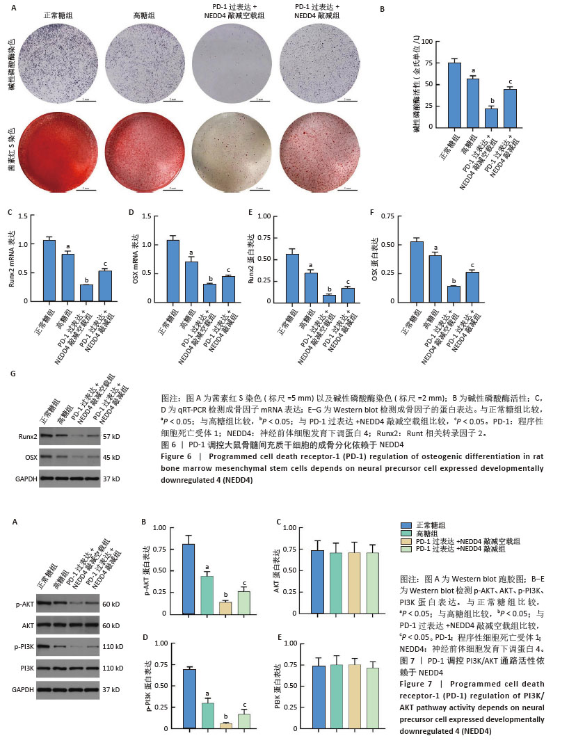
2.6 PD-1调控大鼠骨髓间充质干细胞的成骨分化依赖于NEDD4 茜素红染色和碱性磷酸酶染色显示,PD-1过表达+NEDD4敲减组成骨分化能力较PD-1过表达+NEDD4敲减空载组升高(图6A,B);PD-1过表达+NEDD4敲减组的成骨标志物 Runt相关转录因子2、OSX mRNA和蛋白表达水平较PD-1 过表达+ NEDD4敲减空载组显著升高(P < 0.05)(图6C-G)。 2.7 PD-1调控PI3K/AKT 通路活性依赖于NEDD4 PD-1过表达+NEDD4敲减组p-AKT、p-PI3K蛋白表达较PD-1过表达+NEDD4敲减空载组升高(P < 0.05)(图7A-E)。 "
| [1] 陶恩东,夏伟杰,王福林,等.骨髓间充质干细胞来源的外泌体通过PI3K/AKT信号通路逆转高糖诱导的骨髓间充质干细胞成骨功能障碍[J].温州医科大学学报,2024,54(8):641-649. [2] DU YX, ZHAO YT, SUN YX, et al. Acid sphingomyelinase mediates ferroptosis induced by high glucose via autophagic degradation of GPX4 in type 2 diabetic osteoporosis. Mol Med. 2023;29(1):125. [3] JIN Z, LEE B. Boosting glycolysis to combat fragile bone in type 1 diabetes. Cell Chem Biol. 2023;30(9):1002-1003. [4] CHEN Y, ZHAO W, HU A, et al. Type 2 diabetic mellitus related osteoporosis: focusing on ferroptosis. J Transl Med. 2024;22(1):409. [5] JIA X, ZHANG G, YU D. Application of extracellular vesicles in diabetic osteoporosis. Front Endocrinol (Lausanne). 2024;15: 1466775. [6] OUYANG Z, KANG D, LI K, et al. DEPTOR exacerbates bone-fat imbalance in osteoporosis by transcriptionally modulating BMSC differentiation. Biomed Pharmacother. 2022;151:113164. [7] QIN W, SHANG Q, SHEN G, et al. Restoring bone-fat equilibrium: Baicalin’s impact on P38 MAPK pathway for treating diabetic osteoporosis. Biomed Pharmacother. 2024;175:116571. [8] LI M, XIE Z, LI J, et al. GAS5 protects against osteoporosis by targeting UPF1/SMAD7 axis in osteoblast differentiation. Elife. 2020;9:e59079. [9] 毛钰涵,王甲甲.非编码 RNA 在糖尿病性骨质疏松症中作用的研究进展[J].中国现代医药杂志,2024,26(6):90-94. [10] 魏咸亭,陈宝康,董鑫,等.姜黄素调控HO-1改善高糖环境下骨髓间充质干细胞的成骨分化[J].局解手术学杂志,2024,33(9):783-787. [11] 张锦明,田滢舟,赵玲,等.淫羊藿苷促进骨髓间充质干细胞成骨分化缓解小鼠骨质疏松的机制[J].中国组织工程研究,2022,26(19): 2991-2996. [12] 常俊杰,解强,刘亦冰,等.高糖对骨髓间充质干细胞作用机制的研究进展[J].中国骨质疏松杂志,2022,28(10): 1551-1555. [13] CAI F, LIU Y, LIU K, et al. Diabetes mellitus impairs bone regeneration and biomechanics. J Orthop Surg Res. 2023;18(1):169. [14] KAWADA-HORITANI E, KITA S, OKITA T, et al. Human adipose-derived mesenchymal stem cells prevent type 1 diabetes induced by immune checkpoint blockade. Diabetologia. 2022;65(7):1185-1197. [15] 韩念荣,黄异飞,艾克热木·吾斯曼,等.高糖环境中程序性细胞死亡受体1抑制大鼠骨髓间充质干细胞的成骨分化[J].中国组织工程研究,2025,29(19) 3961-3967. [16] 张万里,白涛,韩念荣,等.程序性细胞死亡受体1影响高糖条件下成骨细胞分化的机制[J].中国组织工程研究,2025,29(17): 3521-3528. [17] LIU R, ZENG LW, LI HF, et al. PD-1 signaling negatively regulates the common cytokine receptor γ chain via MARCH5-mediated ubiquitination and degradation to suppress anti-tumor immunity. Cell Res. 2023;33(12):923-939. [18] XU K, CHU Y, LIU Q, et al. NEDD4 E3 Ligases: Functions and Mechanisms in Bone and Tooth. Int J Mol Sci. 2022;23(17):9937. [19] 范凯健,钮艾雯,吴辉辉.淫羊藿苷通过PI3K/AKT 信号通路促进 MC3T3-E1细胞的增殖分化[J].中南药学,2025,23 (2):417-422. [20] 吴秋,李心乐,张平.力学刺激通过PI3K/Akt通路促进骨质疏松小鼠的脂解和骨生成[J].天津医科大学学报,2024,30(2):144-151. [21] LI B, ZHANG S, YUN X, et al. NEDD4’s effect on osteoblastogenesis potential of bone mesenchymal stem cells in rats concerned with PI3K/Akt pathway. Differentiation. 2025;141:100830. [22] XU W, HU P, WANG J, et al. Neural Precursor Cell-Expressed Developmentally Downregulated Protein 4 (NEDD4)-Mediated Ubiquitination of Glutathione Peroxidase 4 (GPX4): A Key Pathway in High-Glucose-Induced Ferroptosis in Corpus Cavernosum Smooth Muscle Cells. Biomolecules. 2024;14(12):1552. [23] 林园园,马医林,雷烨.利拉鲁肽联合唑来膦酸、碳酸钙D3治疗糖尿病性骨质疏松症的效果及对OPG、Apelin-13的影响[J].临床医学研究与实践,2024,9(21):33-36. [24] ZHANG X, KRISHNAMOORTHY S, TANG CT, et al. Association of Bone Mineral Density and Bone Turnover Markers with the Risk of Diabetes: Hong Kong Osteoporosis Study and Mendelian Randomization. J Bone Miner Res. 2023;38(12):1782-1790. [25] FU F, LUO H, DU Y, et al. AR/PCC herb pair inhibits osteoblast pyroptosis to alleviate diabetes-related osteoporosis by activating Nrf2/Keap1 pathway. J Cell Mol Med. 2023;27(22):3601-3613. [26] ZHANG E, MIRAMINI S, ZHANG L. The impact of osteoporosis and diabetes on fracture healing under different loading conditions. Comput Methods Programs Biomed. 2024;244:107952. [27] FAN D, LU J, YU N, et al. Curcumin Prevents Diabetic Osteoporosis through Promoting Osteogenesis and Angiogenesis Coupling via NF-κB Signaling. Evid Based Complement Alternat Med. 2022;2022:4974343. [28] 阿布都拉·米热合买提,祖丽胡马·热合曼,李包娟,等.高糖高脂诱发骨髓间充质干细胞损伤及铁死亡通路信号异常[J].中国临床药理学杂志,2024,40(17):2508-2512. [29] XU C, WANG Z, LIU YJ, et al. Harnessing GMNP-loaded BMSC-derived EVs to target miR-3064-5p via MEG3 overexpression: Implications for diabetic osteoporosis therapy in rats. Cell Signal. 2024;118:111055. [30] LUO M, ZHAO Z, YI J. Osteogenesis of bone marrow mesenchymal stem cell in hyperglycemia. Front Endocrinol (Lausanne). 2023;14:1150068. [31] LIN K, ZHOU E, SHI T, et al. m6A eraser FTO impairs gemcitabine resistance in pancreatic cancer through influencing NEDD4 mRNA stability by regulating the PTEN/PI3K/AKT pathway. J Exp Clin Cancer Res. 2023;42(1):217. [32] TIAN X, CHEN Y, PENG Z, et al. NEDD4 E3 ubiquitin ligases: Promising biomarkers and therapeutic targets for cancer. Biochem Pharmacol. 2023;214:115641. [33] ZENG R, XIONG Y, LIN Z, et al. E3 Ubiquitin Ligases: Potential Therapeutic Targets for Skeletal Pathology and Degeneration. Stem Cells Int. 2022;2022:6948367. [34] HUANG X, JIE S, LI W, et al. GATA4-activated lncRNA MALAT1 promotes osteogenic differentiation through inhibiting NEDD4-mediated RUNX1 degradation. Cell Death Discov. 2023;9(1):150. [35] GUO Y, WANG Y, LIU H, et al. High glucose environment induces NEDD4 deficiency that impairs angiogenesis and diabetic wound healing. J Dermatol Sci. 2023;112(3):148-157. [36] SUN F, SUN Y, ZHU J, et al. Mesenchymal stem cells-derived small extracellular vesicles alleviate diabetic retinopathy by delivering NEDD4. Stem Cell Res Ther. 2022;13(1):293. [37] YAN Z, RUAN B, WANG S, et al. RNA-binding Protein QKI Inhibits Osteogenic Differentiation Via Suppressing Wnt Pathway. Arch Med Res. 2023;54(5):102853. [38] ZHAO SJ, KONG FQ, JIE J, et al. Macrophage MSR1 promotes BMSC osteogenic differentiation and M2-like polarization by activating PI3K/AKT/GSK3β/β-catenin pathway. Theranostics. 2020;10(1):17-35. [39] ZHANG YL, AN Y, SUN LJ, et al. NADPH-dependent ROS accumulation contributes to the impaired osteogenic differentiation of periodontal ligament stem cells under high glucose conditions. Front Endocrinol (Lausanne). 2023;14:1152845. [40] BAO K, JIAO Y, XING L, et al. The role of wnt signaling in diabetes-induced osteoporosis. Diabetol Metab Syndr. 2023;15(1):84. [41] ZHANG C, BAO LR, YANG YT, et al. Role of M2 Macrophage Exosomes in Osteogenic Differentiation of Mouse Bone Marrow Mesenchymal Stem Cells under High-Glucose and High-Insulin. Sichuan Da Xue Xue Bao Yi Xue Ban. 2022;53(1):63-70. [42] ZHENG HL, XU WN, ZHOU WS, et al. Beraprost ameliorates postmenopausal osteoporosis by regulating Nedd4-induced Runx2 ubiquitination. Cell Death Dis. 2021;12(5):497. [43] 刘岚,魏松,张丞,等.小檗碱通过PI3K/AKT信号通路调控人牙周膜干细胞的增殖和成骨分化[J].中国细胞生物学学报,2024, 46(3):494-501. [44] YANG S, ZHU B, TIAN XY, et al. Exosomes Derived from Human Umbilical Cord Mesenchymal Stem Cells Enhance the Osteoblastic Differentiation of Periodontal Ligament Stem Cells Under High Glucose Conditions Through the PI3K/AKT Signaling Pathway. Biomed Environ Sci. 2022;35(9):811-820. |
| [1] | Chen Haojie, Wang Dai, Shen Shan. Immune inflammatory microenvironment mechanisms in peri-implantitis [J]. Chinese Journal of Tissue Engineering Research, 2026, 30(8): 2054-2062. |
| [2] | Wu Yanting, Li Yu, Liao Jinfeng. Magnesium oxide nanoparticles regulate osteogenesis- and angiogenesis-related gene expressions to promote bone defect healing [J]. Chinese Journal of Tissue Engineering Research, 2026, 30(8): 1885-1895. |
| [3] | Jiang Xinghai, Song Yulin, Li Dejin, Shao Jianmin, Xu Junzhi, Liu Huakai, Wu Yingguo, Shen Yuehui, Feng Sicheng. Vascular endothelial growth factor 165 genes transfected into bone marrow mesenchymal stem cells to construct a vascularized amphiphilic peptide gel module [J]. Chinese Journal of Tissue Engineering Research, 2026, 30(8): 1903-1911. |
| [4] | Liu Anting, Lu Jiangtao, Zhang Wenjie, He Ling, Tang Zongsheng, Chen Xiaoling. Regulation of AMP-activated protein kinase by platelet lysate inhibits cadmium-induced neuronal apoptosis [J]. Chinese Journal of Tissue Engineering Research, 2026, 30(7): 1800-1807. |
| [5] | Hu Xiongke, Liu Shaohua, Tan Qian, Liu Kun, Zhu Guanghui. Shikonin intervention with bone marrow mesenchymal stem cells improves microstructure of femur in aged mice [J]. Chinese Journal of Tissue Engineering Research, 2026, 30(7): 1609-1615. |
| [6] | Yuan Xiaoshuang, Yang Xu, Yang Bo, Chen Xiaoxu, Tian Ting, Wang Feiqing, Li Yanju, Liu Yang, Yang Wenxiu. Effect of conditioned medium of diffuse large B-cell lymphoma cells on proliferation and apoptosis of human bone marrow mesenchymal stem cells [J]. Chinese Journal of Tissue Engineering Research, 2026, 30(7): 1632-1640. |
| [7] | Li Zhenyu, Zhang Siming, Bai Jiaxiang, Zhu Chen. Osthole improves osteogenic differentiation function of bone marrow mesenchymal stem cells under high-glucose conditions [J]. Chinese Journal of Tissue Engineering Research, 2026, 30(7): 1641-1648. |
| [8] | Jin Dongsheng, Zhao Zhanghong, Zhu Ziyin, Zhang Sen, Sun Zuyan, Deng Jiang. Effects of icariin-loaded microsphere-three-dimensional scaffold on osteogenic differentiation of rabbit bone marrow mesenchymal stem cells [J]. Chinese Journal of Tissue Engineering Research, 2026, 30(7): 1658-1668. |
| [9] | Zou Yulian, Chen Chaopei, Huang Haixia, Lan Yuyan, Liu Min, Huang Ting. Resveratrol promotes osteogenic differentiation of bone marrow mesenchymal stem cells in an inflammatory microenvironment [J]. Chinese Journal of Tissue Engineering Research, 2026, 30(7): 1669-1678. |
| [10] | Lyu Guoqing, Aizimaitijiang·Rouzi, Xiong Daohai. Irisin inhibits ferroptosis in human articular chondrocytes: roles and mechanisms [J]. Chinese Journal of Tissue Engineering Research, 2026, 30(6): 1359-1367. |
| [11] | Chen Ju, Zheng Jinchang, Liang Zhen, Huang Chengshuo, Lin Hao, Zeng Li. Effect and mechanism of beta-caryophyllene in mice with osteoarthritis [J]. Chinese Journal of Tissue Engineering Research, 2026, 30(6): 1341-1347. |
| [12] | Wen Guangwei, Zhen Yinghao, Zheng Taikeng, Zhou Shuyi, Mo Guoye, Zhou Tengpeng, Li Haishan, Lai Yiyi. Effects and mechanisms of isoginkgetin on osteoclastogenesis [J]. Chinese Journal of Tissue Engineering Research, 2026, 30(6): 1348-1358. |
| [13] | Peng Zhiwei, Chen Lei, Tong Lei. Luteolin promotes wound healing in diabetic mice: roles and mechanisms [J]. Chinese Journal of Tissue Engineering Research, 2026, 30(6): 1398-1406. |
| [14] | Zhu Kuicheng, Du Chunyan, Zhang Jintao. Mechanism by which hairless gene mutation promotes white adipose tissue browning in hairless mice [J]. Chinese Journal of Tissue Engineering Research, 2026, 30(6): 1424-1430. |
| [15] | Li Hao, Tao Hongcheng, Zeng Ping, Liu Jinfu, Ding Qiang, Niu Chicheng, Huang Kai, Kang Hongyu. Mitogen-activated protein kinase signaling pathway regulates the development of osteoarthritis: guiding targeted therapy with traditional Chinese medicine [J]. Chinese Journal of Tissue Engineering Research, 2026, 30(6): 1476-1485. |
| Viewed | ||||||
|
Full text |
|
|||||
|
Abstract |
|
|||||