Chinese Journal of Tissue Engineering Research ›› 2026, Vol. 30 ›› Issue (7): 1658-1668.doi: 10.12307/2026.591
Previous Articles Next Articles
Effects of icariin-loaded microsphere-three-dimensional scaffold on osteogenic differentiation of rabbit bone marrow mesenchymal stem cells
Jin Dongsheng, Zhao Zhanghong, Zhu Ziyin, Zhang Sen, Sun Zuyan, Deng Jiang
- Third Affiliated Hospital of Zunyi Medical University, Zunyi 563000, Guizhou Province, China
-
Received:2024-12-19Revised:2025-05-11Accepted:2025-05-29Online:2026-03-08Published:2025-08-18 -
Contact:Deng Jiang, Professor, Chief physician, Doctoral supervisor, Third Affiliated Hospital of Zunyi Medical University, Zunyi 563000, Guizhou Province, China -
About author:Jin Dongsheng, Master candidate, Physician, Third Affiliated Hospital of Zunyi Medical University, Zunyi 563000, Guizhou Province, China -
Supported by:National Natural Science Foundation of China, No. 81660367 (to DJ)
CLC Number:
Cite this article
Jin Dongsheng, Zhao Zhanghong, Zhu Ziyin, Zhang Sen, Sun Zuyan, Deng Jiang. Effects of icariin-loaded microsphere-three-dimensional scaffold on osteogenic differentiation of rabbit bone marrow mesenchymal stem cells[J]. Chinese Journal of Tissue Engineering Research, 2026, 30(7): 1658-1668.
share this article
Add to citation manager EndNote|Reference Manager|ProCite|BibTeX|RefWorks
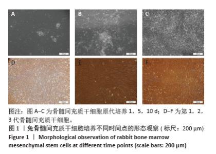
2.1 兔BMSCs的形态 采用全骨髓法获取的兔BMSCs形态呈长梭形,培养至第10天后细胞逐渐增多,簇集生长。经过传代培养后,细胞基本铺满整个T25瓶底,呈漩涡性生长,见图1。"
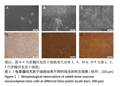

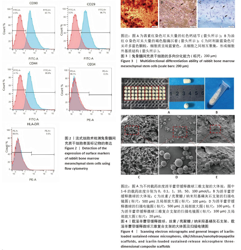
.2 兔BMSCs流式鉴定结果 流式细胞仪检测间充质干细胞表面标记物的表达:CD29(96.7%)、CD44(95.9%)、CD90(97.7%)阳性表达,CD34(0.3%)、HLA-DR(0.5%)阴性表达,符合兔BMSCs的免疫表型,见图2。 2.3 兔BMSCs多向分化能力鉴定 茜素红染色可见红色矿化钙结节;油红O染色可见大量的黄褐色沉着;阿利新蓝染色可见许多蓝色颗粒,细胞质呈现蓝紫色,且细胞之间相互聚集,形成细胞外基质结构,见图3。 2.4 载淫羊藿苷缓释微球、SF/CS/nHA支架、载药微球三维复合支架的扫描电镜及大体图 不同浓度淫羊藿苷缓释微球三维支架在外观上无显著差异,它们都是实心的白色圆柱体,大小可以根据需要塑形(图为SF/CS/nHA-ICA支架制作于96孔板中,高约1 cm,直径约0.5 cm);淫羊藿苷缓释微球低温真空冷冻干燥后为白色粉末。扫描电镜下观察支架为不规则形网状多孔结构,孔径相间,孔壁凹凸不平;微球呈直径不一的球形,高倍镜下观察可见微球表面有细小孔隙,淫羊藿苷缓释微球均匀分布于三维支架中,见图4。 "
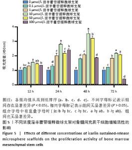
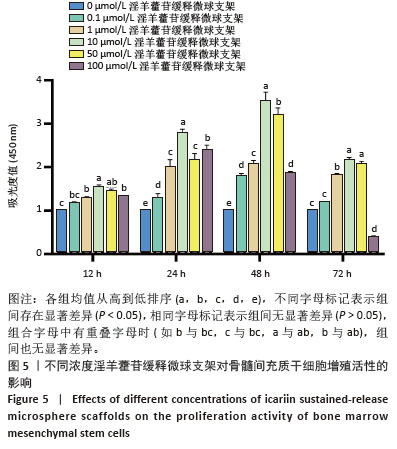
2.5 不同浓度淫羊藿苷缓释微球支架对兔BMSCs增殖能力的影响 培养12 h时,10,50 μmol/L淫羊藿苷缓释微球支架组吸光度值明显高于其余组(P < 0.05),0.1,1,100 μmol/L淫羊藿苷缓释微球支架组之间无明显差异;培养24,48 h时,0.1,1,10,50,100 μmol/L淫羊藿苷缓释微球支架组吸光度值明显高于对照组(P < 0.05),10 μmol/L淫羊藿苷缓释微球支架组最为显著;培养72 h时,1,10,50 μmol/L淫羊藿苷缓释微球支架组吸光度值明显高于对照组(P < 0.05),10,50 μmol/L淫羊藿苷缓释微球支架组之间无明显差异,100 μmol/L淫羊藿苷缓释微球支架组吸光度值明显低于其余组,见图5。"
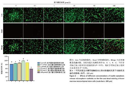
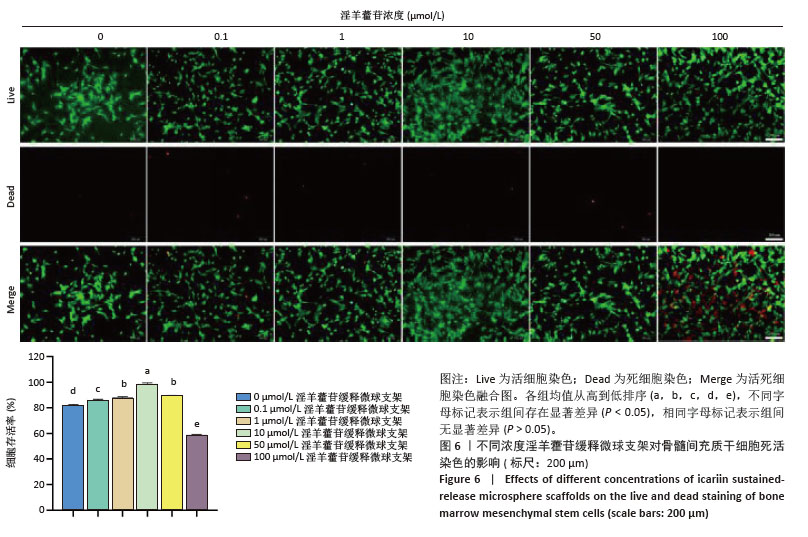
2.6 细胞死活染色结果 荧光显微镜下显示,载药支架浸取液培养48 h后,可见大量存活的骨髓间充质干细胞,为绿色荧光;除100 μmol/L淫羊藿苷缓释微球支架组有大量死细胞外,其他组可见少量死亡细胞,为红色荧光。实验表明,一定浓度范围内的淫羊藿苷缓释微球支架(0.1-50 μmol/L)对BMSCs具有促进增殖作用,高浓度淫羊藿苷缓释微球支架 (100 μmol/L)可能对BMSCs具有一定毒性作用,见图6。"
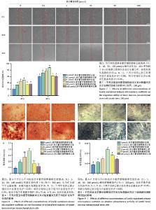
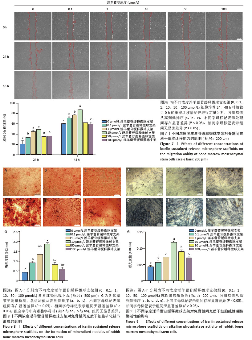
2.7 不同浓度淫羊藿苷缓释微球支架对BMSCs迁移能力的影响 细胞划痕实验显示:培养24 h时,0.1,10 μmol/L淫羊藿苷缓释微球支架组细胞迁移率较其余组明显(P < 0.05),而0.1,10 μmol/L淫羊藿苷缓释微球支架组间无差异(P > 0.05);培养48 h 时,10 μmol/L淫羊藿苷缓释微球支架组划痕区基本被迁移的细胞填满,而其他组虽较于24 h迁移的细胞增多,但10 μmol/L淫羊藿苷缓释微球支架组细胞迁移区域最大,说明淫羊藿苷可以促进骨髓间充质干细胞的迁移,且10 μmol/L为最佳缓释浓度,见图7。 2.8 茜素红染色结果 成骨诱导14 d后,茜素红染色显示,淫羊藿苷缓释微球支架组矿化结节数量均多于对照组。茜素红半定量检测结果显示,在0.1-10 μmol/L浓度范围内,随淫羊藿苷浓度增加,BMSCs形成的矿化结节数量越多,并与淫羊藿苷浓度呈正比关系,说明淫羊藿苷对BMSCs的成骨能力有促进作用;当淫羊藿苷浓度增加至50,100 μmol/L时,其成骨促进作用相较于0.1-10 μmol/L浓度有所降低,且50,100 μmol/L淫羊藿苷缓释微球支架组细胞状态变差(细胞脱落、翻边),说明高浓度淫羊藿苷(50,100 μmol/L)可能抑制细胞生长及抑制成骨分化,见图8A-G。 2.9 碱性磷酸酶染色结果 成骨诱导7 d后,碱性磷酸酶染色显示,不同浓度淫羊藿苷缓释微球支架均增强了BMSCs的成骨分化能力,当载药浓度升至10 μmol/L时,BMSCs的成骨分化能力更为显著,见图9A-F;碱性磷酸酶活性检测结果显示,不同浓度淫羊藿苷缓释微球支架组BMSCs的碱性磷酸酶活性均出现了显著上升(P < 0.05),当淫羊藿苷浓度增加至10 μmol/L,碱性磷酸酶活力上升最为显著(P < 0.05),见图9G。 "

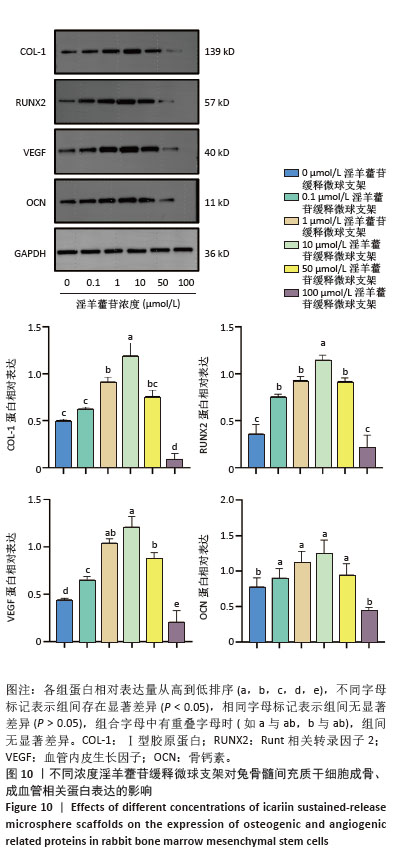
2.10 不同浓度淫羊藿苷缓释微球支架对BMSCs成骨及成血管相关蛋白表达的影响 成骨诱导14 d后,与对照组相比,0.1-50 μmol/L淫羊藿苷缓释微球支架组Ⅰ型胶原、Runt相关转录因子2、骨钙素、血管内皮生长因子蛋白表达量均明显增强,10 μmol/L最为显著,差异有显著性意义(P < 0.05),100 μmol/L淫羊藿苷缓释微球支架组Ⅰ型胶原、Runt相关转录因子2、骨钙素、血管内皮生长因子蛋白表达量低于对照组,见图10。 "
| [1] RODDY E, DEBAUN MR, DAOUD-GRAY A, et al. Treatment of critical-sized bone defects: clinical and tissue engineering perspectives. Eur J Orthop Surg Traumatol. 2018;28(3):351-362. [2] 倪建光,刘丹平.骨组织工程支架材料的研究进展[J].中国组织工程研究与临床康复,2009,13(8):1525-1528. [3] 谭嘉,陈国平,郝永强.生物3D打印的关键技术及骨科应用进展[J].中华骨科杂志,2020,40(2):110-118. [4] ZHOU M, YIN Y, ZHAO J, et al. Applications of microalga-powered microrobots in targeted drug delivery. Biomater Sci. 2023;11(23): 7512-7530. [5] HUANG H, LIN Y, JIANG Y, et al. Recombinant protein drugs-based intra articular drug delivery systems for osteoarthritis therapy. Eur J Pharm Biopharm. 2023;183:33-46. [6] 叶鹏,田仁元,黄文良,等.丝素/壳聚糖/纳米羟基磷灰石构建的骨组织工程支架[J].中国组织工程研究,2013,17(29):5269-5274. [7] 肖红利,黄文良,能坤,等.丝素蛋白壳聚糖和纳米羟基磷灰石制备骨软骨梯度孔径支架初探[J].中华老年医学杂志,2018,37(7): 816-820. [8] RUAN SQ, DENG J, YAN L, et al. Composite scaffolds loaded with bone mesenchymal stem cells promote the repair of radial bone defects in rabbit model. Biomed Pharmacother. 2018;97:600-606. [9] ZHANG J, MAO Y, RAO J. The SPI1/SMAD5 cascade in the promoting effect of icariin on osteogenic differentiation of MC3T3-E1 cells: a mechanism study. J Orthop Surg Res. 2024;19(1):444. [10] CHEN M, CUI Y, LI H, et al. Icariin Promotes the Osteogenic Action of BMP2 by Activating the cAMP Signaling Pathway. Molecules. 2019; 24(21):3875. [11] ZU Y, MU Y, LI Q, et al. Icariin alleviates osteoarthritis by inhibiting NLRP3-mediated pyroptosis. J Orthop Surg Res. 2019;14(1):307. [12] 程琳燕,金晓丽,陈煊威,等.淫羊藿苷通过RANKL-p38/ERK-NFAT通路抑制硫代乙酰胺诱导的破骨分化[J].中国中药杂志,2022, 47(21):5882-5889. [13] 梅杰,何强,孙欣,等.淫羊藿苷促进成骨细胞增殖分化的非核效应信号通路[J].中国组织工程研究,2023,27(20):3129-3135. [14] 李时斌,夏天,章晓云,等.淫羊藿活性单体成分调控骨质疏松症相关信号通路影响骨吸收与骨形成的稳态[J].中国组织工程研究, 2022,26(11):1772-1779. [15] 辛红美,许洁,汪长东.淫羊藿苷促进MC3T3-E1成骨分化通过Hedgehog信号通路[J].中国药理学通报,2020,36(5):616-620. [16] XIE Y, SUN W, YAN F, et al. Icariin-loaded porous scaffolds for bone regeneration through the regulation of the coupling process of osteogenesis and osteoclastic activity. Int J Nanomedicine. 2019;14: 6019-6033. [17] ZHANG X, LIN X, LIU T, et al. Osteogenic Enhancement Between Icariin and Bone Morphogenetic Protein 2: A Potential Osteogenic Compound for Bone Tissue Engineering. Front Pharmacol. 2019;10:201. [18] MOHAMMADZADEH M, ZAREI M, ABBASI H, et al. Promoting osteogenesis and bone regeneration employing icariin-loaded nanoplatforms. J Biol Eng. 2024;18(1):29. [19] LIU N, HUANG S, GUO F, et al. Calcium phosphate cement with icariin-loaded gelatin microspheres as a local drug delivery system for bone regeneration. Biomed Eng Online. 2022;21(1):89. [20] ZOU L, HU L, PAN P, et al. Icariin-releasing 3D printed scaffold for bone regeneration. Compos B Eng. 2022;232:109625. [21] 杨治航,孙祖延,黄文良,等.神经生长因子促进兔骨髓间充质干细胞软骨分化并抑制肥大分化[J].中国组织工程研究,2025,29(7): 1336-1342. [22] 李飞非,王布雨,杨治航,等.生长分化因子5诱导兔骨髓间充质干细胞的软骨分化[J].中国组织工程研究,2024,28(13):1976-1982. [23] XIAO H, HUANG W, XIONG K, et al. Osteochondral repair using scaffolds with gradient pore sizes constructed with silk fibroin, chitosan, and nano-hydroxyapatite. Int J Nanomedicine. 2019;14:2011-2027. [24] TIN TIN HTAR M, MADHAVA H, BALMER P, et al. A review of the impact of pneumococcal polysaccharide conjugate vaccine (7-valent) on pneumococcal meningitis. Adv Ther. 2013;30(8):748-762. [25] KRAUS KH, KIRKER‐HEAD C. Mesenchymal stem cells and bone regeneration. Vet Surg. 2006;35(3):232-242. [26] WEN Z, ZHENG S, ZHOU C, et al. Repair mechanisms of bone marrow mesenchymal stem cells in myocardial infarction. J Cell Mol Med. 2011;15(5):1032-1043. [27] WADA N, GRONTHOS S, BARTOLD PM. Immunomodulatory effects of stem cells. Periodontol 2000. 2013;63(1):198-216. [28] ABO-ZAID AE, EL RAZZAK MYA, SARHAN NI, et al. Evaluation of bone regeneration using human derived-gingival mesenchymal stem cells loaded on beta tricalcium phosphate and hyaluronic acid: An experimental study. Tanta Dent J. 2024;21(1):60-65. [29] SEONG JM, KIM BC, PARK JH, et al. Stem cells in bone tissue engineering. Biomed Mater. 2010;5(6):062001. [30] AISAITI A, AIERXIDING S, SHOUKEER K, et al. Mesenchymal stem cells for peripheral nerve injury and regeneration: a bibliometric and visualization study. Front Neurol. 2024;15:1420402. [31] LI M, YU B, WANG S, et al. Microenvironment-responsive nanocarriers for targeted bone disease therapy. Nano Today. 2023;50:101838. [32] LI X, SHU X, SHI Y, et al. MOFs and bone: Application of MOFs in bone tissue engineering and bone diseases. Chin Chem Lett. 2023;34(7): 107986. [33] 张志荣,董尔丹,吴镭,等.生物大分子药物递送系统研究现状与前沿方向[J].中国基础科学,2014,16(5):3-8. [34] 李秀英,曾凡,赵曜,等.脂质体药物递送系统的研究进展[J].中国新药杂志,2014,23(16):1904-1911,1917. [35] CHEN X, DONG J, MA S, et al. Targeting agents used in specific bone-targeting drug delivery systems: A review. Sci Adv Mater. 2022;14(4): 613-621. [36] 张铭笑,孙春萌.骨靶向药物递送系统研究进展[J].药学进展, 2023,47(8):637-644. [37] 吴梅剑宗,秦佳佳,刘海全.不同浓度淫羊藿苷对大鼠脂肪干细胞成骨分化相关因子的体外研究[J].北京中医药大学学报,2022, 45(12):1249-1256. [38] 吴迪,米甜,王贺东,等.淫羊藿素的药理作用及分子机制研究进展[J].医学研究与教育,2023,40(6):1-11. [39] 姜涛,凌翠敏,陈庆真,等.淫羊藿苷通过提高自噬促进成骨细胞分化防治骨质疏松[J].中国组织工程研究,2021,25(17):2643-2649. [40] MA HP, MA XN, GE BF, et al. Icariin attenuates hypoxia-induced oxidative stress and apoptosis in osteoblasts and preserves their osteogenic differentiation potential in vitro. Cell Prolif. 2014;47(6): 527-539. [41] 四川大学.一种具有缓释和促进成骨功能的3D打印骨组织工程支架及其制备方法与应用: CN202010801668.1[P].2021-11-30. [42] 何思良.水凝胶/微球/淫羊藿苷复合载药体系的制备及性能研究[D].湘潭: 湖南科技大学,2016. [43] 薛鹏,杜斌,王礼宁,等.可控释淫羊藿苷-β-磷酸三钙复合支架的制备[J].中国组织工程研究,2018,22(6):865-870. [44] 张孟伟.淫羊藿苷/多孔镁合金新型支架通过Wnt/β-catenin信号通路修复大鼠膝关节软骨缺损的实验研究[D].汕头:汕头大学, 2022. [45] 刘琪,李林臻,张君涛.淫羊藿苷促进关节软骨损伤修复作用机制研究进展[J].药物评价研究,2024,47(6):1393-1399. |
| [1] | Jiang Xinghai, Song Yulin, Li Dejin, Shao Jianmin, Xu Junzhi, Liu Huakai, Wu Yingguo, Shen Yuehui, Feng Sicheng. Vascular endothelial growth factor 165 genes transfected into bone marrow mesenchymal stem cells to construct a vascularized amphiphilic peptide gel module [J]. Chinese Journal of Tissue Engineering Research, 2026, 30(8): 1903-1911. |
| [2] | Wang Qisa, Lu Yuzheng, Han Xiufeng, Zhao Wenling, Shi Haitao, Xu Zhe. Cytocompatibility of 3D printed methyl acrylated hyaluronic acid/decellularized skin hydrogel scaffolds [J]. Chinese Journal of Tissue Engineering Research, 2026, 30(8): 1912-1920. |
| [3] | Sun Lei, Zhang Qi, Zhang Yu. Pro-osteoblastic effect of chlorogenic acid protein microsphere/polycaprolactone electrospinning membrane [J]. Chinese Journal of Tissue Engineering Research, 2026, 30(8): 1877-1884. |
| [4] | Wu Yanting, Li Yu, Liao Jinfeng. Magnesium oxide nanoparticles regulate osteogenesis- and angiogenesis-related gene expressions to promote bone defect healing [J]. Chinese Journal of Tissue Engineering Research, 2026, 30(8): 1885-1895. |
| [5] | Hu Xiongke, Liu Shaohua, Tan Qian, Liu Kun, Zhu Guanghui. Shikonin intervention with bone marrow mesenchymal stem cells improves microstructure of femur in aged mice [J]. Chinese Journal of Tissue Engineering Research, 2026, 30(7): 1609-1615. |
| [6] | Yuan Xiaoshuang, Yang Xu, Yang Bo, Chen Xiaoxu, Tian Ting, Wang Feiqing, Li Yanju, Liu Yang, Yang Wenxiu. Effect of conditioned medium of diffuse large B-cell lymphoma cells on proliferation and apoptosis of human bone marrow mesenchymal stem cells [J]. Chinese Journal of Tissue Engineering Research, 2026, 30(7): 1632-1640. |
| [7] | Li Zhenyu, Zhang Siming, Bai Jiaxiang, Zhu Chen. Osthole improves osteogenic differentiation function of bone marrow mesenchymal stem cells under high-glucose conditions [J]. Chinese Journal of Tissue Engineering Research, 2026, 30(7): 1641-1648. |
| [8] | Han Nianrong, Huang Yifei, Akram · Osman, Liu Yanlu, Hu Wei . Programmed cell death receptor-1 suppresses osteogenic differentiation of rat bone marrow mesenchymal stem cells in a high-glucose microenvironment [J]. Chinese Journal of Tissue Engineering Research, 2026, 30(7): 1649-1657. |
| [9] | Zou Yulian, Chen Chaopei, Huang Haixia, Lan Yuyan, Liu Min, Huang Ting. Resveratrol promotes osteogenic differentiation of bone marrow mesenchymal stem cells in an inflammatory microenvironment [J]. Chinese Journal of Tissue Engineering Research, 2026, 30(7): 1669-1678. |
| [10] | Li Linzhen, Jiao Hongzhuo, Chen Weinan, Zhang Mingzhe, Wang Jianlong, Zhang Juntao. Effect of icariin-containing serum on lipopolysaccharide-induced inflammatory damage in human chondrocytes [J]. Chinese Journal of Tissue Engineering Research, 2026, 30(6): 1368-1374. |
| [11] | Huang Liuyan, Zhang Wenxi, Chen Shuwen, Yu Shimei, Dai Zhong, Zuo Changqing. Forskolin promotes C2C12 myoblast differentiation via regulating the ERK and Akt signaling pathways [J]. Chinese Journal of Tissue Engineering Research, 2026, 30(5): 1114-1121. |
| [12] | Zhou Zixiang, Zhao Baoxiang. Research progress in the relationship between nontraumatic necrosis of the femoral head and lipid metabolism and its treatment [J]. Chinese Journal of Tissue Engineering Research, 2026, 30(3): 680-690. |
| [13] | Yan Qiquan, Yang Libin, Li Mengjun, Ni Yazhuo, Chen Keying, Xu Bo, Li Yaoyang, Ma Shiqing, Li Rui, Li Jianwen. Preparation and antibacterial properties of porcine small intestinal submucosal composite nanohydroxyapatite bioscaffold loaded with antimicrobial peptide KR-12-a5 [J]. Chinese Journal of Tissue Engineering Research, 2026, 30(2): 384-394. |
| [14] | Chen Qiheng, Weng Tujun, Peng Jiang. Effect of dimethylglyoxal glycine on osteogenic, adipogenesis differentiation, and mitophagy of human bone marrow mesenchymal stem cells [J]. Chinese Journal of Tissue Engineering Research, 2026, 30(1): 50-57. |
| [15] | Zhang Tingting, Li Yalong, Yue Haodi, Li Yanjun, Geng Xiwen, Zhang Yuwei, Liu Xiaozhuan. Protection of exosomes derived from bone marrow mesenchymal stem cells of different mouse ages on radiation-induced lung injury [J]. Chinese Journal of Tissue Engineering Research, 2026, 30(1): 1-9. |
| Viewed | ||||||
|
Full text |
|
|||||
|
Abstract |
|
|||||