Chinese Journal of Tissue Engineering Research ›› 2026, Vol. 30 ›› Issue (33): 8712-8720.doi: 10.12307/2026.465
Previous Articles Next Articles
Mechanisms of secondary demyelinating injury and inflammatory response in mice with acute ischemic stroke
Zhang Ziwei1, Pu Meng1, Tang Yibin1, Zheng Lulu1, Liang Yajie1, Liu Jian1, Chen Ying1, Wang Qing1, Li Yanqing1, Li Yanhua1, 2
- 1The Key Research Laboratory of Benefiting Qi for Acting Blood Circulation Method to Treat Multiple Sclerosis, State Administration of Traditional Chinese Medicine/Neurobiology Research Center, Shanxi University of Chinese Medicine, Jinzhong 030619, Shanxi Province, China; 2School of Medicine, Shanxi Datong University, Datong 037009, Shanxi Province, China
-
Received:2025-10-17Revised:2026-02-27Online:2026-11-28Published:2026-06-15 -
Contact:Wang Qing, Associate professor, Master’s supervisor, The Key Research Laboratory of Benefiting Qi for Acting Blood Circulation Method to Treat Multiple Sclerosis, State Administration of Traditional Chinese Medicine/Neurobiology Research Center, Shanxi University of Chinese Medicine, Jinzhong 030619, Shanxi Province, China -
About author:Zhang Ziwei, MS, The Key Research Laboratory of Benefiting Qi for Acting Blood Circulation Method to Treat Multiple Sclerosis, State Administration of Traditional Chinese Medicine/Neurobiology Research Center, Shanxi University of Chinese Medicine, Jinzhong 030619, Shanxi Province, China -
Supported by:Shanxi Province Research Support Program for Returning Overseas Scholars, No. 2022-165 (to WQ); Shanxi Provincial Administration of Traditional Chinese Medicine Innovation Team, No. zyytd2024039 (to WQ); Shanxi Provincial Administration of Traditional Chinese Medicine Research Project, No. 2023ZYYB040 (to WQ)

CLC Number:
Cite this article
Zhang Ziwei, Pu Meng, Tang Yibin, Zheng Lulu, Liang Yajie, Liu Jian, Chen Ying, Wang Qing, Li Yanqing, Li Yanhua. Mechanisms of secondary demyelinating injury and inflammatory response in mice with acute ischemic stroke[J]. Chinese Journal of Tissue Engineering Research, 2026, 30(33): 8712-8720.
share this article
Add to citation manager EndNote|Reference Manager|ProCite|BibTeX|RefWorks
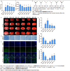
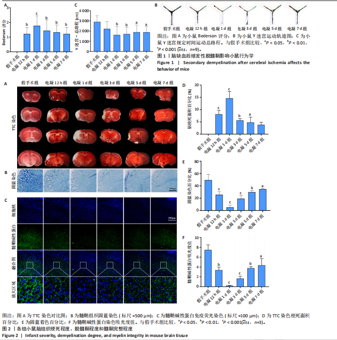
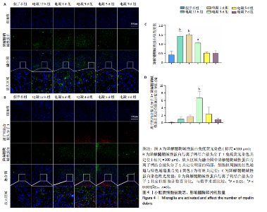
2.1 实验动物数量分析 共54只昆明小鼠参与实验,其中9只进行假手术,45只采用电凝法制备模型,在术后恢复过程中,所有小鼠的生命体征良好,45只小鼠成功构建远端大脑中动脉闭塞模型,54只小鼠均进入结果分析。 2.2 缺血后继发性脱髓鞘影响小鼠行为学 2.2.1 Bederson评分 Bederson评分用来反映小鼠的神经功能缺损情况。如图1A所示,假手术组小鼠未出现神经功能缺损,Bederson评分为0;以假手术组为对照,电凝12 h,1 d,3 d,5 d,7 d组小鼠Bederson评分有不同程度的升高(P < 0.01,P < 0.001,P < 0.01,P < 0.01,P < 0.01)。电凝1 d组的小鼠行为学评分最高,提示小鼠神经功能缺损在第1天达到峰值,后续几天有所恢复。 通过Bederson评分反映小鼠的神经功能缺损情况从而评价小鼠的运动功能,而运动功能结果的异常会干扰认知功能测试的结果。在此实验中,Bederson评分结果显示小鼠运动功能受损,而接下来的Y迷宫实验结果不能判断小鼠在认知功能方面的差异性,只能将Y迷宫实验结果作为评判运动功能的辅助结论。 2.2.2 Y迷宫实验结果 如图1B、C所示,选取总距离作为小鼠运动量的评价标准,以假手术组为对照,电凝组均出现不同程度的运动量减少;其中电凝1,3,5,7 d组的运动量出现显著性差异(P < 0.01,P < 0.01,P < 0.05,P < 0.05),电凝1 d组的小鼠运动量最少。Y迷宫结果与Bederson评分结果结合判断,提示小鼠的运动功能在第1天受累最严重,后续几天有所恢复。 2.3 脑梗死程度、脱髓鞘程度和髓鞘完整程度 2.3.1 脑梗死程度 TTC染色用来评估小鼠的脑梗死程度,红色为非梗死区域、白色为梗死区域。如图2A、D所示,以假手术组为对照,各电凝组均出现不同程度的梗死,电凝12 h组、电凝1,3,5 d组出现显著性差异(P < 0.001,P < 0.001,P < 0.01,P < 0.05),其中电凝1 d组的梗死面积达到峰值,电凝7 d组的小鼠梗死面积没有显著性差异(P=0.056 8)。TTC染色结果表明小鼠的缺血程度在第1天受累最严重,波及缺血侧大部分大脑皮质,后续几天有所恢复。 由于脱髓鞘受缺血程度影响,接下来的固蓝染色及免疫荧光染色将以TTC染色峰值组(1 d组)的皮质梗死面积区域作为参照进行重点分析。 2.3.2 脱髓鞘程度 固蓝染色用来评估小鼠的脱髓鞘程度,蓝色着色越深代表髓鞘结构越完整。如图2B、E所示,以假手术组为对照,电凝12 h组、电凝1,3,5,7 d组受缺血累及的皮质区域固蓝着色均有不同程度减少(P < 0.001,P < 0.001,P < 0.001,P < 0.01,P < 0.05),电凝1 d组着色最少,固蓝染色趋势与TTC染色相同。但值得注意的是:第7天时,在TTC染色没有显著性差异的情况下,固蓝染色仍然有显著性差异,由于髓鞘修复与少突胶质细胞前体细胞的分化相关,但是少突胶质细胞前体细胞分化能力有限,通常只能补充生理状态下的髓鞘修复,而不能满足病理状态下的大面积髓鞘修复,这提示髓鞘严重受到缺血程度影响,髓鞘恢复程度不良。 2.3.3 髓鞘完整程度 髓鞘碱性蛋白染色用来评估小鼠的髓鞘完整程度。如图2C、F所示,以假手术组为对照,各电凝组吸光度值均降低(P < 0.05),电凝1 d组吸光度值最低。髓鞘碱性蛋白染色与固蓝染色趋势相同,提示各电凝组受累及的皮质区域出现脱髓鞘现象。 2.4 脑梗死引发炎症反应 2.4.1 小鼠梗死侧半脑出现炎症反应 如图3A所示,以假手术组为对照,电凝组白细胞介素6水平呈现上升趋势,在1 d达到峰值(P < 0.001),在5-7 d时无显著差异;如图3B所示,以假手术组为对照,电凝组肿瘤坏死因子α水平呈现上升趋势,在3 d达到峰值(P < 0.001),在7 d时无显著差异;如图3C所示,以假手术组为对照,电凝组白细胞介素1β水平呈现上升趋势,在1-5 d达到峰值(P < 0.001),在7 d时无显著差异;如图3D所示,以假手术组为对照,电凝组干扰素γ水平呈现上升趋势,在3 d达到峰值(P < 0.001),在7 d时仍然具有显著性差异(P < 0.01);如图3E所示,与假手术组相比,电凝组白细胞介素10水平在1 d出现显著性差异(P < 0.05),其余各时间点均未出现显著性差异;各项结果提示小鼠梗死半脑出现炎症反应。 2.4.2 小胶质细胞被激活 离子钙结合接头分子1是小胶质细胞标志物,染色结果如图3F、G所示,以假手术组为对照,各电凝组吸光度值增加,电凝3-5 d组增高明显(P < 0.001),提示小胶质细胞被大量激活;电凝7 d组相较于3-5 d组,吸光度值呈现减少趋势,但以假手术组为对照,仍然具有统计学差异(P < 0.05)。 2.5 小胶质细胞对髓鞘碎片的影响 为了探究小胶质细胞与脱髓鞘的关系,分别对小鼠半脑梗死区进行了荧光降解髓鞘碱性蛋白单染以及降解髓鞘碱性蛋白-离子钙结合接头分子1共染。 降解髓鞘碱性蛋白是髓鞘碎片标志物,代表脱髓鞘的严重程度。如图4A、C所示,以假手术组为对照,各电凝组吸光度值整体呈现升高趋势,电凝12 h组、电凝1,3 d组降解髓鞘碱性蛋白吸光度值升高趋势明显(P < 0.001,P < 0.001,P < 0.01),电凝5,7 d组降解髓鞘碱性蛋白吸光度值已无显著差异(P=0.893,P=0.940 8)。降解髓鞘碱性蛋白单染结果提示模型小鼠出现脱髓鞘现象;脱髓鞘趋势表明髓鞘碎片被清除,可能与机体自身具备的清除作用相关。"
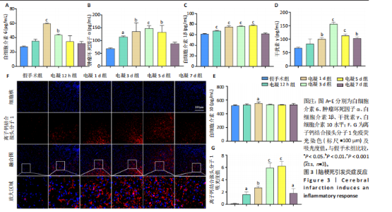
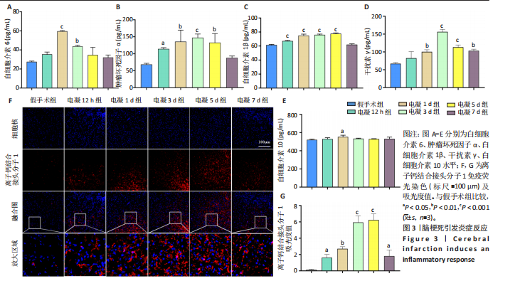
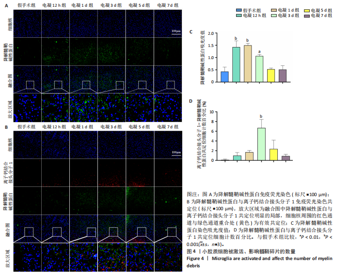
降解髓鞘碱性蛋白-离子钙结合接头分子1共染结果如图4B、D所示,以假手术组为对照,降解髓鞘碱性蛋白-离子钙结合接头分子1共定位数量百分比在电凝3 d时出现显著性差异(P < 0.001),电凝12 h组、电凝1,5,7 d组未出现显著性差异(P=0.780 2,P=0.308 8,P=0.093 7,P=0.835 7)。综合降解髓鞘碱性蛋白单染、降解髓鞘碱性蛋白-离子钙结合接头分子1共染结果分析,提示小胶质细胞对髓鞘碎片数量有影响,髓鞘碎片清除可能与小胶质细胞的吞噬、清除作用有关。 2.6 炎症反应激活Janus激酶2-信号转导与转录激活因子3信号通路 离子钙结合接头分子1-Janus激酶2共染结果如图5A、C所示,以假手术组为对照,电凝1 d组的共定位数量百分比增多,出现显著性差异(P < 0.01)。离子钙结合接头分子1-信号转导与转录激活因子3共染结果如图5B、D所示,以假手术组为对照,电凝1,3 d组的共定位数量百分比增多,出现显著性差异(P < 0.01)。两项共定位结果提示小胶质细胞中Janus激酶2-信号转导与转录激活因子3信号通路被激活。"
"
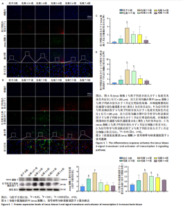
2.7 Janus激酶2、信号转导与转录激活因子3蛋白表达上调 如图6所示,以假手术组为对照,Janus激酶2相对蛋白表达量在电凝12 h组、1 d组呈现显著性差异(P < 0.01和P < 0.001),信号转导与转录激活因子3相对蛋白表达量在电凝1 ,3 ,5 d组呈现显著性差异(P < 0.01,P < 0.05和P < 0.05)。"

| [1] SHI H, HU X, LEAK RK, et al. Demyelination as a rational therapeutic target for ischemic or traumatic brain injury. Exp Neurol. 2015;272:17-25. [2] LI S, RAO JH, LAN XY, et al. White matter demyelination predates axonal injury after ischemic stroke in cynomolgus monkeys. Exp Neurol. 2021;340:113655. [3] LI Y, SU P, CHEN Y, et al. The Eph receptor A4 plays a role in demyelination and depression-related behavior. J Clin Invest. 2022;132(10):e161559. [4] HUANG S, REN C, LUO Y, et al. New insights into the roles of oligodendrocytes regulation in ischemic stroke recovery. Neurobiol Dis. 2023;184:106200. [5] RAFFAELE S, CLAUSEN BH, MANNELLA FC, et al. Characterisation of GPR17-expressing oligodendrocyte precursors in human ischaemic lesions and correlation with reactive glial responses. J Pathol. 2025;265(2):226-243. [6] SHEN Z, XIANG M, CHEN C, et al. Glutamate excitotoxicity: Potential therapeutic target for ischemic stroke. Biomed Pharmacother. 2022;151:113125. [7] 李朝唯,孔令雷,秦雪梅,等.基于铁死亡与神经炎症调节神经退行性疾病的靶点及药物研究进展[J].药学学报,2025,60(5):1325-1343. [8] XU B, SHIMAUCHI-OHTAKI H, YOSHIMOTO Y, et al. Transplanted human iPSC-derived vascular endothelial cells promote functional recovery by recruitment of regulatory T cells to ischemic white matter in the brain. J Neuroinflammation. 2023;20(1):11. [9] ZHANG Y, LI J, ZHAO Y, et al. Arresting the bad seed: HDAC3 regulates proliferation of different microglia after ischemic stroke. Sci Adv. 2024;10(10):eade6900. [10] CAO Q, CHEN J, ZHANG Z, et al. Astrocytic CXCL5 hinders microglial phagocytosis of myelin debris and aggravates white matter injury in chronic cerebral ischemia. J Neuroinflammation. 2023;20(1):105. [11] MERIGHI S, NIGRO M, TRAVAGLI A, et al. Microglia and Alzheimer’s Disease. Int J Mol Sci. 2022;23(21):12990. [12] BECCARI S, SIERRA-TORRE V, VALERO J, et al. Microglial phagocytosis dysfunction in stroke is driven by energy depletion and induction of autophagy. Autophagy. 2023;19(7):1952-1981. [13] 仝国栋,朱卿昊,王军,等.脑型疟炎症微环境诱导星形胶质细胞活化及对神经元损伤的研究[J].中国寄生虫学与寄生虫病杂志,2024,42(2):160-168. [14] 曹兴兵.EMSCs-sEVs、ETM-sEVs和AS-sEVs调控炎症微环境促进脊髓损伤修复的机制研究[D].南京:南京医科大学,2023. [15] 吴文成.炎症微环境下外泌体介导的小胶质细胞调控OPCs分化的作用机制研究[D].西安:陕西师范大学,2022. [16] 梁彦峰,郝鹏,段红梅,等.成年小鼠缺血性脑卒中后病理及行为功能的变化[J].中国组织工程研究,2020,24(35):5625-5631. [17] WANG L, YAO C, CHEN J, et al. γδ T Cell in Cerebral Ischemic Stroke: Characteristic, Immunity-Inflammatory Role, and Therapy. Front Neurol. 2022;13:842212. [18] SHEN H, PEI H, ZHAI L, et al. Salvianolic acid C improves cerebral ischemia reperfusion injury through suppressing microglial cell M1 polarization and promoting cerebral angiogenesis. Int Immunopharmacol. 2022;110:109021. [19] ORIHUELA R, MCPHERSON CA, HARRY GJ. Microglial M1/M2 polarization and metabolic states. Br J Pharmacol. 2016;173(4):649-665. [20] MA H, LI H, ZHANG Y, et al. Microglia Exhibit Distinct Heterogeneity Rather than M1/M2 Polarization within the Early Stage of Acute Ischemic Stroke. Aging Dis. 2023;14(6):2284-2302. [21] LIAN L, ZHANG Y, LIU L, et al. Neuroinflammation in Ischemic Stroke: Focus on MicroRNA-mediated Polarization of Microglia. Front Mol Neurosci. 2021;13:612439. [22] PLANAS AM. Role of microglia in stroke. Glia. 2024;72(6):1016-1053. [23] CIPRIANI R, DOMERQ M, MARTÍN A, et al. Role of Microglia in Stroke. Adv Neurobiol. 2024;37:405-422. [24] POPESCU AS, BUTLER CA, ALLENDORF DH, et al. Alzheimer’s disease-associated R47H TREM2 increases, but wild-type TREM2 decreases, microglial phagocytosis of synaptosomes and neuronal loss. Glia. 2023;71(4):974-990. [25] SHIMIZU T, SCHUTT CR, IZUMI Y, et al. Direct activation of microglia by β-glucosylceramide causes phagocytosis of neurons that exacerbates Gaucher disease. Immunity. 2023;56(2):307-319.e8. [26] PAMPUSCENKO K, JANKEVICIUTE S, MORKUNIENE R, et al. S100A9 protein activates microglia and stimulates phagocytosis, resulting in synaptic and neuronal loss. Neurobiol Dis. 2025;206:106817. [27] XING C, LV J, ZHU Z, et al. Regulation of microglia related neuroinflammation contributes to the protective effect of Gelsevirine on ischemic stroke. Front Immunol. 2023;14:1164278. [28] 童雨婷,刘新娟,杨光,等.金松双黄酮抑制JAK2/STAT3通路对脂多糖诱导的BV2小胶质细胞炎症反应的影响[J].上海中医药杂志,2024,58(5): 73-77+100. [29] 刘伟,方军,张抗抗.二氢杨梅素通过JAK2/STAT3通路调控小胶质细胞活化对异氟醚所致成年小鼠早期认知功能障碍的影响[J].中国免疫学杂志, 2023,39(10):2154-2159. [30] 王英,巩子汉,梁文青,等.基于JAK2/STAT3信号通路探讨温阳解郁方调节小鼠海马小胶质细胞激活的机制[J].中国实验方剂学杂志,2025,31(8):88-96. [31] 王棒,吴娟,陈硕硕,等.白细胞介素-1β通过IL-6/JAK2/STAT3轴促进脑胶质瘤细胞的侵袭[J].中国实验诊断学,2023,27(5):596-602. [32] 张日云,吴凤兰,陈月桥,等.基于IL-6/JAK2/STAT3轴探讨解毒化瘀颗粒对急性肝衰竭炎症反应的作用机制[J/OL].辽宁中医杂志,1-16[2025-08-25].https://link.cnki.net/urlid/21.1128.R.20250819.1508.002. [33] HUA M, GAO P, FANG F, et al. IL-6 enhances the phagocytic function of mouse alveolar macrophages by activating the JAK2/STAT3 signaling pathway. Xi Bao Yu Fen Zi Mian Yi Xue Za Zhi. 2024;40(1):13-18. [34] HE GL, LUO Z, SHEN TT, et al. Inhibition of STAT3- and MAPK-dependent PGE synthesis ameliorates phagocytosis of fibrillar β-amyloid peptide (1-42) via EP2 receptor in EMF-stimulated N9 microglial cells. J Neuroinflammation. 2016;13(1):296. [35] NAKAMURA K, AGO T. Pericyte-Mediated Molecular Mechanisms Underlying Tissue Repair and Functional Recovery after Ischemic Stroke. J Atheroscler Thromb. 2023;30(9):1085-1094. [36] CUI MY, FU YQ, LI ZL, et al. Neuregulin-1/PI3K signaling effects on oligodendrocyte proliferation, remyelination and behaviors deficit in a male mouse model of ischemic stroke. Exp Neurol. 2023;362:114323. [37] LI J, WANG C, ZHANG Y, et al. Orchestrating the frontline: HDAC3-miKO recruits macrophage reinforcements for accelerated myelin debris clearance after stroke. Theranostics. 2025;15(2):632-655. [38] LIAO Y, CHENG J, KONG X, et al. HDAC3 inhibition ameliorates ischemia/reperfusion-induced brain injury by regulating the microglial cGAS-STING pathway. Theranostics. 2020;10(21):9644-9662. [39] 李曼菁.基于“小胶质细胞-神经干细胞”单元的双氢青蒿素促进EAE髓鞘再生的机制研究[D].北京:中国中医科学院,2023. [40] 雷楚.不同来源BM-MSCs对脱髓鞘模型髓鞘修复及情感障碍影响及机制的初步研究[D].重庆:第三军医大学,2016. |
| [1] | Wang Zheng, Cheng Ji, Yu Jinlong, Liu Wenhong, Wang Zhaohong, Zhou Luxing. Progress and future perspectives on the application of hydrogel materials in stroke therapy [J]. Chinese Journal of Tissue Engineering Research, 2026, 30(8): 2081-2090. |
| [2] | Xia Linfeng, Wang Lu, Long Qianfa, Tang Rongwu, Luo Haodong, Tang Yi, Zhong Jun, Liu Yang. Human umbilical cord mesenchymal stem cell-derived exosomes alleviate blood-brain barrier damage in mice with septic encephalopathy [J]. Chinese Journal of Tissue Engineering Research, 2026, 30(7): 1711-1719. |
| [3] | Cui Lianxu, Li Haomin, Xu Junrong, Tan Baodong, Lu Dahong, Peng Siwei, Wang Jinhui. Effect of umbilical cord mesenchymal stem cell conditioned medium on tissue repair after traumatic craniocerebral injury in miniature pigs [J]. Chinese Journal of Tissue Engineering Research, 2026, 30(7): 1730-1735. |
| [4] | Tao Daiju, Su Haiyu, Wang Yuqi, Shen Zhiqiang, He Bo . Construction and identification of stable PC12 cell lines with high/low expression of miR-122-5p [J]. Chinese Journal of Tissue Engineering Research, 2026, 30(7): 1790-1799. |
| [5] | Yin Yongcheng, Zhao Xiangrui, Yang Zhijie, Li Zheng, Li Fang, Ning Bin. Effect and mechanism of peroxiredoxin 1 in microglial inflammation after spinal cord injury [J]. Chinese Journal of Tissue Engineering Research, 2026, 30(5): 1106-1113. |
| [6] | Liu Yu, Lei Senlin, Zhou Jintao, Liu Hui, Li Xianhui. Mechanisms by which aerobic and resistance exercises improve obesity-related cognitive impairment [J]. Chinese Journal of Tissue Engineering Research, 2026, 30(5): 1171-1183. |
| [7] | Yang Jiao, Wang Zhifeng, Shi Ting, Xu Shuangfeng, Tian Daman, Lan Junfeng, He Pengfen, Hao Zhihui, Pan Pan, Wang Jian. Electroacupuncture regulates mitochondrial copper homeostasis to modulate neuroinflammatory injury in rats with ischemic stroke [J]. Chinese Journal of Tissue Engineering Research, 2026, 30(36): 9519-9525. |
| [8] | Su Xu, Zhang Xiaoxi, Yang Yaqing, Fu Zhenyi, Liu Jiaxin. Mechanism by which non-apoptotic regulated cell death induces neuronal injury in ischemic stroke [J]. Chinese Journal of Tissue Engineering Research, 2026, 30(35): 9281-9293. |
| [9] | Li Xiaoyan, Li Jinglin, Zhang Qiujuan, Zhang Xiaolina, Yang Li. MicroRNA-23a-3p improves neurological function in mice with traumatic brain injury by regulating microglial polarization [J]. Chinese Journal of Tissue Engineering Research, 2026, 30(35): 9206-9216. |
| [10] | . Regional specificity of brain organoids and their application in ischemic stroke modeling and drug development [J]. Chinese Journal of Tissue Engineering Research, 2026, 30(34): 9008-9016. |
| [11] | Shi Linjuan, Xu Xiaoshan, Zhai Chuntao, Li Wei, Tian Yuefeng. Herbal cake-separated moxibustion improves cognitive function in immunosuppressed rats [J]. Chinese Journal of Tissue Engineering Research, 2026, 30(33): 8744-8750. |
| [12] | Zheng Ying, Li Mengyao, Zheng Fanfan, He Zhao, Zhang Ning, Zou Jialun, Li Youlei, Gao Feng. Mechanism of action of extracellular vesicles loaded with biomaterials in repairing spinal cord injury [J]. Chinese Journal of Tissue Engineering Research, 2026, 30(32): 8544-8554. |
| [13] | Wang Degang, Mei Junhua, Wang Junli, Zheng Li, Chen Guohua. Bibliometric and visualization analysis of the mechanism of osteogenic factors and neurotransmitters in the bone-brain axis [J]. Chinese Journal of Tissue Engineering Research, 2026, 30(29): 7724-7731. |
| [14] | Gao Hongmei, Zhang Kun, Xiao Dongjie, Liu Hua. Human adipose multilineage-differentiating stress-enduring cells on treatment of ischemic stroke in rats [J]. Chinese Journal of Tissue Engineering Research, 2026, 30(19): 4853-4859. |
| [15] | Wu Xue, Zhang Linao, Luo Shifang, Liu Feifan, Wan Yan, Bai Yuanmei, Cao Julin, Xie Yuhuan, Guo Peixin. Dandeng Tongnao soft capsules against ischemic stroke: fingerprinting and network pharmacological analysis of efficacy and mechanism of action [J]. Chinese Journal of Tissue Engineering Research, 2026, 30(17): 4517-4528. |
| Viewed | ||||||
|
Full text |
|
|||||
|
Abstract |
|
|||||