Chinese Journal of Tissue Engineering Research ›› 2026, Vol. 30 ›› Issue (35): 9206-9216.doi: 10.12307/2026.281
Previous Articles Next Articles
MicroRNA-23a-3p improves neurological function in mice with traumatic brain injury by regulating microglial polarization
Li Xiaoyan1, Li Jinglin1, Zhang Qiujuan1, Zhang Xiaolina2, Yang Li1
- 1Department of Emergency Medicine, ²Department of Pain Management, First Affiliated Hospital of Kunming Medical University, Kunming 650000, Yunnan Province, China
-
Received:2025-09-17Revised:2026-01-26Online:2026-12-18Published:2026-04-28 -
Contact:Yang Li, MD, Department of Emergency Medicine, First Affiliated Hospital of Kunming Medical University, Kunming 650000, Yunnan Province, China -
About author:Li Xiaoyan, MS, Department of Emergency Medicine, First Affiliated Hospital of Kunming Medical University, Kunming 650000, Yunnan Province, China Li Jinglin, MS, Department of Emergency Medicine, First Affiliated Hospital of Kunming Medical University, Kunming 650000, Yunnan Province, China Li Xiaoyan and Li Jinglin contributed equally to this article. -
Supported by:Young Scientists Fund of Basic Research Special Program of Yunnan Provincial Science and Technology Department, No. 202301AU070164 (to YL); Basic Research Special Program of Yunnan Provincial Science and Technology Department (General Program), No. 202401AT070067 (to ZXLN)
CLC Number:
Cite this article
Li Xiaoyan, Li Jinglin, Zhang Qiujuan, Zhang Xiaolina, Yang Li. MicroRNA-23a-3p improves neurological function in mice with traumatic brain injury by regulating microglial polarization[J]. Chinese Journal of Tissue Engineering Research, 2026, 30(35): 9206-9216.
share this article
Add to citation manager EndNote|Reference Manager|ProCite|BibTeX|RefWorks

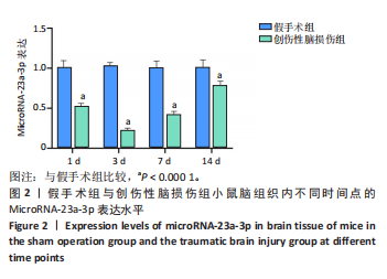
2.1 实验动物数量分析 研究共纳入80只C57BL/6J小鼠,分别观察4个时间点共10组,实验过程中有5组各死亡1只小鼠,整体脱失率在预设冗余范围内。最终各组均保证6只动物纳入结果统计分析。 2.2 MicroRNA-23a-3p在创伤性脑损伤后小鼠脑组织中的表达变化 RT-qPCR检测结果发现,假手术组小鼠在不同观测时间点(损伤后1,3,7和14 d)脑组织中MicroRNA-23a-3p的表达水平维持稳定,其相对表达量波动范围在0.98-1.16之间,提示其处于稳定表达状态。与假手术组相比,创伤性脑损伤组小鼠在损伤后早期MicroRNA-23a-3p表达即出现显著下降,伤后1 d表达量明显下降至0.42±0.07,并在第3天达到最低值0.35±0.04。值得注意的是,损伤后第7天检测到表达量开始上升,恢复至0.47±0.05,至第14天则回升至0.87±0.11,表达量呈逐渐上升趋势,但仍低于假手术组。提示创伤性脑损伤后小鼠脑组织内MicroRNA-23a-3p的表达水平下降,在损伤后7 d其表达逐渐升高,它的表达变化呈现先降低后升高的“V型”曲线,见图2。"
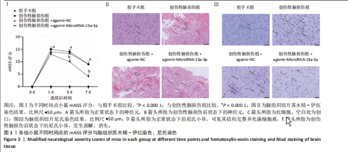
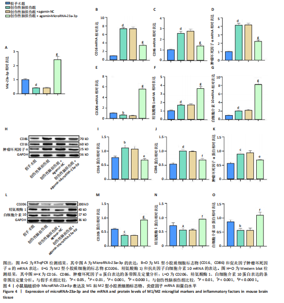
2.3 MicroRNA-23a-3p干预对创伤性脑损伤小鼠神经功能及脑组织病理学改变的影响 2.3.1 上调MicroRNA-23a-3p后创伤性脑损伤小鼠mNSS评分下降 各组小鼠在创伤性脑损伤后1,3和7 d分别进行了mNSS评分,结果表明,创伤性脑损伤后1 d,创伤性脑损伤组小鼠mNSS评分较假手术组显著升高,而在损伤后3 d和7 d时,创伤性脑损伤组小鼠mNSS评分逐渐下降,但仍高于假手术组(P < 0.000 1)。对MicroRNA-23a-3p表达进行干扰,发现各时间点创伤性脑损伤+agomir-NC组小鼠mNSS评分与创伤性脑损伤组相比无明显差异,创伤性脑损伤+agomir-MicroRNA-23a-3p组小鼠各时间点mNSS评分显著低于创伤性脑损伤组(P < 0.000 1),见图3A。 2.3.2 上调MicroRNA-23a-3p后创伤性脑损伤小鼠脑组织结构破坏减轻 苏木精-伊红染色结果显示,假手术组脑组织神经元细胞核清晰完整且居中,细胞膜结构完整,无明显出血或脑水肿;组织结构完整,细胞排列规则,轮廓分明,细胞质清晰。创伤性脑损伤组脑组织损伤严重,皮质有明显创口,出现明显的间质水肿、出血和神经元丢失;神经元肿胀、排列紊乱,脑组织出现坏死,组织和细胞结构变形,红细胞在受损区域内及周围聚集。创伤性脑损伤+agomir-NC组病理变化与创伤性脑损伤组相似,出现大面积细胞死亡,伴随核固缩和碎片化现象,细胞密度下降,伴有出血、水肿和炎症细胞浸润,神经元肿胀和排列紊乱,组织结构破坏严重,坏死细胞数量增多。与创伤性脑损伤组相比,创伤性脑损伤+agomir-MicroRNA-23a-3p组脑组织细胞结构较为完整,脑组织损伤和神经元丧失较创伤性脑损伤组好转,皮质有创口但基本愈合,细胞排列规则,轮廓完整,核居中于细胞中央,细胞质清晰,水肿和坏死面积明显小于创伤性脑损伤组,出血现象减少,视野中红细胞数量较创伤性脑损伤组显著降低,见图3B。 2.3.3 上调MicroRNA-23a-3p后创伤性脑损伤小鼠神经元破坏减轻 尼氏染色结果显示,假手术组中神经元边界清晰,细胞体较大、胞浆丰富且尼氏小体含量高,尼氏小体呈现深蓝色或紫蓝色,颗粒状或虎斑样分布,排列密集,充满细胞质,结构完整。创伤性脑损伤组与创伤性脑损伤+agomir-NC组神经元形态变化相似,显示神经元受到损伤,表现为胞体缩小、胞核浓缩,核偏位;尼氏小体数量减少,发生溶解或消失,不规则的斑块或颗粒状结构被破坏,从细胞质边缘向细胞核周围聚集,形成中央性染色质溶解。与创伤性脑损伤组相比,创伤性脑损伤+agomir-MicroRNA-23a-3p组尼氏小体的形态基本恢复,呈现不规则的斑块或颗粒状结构,充满细胞质,结构完整,见图3C。 2.4 上调MicroRNA-23a-3p促进创伤性脑损伤小鼠小胶质细胞由M1型向M2型极化 2.4.1 M1型小胶质细胞标记物mRNA表达下调,M2型小胶质细胞标记物mRNA表达上调 (1) MicroRNA-23a-3p的表达:RT-qPCR结果显示,创伤性脑损伤组小鼠脑组织损伤侧皮质内MicroRNA-23a-3p的表达水平显著低于假手术组(P < 0.000 1),创伤性脑损伤+agomir-NC组中MicroRNA-23a-3p表达水平接近于创伤性脑损伤组,创伤性脑损伤+agomir-MicroRNA-23a-3p组中MicroRNA-23a-3p的表达量较创伤性脑损伤组显著升高(P < 0.000 1),见图4A。 (2) CD16、CD86及肿瘤坏死因子α mRNA的表达:进一步检测各组小鼠脑组织中M1型小胶质细胞标志物(CD16、CD86)及促炎细胞因子肿瘤坏死因子α的mRNA表达水平,发现上述指标表达水平在创伤性脑损伤组小鼠脑组织中均较假手术组升高(P < 0.000 1);在创伤性脑损伤+agomir-NC组脑组织中上述因子表达水平与创伤性脑损伤组相比差异无显著性意义;上调MicroRNA-23a-3p表达后,小鼠脑组织中CD16、CD86及肿瘤坏死因子α的mRNA表达水平较创伤性脑损伤组显著降低(P < 0.000 1),见图4B-D。 (3)CD206、精氨酸酶1及白细胞介素10 mRNA的表达:与假手术组相比,创伤性脑损伤组小鼠脑组织中M2型小胶质细胞的标志物(CD206)的mRNA表达水平较假手术组显著下降(P < 0.01),精氨酸酶1和抗炎因子白细胞介素10的mRNA表达水平显著升高(P < 0.000 1);创伤性脑损伤+agomir-NC组上述因子mRNA表达水平与创伤性脑损伤组相比差异无显著性意义;上调MicroRNA-23a-3p表达后,小鼠脑组织CD206、精氨酸酶1、白细胞介素10的表达水平较创伤性脑损伤组显著升高(P < 0.000 1 ),见图4E-G。"

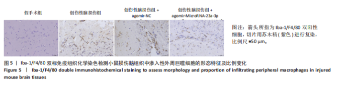
2.4.2 M1型小胶质细胞标记物蛋白表达下调,M2型小胶质细胞标记物蛋白表达上调 (1) CD16、CD86、肿瘤坏死因子α蛋白的表达:Western Blot结果显示,与假手术组相比,创伤性脑损伤组小鼠脑组织中CD16、CD86、肿瘤坏死因子α的蛋白表达水平显著升高(P < 0.01),提示创伤性脑损伤后小胶质细胞M1型极化增加;创伤性脑损伤+agomir-NC组上述因子蛋白表达水平与创伤性脑损伤组相比无显著差异;而创伤性脑损伤+agomir-MicroRNA-23a-3p组中小胶质细胞M1型标志物蛋白表达水平较创伤性脑损伤组显著降低(P < 0.01),表明MicroRNA-23a-3p表达上调可抑制创伤性脑损伤诱导的小胶质细胞M1型极化。见图4H-K。 (2) CD206、精氨酸酶1、白细胞介素10的蛋白表达:创伤性脑损伤组小鼠脑组织中CD206、精氨酸酶1、白细胞介素10的蛋白表达水平较假手术组显著下降(P < 0.05);上述因子在创伤性脑损伤+agomir-NC组脑组织中的表达水平与创伤性脑损伤组相比差异无显著性意义;MicroRNA-23a-3p表达上调后,小鼠脑组织中CD206、精氨酸酶1、白细胞介素10表达水平较创伤性脑损伤组显著上调(P < 0.001),提示MicroRNA-23a-3p表达上调可促进创伤性脑损伤后小胶质细胞向M2型极化,图4L-O。 2.5 MicroRNA-23a-3p上调促进创伤性脑损伤小鼠小胶质细胞M2型极化 为评估MicroRNA-23a-3p对小胶质细胞形态的影响,采用Iba-1与F4/80双标免疫组织化学染色识别脑组织中渗入性外周来源的巨噬细胞,区分其与中枢内源性小胶质细胞的来源差异,在检测细胞类型与活化状态的同时,观察其在损伤区域的分布情况与细胞间相互作用。结果显示,假手术组中未见F4/80?细胞,在创伤性脑损伤组小鼠脑组织损伤区域,Iba-1?/F4/80?细胞大量聚集,细胞体积增大、轮廓不规则,伴有明显的假足形成,呈典型M1型表型,提示外周来源巨噬细胞在损伤后大量渗入中枢神经系统并发生活化。创伤性脑损伤+ agomir-NC组中Iba-1?/F4/80?细胞的数量及形态与创伤性脑损伤组相比无显著差异。相比于创伤性脑损伤组,创伤性脑损伤+ agomir-microRNA-23a-3p组中Iba-1?/F4/80?细胞数量明显减少,呈现出细胞体积减小、胞体形态圆润、伪足数量减少等M2型极化的特征,提示microRNA-23a-3p的上调在一定程度上抑制了渗入性巨噬细胞的促炎活化表型,见图5。"
"

| [1] KUMARI N, BAGRI K, KUMARI S, et al. Traumatic Brian Injury (TBI) unraveled: molecular disruptions and therapeutic avenues. Inflammopharmacology. 2025;33(8):4323-4334. [2] MANLEY GT, MAAS AI.Traumatic brain injury: an international knowledge-based approach. Jama. 2013;310(5):473-474. [3] SPITZ G, HICKS AJ, MCDONALD SJ, et al. Plasma biomarkers in chronic single moderate-severe traumatic brain injury. Brain. 2024;147(11): 3690-3701. [4] SCHIFF ND, GIACINO JT, BUTSON CR, et al. Thalamic deep brain stimulation in traumatic brain injury: a phase 1, randomized feasibility study. Nat Med. 2023;29(12):3162-3174. [5] ZHONG H, FENG Y, SHEN J, et al. Global Burden of Traumatic Brain Injury in 204 Countries and Territories From 1990 to 2021. Am J Prev Med. 2025; 68(4):754-763. [6] ZENG Q, YOU G, LI W, et al. By inhibiting pyroptosis to reduce neuroinflammation, PEG-bHb may prevent the development of secondary injury after traumatic brain injury. Exp Neurol. 2025;394:115447. [7] JACQUENS A, NEEDHAM EJ, ZANIER ER, et al. Neuro-Inflammation Modulation and Post-Traumatic Brain Injury Lesions: From Bench to Bed-Side. Int J Mol Sci. 2022;23(19):11193. [8] HAN Y, GU J, XU M, et al. Intraoperative application of an antioxidant nanoparticle-hydrogel targeting microglia regulates neuroinflammation in traumatic brain injury. J Nanobiotechnology. 2025;23(1):599-604. [9] ZOU W, LV Y, LI L, et al. FOXQ1 Regulates Brain Endothelial Mitochondrial Function by Orchestrating Calcium Signaling and Cristae Morphology. Adv Sci (Weinh). 2025;30:e03082. [10] SOLIMAN E, LEONARD J, BASSO EKG, et al. Efferocytosis is restricted by axon guidance molecule EphA4 via ERK/Stat6/MERTK signaling following brain injury. J Neuroinflammation. 2023;20(1):256. [11] WANG W, LU G, GUO P, et al. Selenized neural stem cell-derived exosomes: A neotype therapeutic agent for traumatic injuries of the central nervous system. Cell Rep Med. 2025;6(9):102319. [12] BANOEI MM, HUTCHISON J, PANENKA W, et al. Metabolomic in severe traumatic brain injury: exploring primary, secondary injuries, diagnosis, and severity. Crit Care. 2025;29(1):26-37. [13] LI X. Research Progress on Prognostic Markers of Neuroinflammation in Traumatic Head Injury. Mol Neurobiol. 2025;62(6):7846-7863. [14] DAS M, MOHAPATRA S, MOHAPATRA SS. New perspectives on central and peripheral immune responses to acute traumatic brain injury. J Neuroinflammation. 2012;9:236-242. [15] LOANE DJ, KUMAR A. Microglia in the TBI brain: The good, the bad, and the dysregulated. Exp Neurol. 2016;275:316-327. [16] PRINZ M, PRILLER J. Microglia and brain macrophages in the molecular age: from origin to neuropsychiatric disease. Nat Rev Neurosci. 2014;15(5):300-512. [17] SHI L, LIU S, CHEN J, et al. Microglial polarization pathways and therapeutic drugs targeting activated microglia in traumatic brain injury. Neural Regen Res. 2026;21(1):39-56. [18] DELAGE C, TAIB T, MAMMA C, et al. Traumatic Brain Injury: An Age-Dependent View of Post-Traumatic Neuroinflammation and Its Treatment. Pharmaceutics. 2021;13(10):82-93. [19] FAN Z, JIA M, ZHOU J, et al. Pharmacological targeting cGAS/STING/NF-κB axis by tryptanthrin induces microglia polarization toward M2 phenotype and promotes functional recovery in a mouse model of spinal cord injury. Neural Regen Res. 2025;20(11):3287-3301. [20] LONG Y, LI XQ, DENG J, et al. Modulating the polarization phenotype of microglia - A valuable strategy for central nervous system diseases. Ageing Res Rev. 2024;93:102160. [21] SHANG R, LEE S, Senavirathne G, et al. microRNAs in action: biogenesis, function and regulation. Nat Rev Genet. 2023;24(12):816-833. [22] EL-ASHMAWY NE, KHEDR EG, DARWISH RT, et al. Competing endogenous RNAs network and therapeutic implications: New horizons in disease research. Biochim Biophys Acta Gene Regul Mech. 2025;1868(1):195073. [23] MA S, LIU M, XU Z, et al. A double feedback loop mediated by microRNA-23a/27a/24-2 regulates M1 versus M2 macrophage polarization and thus regulates cancer progression. Oncotarget. 2016;7(12):13502-13519. [24] SABIRZHANOV B, ZHAO Z, STOICA BA, et al. Downregulation of miR-23a and miR-27a following experimental traumatic brain injury induces neuronal cell death through activation of proapoptotic Bcl-2 proteins. J Neurosci. 2014;34(30):10055-10071. [25] SUN Q, WANG B, LI M. MicroRNA-23a-3p targeting of HMGB1 inhibits LPS-induced inflammation in murine macrophages in vitro. Exp Ther Med. 2022;23(5):322-329. [26] WANG J, HU M, LI L. Clinical Values of miR-23a-3p in Oral Lichen Planus and Its Role in Keratinocyte Proliferation and Inflammatory Response. J Inflamm Res. 2021;14:5013-5021. [27] DONG C, CHEN M, CAI B, et al. Mesenchymal Stem Cell-Derived Exosomes Improved Cerebral Infarction via Transferring miR-23a-3p to Activate Microglia. Neuromolecular Med. 2022;24(3):290-298. [28] YANG DD, WAN XD, CHEN AD, et al. Characteristics of traumatic brain injury models: from macroscopic blood flow changes to microscopic mitochondrial changes. Neural Regen Res. 2023;18(10):2268-2277. [29] SIEBOLD L, OBENAUS A, GOYAL R. Criteria to define mild, moderate, and severe traumatic brain injury in the mouse controlled cortical impact model. Exp Neurol. 2018;310:48-57. [30] 于永鹏,董霞.RohA信号通路介导miR-133b调控脑缺血再灌注后血脑屏障通透性[J].生物医学,2024,14(3):426-434. [31] WANG X, QIAN J, LI Y, et al. Protective effects of forsythoside A against severe acute pancreatitis- induced brain injury in mice. Biomed Pharmacother. 2024;178:117301. [32] SAMAEEKIA R, ADORNO-CRUZ V, BOCKHORN J, et al. miR-206 Inhibits Stemness and Metastasis of Breast Cancer by Targeting MKL1/IL11 Pathway. Clin Cancer Res. 2017;23(4):1091-1103. [33] CÁCERES E, OLIVELLA JC, DI NAPOLI M, et al. Immune Response in Traumatic Brain Injury. Curr Neurol Neurosci Rep. 2024;24(12):593-609. [34] WU H, ZHENG J, XU S, et al.Mer regulates microglial/macrophage M1/M2 polarization and alleviates neuroinflammation following traumatic brain injury. J Neuroinflammation. 2021;18(1):2. [35] LI N, LU W, TANG L, et al. Microglia in Post-Traumatic Brain Injury (TBI) Cognitive Impairment: From Pathological Changes to Therapeutic Approaches. CNS Neurosci Ther. 2025;31(8):e70568. [36] ZIGMOND RE, ECHEVARRIA FD. Macrophage biology in the peripheral nervous system after injury. Prog Neurobiol. 2019;173:102-121. [37] MAVROUDIS I, BALMUS IM, CIOBICA A, et al. The Role of Microglial Exosomes and miR-124-3p in Neuroinflammation and Neuronal Repair after Traumatic Brain Injury. Life (Basel). 2023;13(9):1924. [38] LUO F, LI X, ZHU Z. Inhibition of neurokinin B promotes functional recovery in traumatic brain injury by increasing M2 microglia. Neuropeptides. 2025;113:102549. [39] WANG WX, VISAVADIYA NP, PANDYA JD, et al. Mitochondria-associated microRNAs in rat hippocampus following traumatic brain injury. Exp Neurol. 2015;265:84-93. [40] LI Y, SUN M, WANG X, et al. Dental stem cell-derived extracellular vesicles transfer miR-330-5p to treat traumatic brain injury by regulating microglia polarization. Int J Oral Sci. 2022;14(1):44-49. [41] KISHORE A, PETREK M. Roles of Macrophage Polarization and Macrophage-Derived miRNAs in Pulmonary Fibrosis. Front Immunol. 2021;12:678457. [42] ESSANDOH K, LI Y, HUO J, FAN GC. MiRNA-Mediated Macrophage Polarization and its Potential Role in the Regulation of Inflammatory Response. Shock. 2016;46(2):122-131. [43] SABIRZHANOV B, MAKAREVICH O, BARRETT J, et al. Down-Regulation of miR-23a-3p Mediates Irradiation-Induced Neuronal Apoptosis. Int J Mol Sci. 2020;21(10):3695. [44] SHI Z, MAO L, CHEN S, et al. Reversing Persistent PTEN Activation after Traumatic Brain Injury Fuels Long-Term Axonal Regeneration via Akt/mTORC1 Signaling Cascade. Adv Sci (Weinh). 2025;12(6):e2410136. [45] LI Z, XU R, ZHU X, et al. MicroRNA-23a-3p improves traumatic brain injury through modulating the neurological apoptosis and inflammation response in mice. Cell Cycle. 2020;19(1):24-29. [46] TIANG T, SUN L, ZHU J, et al. MicroRNA-23a-3p promotes macrophage M1 polarization and aggravates lipopolysaccharide-induced acute lung injury by regulating PLK1/STAT1/STAT3 signalling. Int J Exp Pathol. 2022;103(5):198-207. [47] PAN Y, CENGIZ R, KLUIVER J, et al.Pinpointing Functionally Relevant miRNAs in Classical Hodgkin Lymphoma Pathogenesis.Cancers (Basel). 2024;16(6):1126. [48] KALRA S, MALIK R, SINGH G, et al. Pathogenesis and management of traumatic brain injury (TBI): role of neuroinflammation and anti-inflammatory drugs. Inflammopharmacology. 2022;30(4):1153-1166. [49] SMAIL SW, KARIMIAN A, ABDOLMALEKI A, et al. Macrophages’ Functions in the Central and Peripheral Nervous Regeneration. Regen Eng Transl Med. 2024;11(2):312-326. [50] SHI L, KIDDER K, BIAN Z, et al. SIRPα sequesters SHP-2 to promote IL-4 and IL-13 signaling and the alternative activation of macrophages. Sci Signal. 2021;14(702):eabb3966. [51] EDIN S, WIKBERG ML, RUTEGÅRD J, et al. Phenotypic skewing of macrophages in vitro by secreted factors from colorectal cancer cells. PLoS One. 2013;8(9):e74982. [52] TRAVNICKOVA J, NHIM S, ABDELLAOUI N, et al. Macrophage morphological plasticity and migration is Rac signalling and MMP9 dependant. Sci Rep. 2021;11(1):10123. [53] MCWHORTER FY, DAVIS CT, LIU WF. Physical and mechanical regulation of macrophage phenotype and function. Cell Mol Life Sci. 2015;72(7):1303-1316. [54] KESSELS S, TRIPPAERS C, MERTENS M, et al. Cytoskeletal control in adult microglia is essential to restore neurodevelopmental synaptic and cognitive deficits. Sci Adv. 2025;11(35):eadw0128. [55] 贺琦,王燕.miRNA-23a-3p和TLR4在急性脑出血患者血清中的表达及临床意义[J].海南医学,2023,34(15):2135-2139. [56] QIN D, WANG C, LI D, et al. Exosomal miR-23a-3p derived from human umbilical cord mesenchymal stem cells promotes remyelination in central nervous system demyelinating diseases by targeting Tbr1/Wnt pathway.J Biol Chem. 2024;300(1):105487. [57] MAAS AIR, MENON DK, MANLEY GT, et al. Traumatic brain injury: progress and challenges in prevention, clinical care, and research. Lancet Neurol. 2022;21(11):1004-1060. [58] MCDONALD BZ, TARUDJI AW, ZHANG H, et al. Traumatic brain injury heterogeneity affects cell death and autophagy. Exp Brain Res. 2024; 242(7):1645-1658. [59] SAIDU UF, BULAMA I, ONU A, et al. Experimental animal models in traumatic brain injury research: a comprehensive review of methods and outlook. International Journal of Scientific Reports. 2024;10(6):206-214. [60] FESHARAKI-ZADEH A, DATTA D. An overview of preclinical models of traumatic brain injury (TBI): relevance to pathophysiological mechanisms. Front Cell Neurosci. 2024;18:1371213. [61] ZHANG YP, CAI J, SHIELDS LB, et al. Traumatic brain injury using mouse models. Transl Stroke Res. 2014;5(4):454-471. [62] YANG X, CHEN L, PU J, et al. Guideline of clinical neurorestorative treatment for brain trauma (2022 China version). Journal of Neurorestoratology. 2022;10(2):100005. |
| [1] | Cui Lianxu, Li Haomin, Xu Junrong, Tan Baodong, Lu Dahong, Peng Siwei, Wang Jinhui. Effect of umbilical cord mesenchymal stem cell conditioned medium on tissue repair after traumatic craniocerebral injury in miniature pigs [J]. Chinese Journal of Tissue Engineering Research, 2026, 30(7): 1730-1735. |
| [2] | Xun Chong, Wang Qiang, Li Changzhou, Liu Xiaofeng. Potential molecular targets and therapeutic mechanisms underlying transplantation of autologous bone marrow stem cells for the treatment of spinal cord injury based on bioinformatics [J]. Chinese Journal of Tissue Engineering Research, 2020, 24(31): 4927-4933. |
| Viewed | ||||||
|
Full text |
|
|||||
|
Abstract |
|
|||||