Chinese Journal of Tissue Engineering Research ›› 2026, Vol. 30 ›› Issue (19): 4853-4859.doi: 10.12307/2026.674
Previous Articles Next Articles
Human adipose multilineage-differentiating stress-enduring cells on treatment of ischemic stroke in rats
Gao Hongmei1, Zhang Kun1, 2, Xiao Dongjie1, 2, Liu Hua1, 2
- 1Cell Therapy Center, Central Hospital Affiliated to Shandong First Medical University, Jinan 250013, Shandong Province, China; 2Shandong Research Center of Transplantation and Tissue Engineering Technology, Jinan 250013, Shandong Province, China
-
Received:2025-05-26Accepted:2025-09-18Online:2026-07-08Published:2026-02-13 -
Contact:Liu Hua, MD, Senior technologist, Cell Therapy Center, Central Hospital Affiliated to Shandong First Medical University, Jinan 250013, Shandong Province, China; Shandong Research Center of Transplantation and Tissue Engineering Technology, Jinan 250013, Shandong Province, China -
About author:Gao Hongmei, MS, Associate senior technologist, Cell Therapy Center, Central Hospital Affiliated to Shandong First Medical University, Jinan 250013, Shandong Province, China -
Supported by:Jinan Science and Technology Development Plan, No. 201907053 (to LH)
CLC Number:
Cite this article
Gao Hongmei, Zhang Kun, Xiao Dongjie, Liu Hua. Human adipose multilineage-differentiating stress-enduring cells on treatment of ischemic stroke in rats[J]. Chinese Journal of Tissue Engineering Research, 2026, 30(19): 4853-4859.
share this article
Add to citation manager EndNote|Reference Manager|ProCite|BibTeX|RefWorks
2.1 磁珠分选脂肪Muse细胞 脂肪间充质干细胞经流式细胞仪检测CD105、CD73高表达,表达量 > 95%,CD45及HLA-DR低表达,表达量小于< 5%(图1A)。脂肪间充质干细胞消化4 h后,经磁珠分选获得SSEA3阳性的Muse细胞。流式细胞仪检测结果显示,脂肪间充质干细胞SSEA3阳性表达率为5.07%,长时间消化后的脂肪间充质干细胞SSEA3阳性表达率为14.6%,经磁珠分选后,阳性筛选的脂肪间充质干细胞SSEA3阳性表达率为80.0%,阴性筛选的脂肪间充质干细胞SSEA3阳性表达率为5.08%(图1B)。磁珠分选,荧光显微镜下观察可看到阳性筛选的脂肪间充质干细胞呈红色荧光,而阴性筛选的脂肪间充质干细胞几乎不表达荧光(图1C)。 "

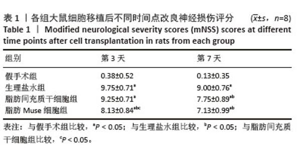
2.2 脂肪Muse细胞促进大脑中动脉阻塞大鼠神经功能恢复 以大鼠麻醉清醒后24 h行走时向对侧转圈(2分)及行走时向对侧倾倒(3分)作为模型鼠。造模时死亡4只,行为学评分不符合要求以及移植、观察过程中死亡5只,死亡大鼠以新鼠补充,确保各组实验动物为16只,最终有48只大脑中动脉阻塞大鼠,16只假手术大鼠进入结果分析。 神经功能评分结果显示,与假手术组相比,生理盐水组及细胞移植组第3,7天mNSS评分均显著升高(P < 0.05);与生理盐水组相比,脂肪间充质干细胞移植后第7天mNSS评分显著降低(P < 0.05),脂肪Muse细胞移植后第3,7天mNSS评分均显著降低(P < 0.05);与脂肪间充质干细胞组相比,脂肪Muse细胞组移植后第3天mNSS评分显著降低(P < 0.05),见表1。"

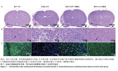
2.3 脂肪Muse细胞减轻脑组织神经细胞损伤 脂肪间充质干细胞以及脂肪Muse细胞移植后3 d,苏木精-伊红染色显示假手术组脑组织结构清晰完整,神经细胞排列有序,细胞周围间隙致密无水肿;生理盐水组大鼠损伤以左侧大脑皮质为主,神经细胞消失、排列紊乱,细胞形态异常,间质水肿疏松;相对生理盐水组,脂肪间充质干细胞以及脂肪Muse细胞组脑皮质损伤区神经细胞形态好转,损伤部位空泡减少,病理学改变减轻;脂肪Muse细胞组脑组织结构较脂肪间充质干细胞组更为完整、正常,见图2A,B。 移植后3 d,微管相关蛋白2免疫荧光染色结果显示,生理盐水组大鼠脑组织皮质损伤区域微管相关蛋白2基本丢失,脂肪间充质干细胞组及脂肪Muse细胞组损伤区微管相关蛋白2表达增加;相比脂肪间充质干细胞组,脂肪Muse细胞组抑制微管相关蛋白2丢失更为显著,神经元结构及形态更为完整,见图2C(绿色荧光为微管相关蛋白2,蓝色为细胞核)。"
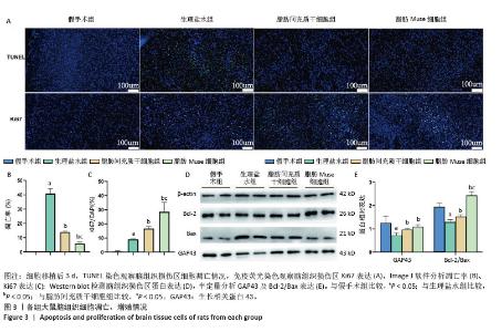
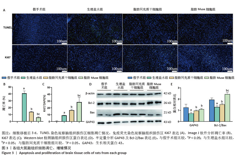
2.4 脂肪Muse细胞抑制神经细胞凋亡、促进神经细胞再生 脂肪间充质干细胞以及脂肪Muse细胞移植后3 d,TUNEL染色检测脑组织细胞凋亡情况,结果显示,正常大鼠脑组织基本检测不到凋亡细胞,生理盐水组损伤区域神经细胞凋亡显著增加(P < 0.05),脂肪间充质干细胞及脂肪Muse细胞移植后损伤区域神经细胞凋亡减少(P < 0.05),与脂肪间充质干细胞组相比,脂肪Muse细胞组神经细胞凋亡明显减少(P < 0.05),见图3。 脂肪间充质干细胞和脂肪Muse细胞移植后3 d,采用Ki67免疫荧光染色对大鼠损伤区域皮质层细胞增殖情况进行分析,结果显示假手术组大鼠脑皮质层未检测到增殖细胞;生理盐水组大鼠损伤区脑皮质层可见ki67阳性细胞;细胞移植组Ki67阳性细胞明显高于生理盐水组(P < 0.05),以脂肪Muse细胞组更为显著(P < 0.05)。 提取细胞移植后第3天脑皮质损伤区蛋白,Western blot检测凋亡相关蛋白及神经相关蛋白生长相关蛋白43表达,结果显示与假手术组相比,生理盐水组Bcl-2/Bax比值、生长相关蛋白43表达下降(P < 0.05);与生理盐水组相比,细胞移植组Bcl-2/Bax比值及生长相关蛋白43表达上升(P < 0.05),其中脂肪Muse细胞组Bcl-2/Bax比值高于脂肪间充质干细胞组。"
| [1] HUANG S, LIU L, TANG X, et al. Research progress on the role of hormones in ischemic stroke. Front Immunol. 2022;13:1062977. [2] 乔玥,李传辉,赵文博.急性缺血性脑卒中的再灌注治疗的现状与未来[J].首都医科大学学报,2025,46(1):68-70. [3] RUST R, NIH LR, LIBERALE L, et al. Brain repair mechanisms after cell therapy for stroke. Brain. 2024;147(10):3286-3305. [4] EJMA M, MADETKO N, BRZECKA A, et al. The Role of Stem Cells in the Therapy of Stroke.Curr Neuropharmacol. 2022;20(3):630-647. [5] CHUNG JW, CHANG WH, BANG OY, et al. Efficacy and Safety of Intravenous Mesenchymal Stem Cells for Ischemic Stroke. Neurology. 2021;96(7):e1012-e1023. [6] LAI S, GUO Z. Stem cell therapies for chronic obstructive pulmonary disease: mesenchymal stem cells as a promising treatment option. Stem Cell Res Ther. 2024;15(1):312. [7] TIAN H, TIAN F, MA D, et al. Priming and Combined Strategies for the Application of Mesenchymal Stem Cells in Ischemic Stroke: A Promising Approach. Mol Neurobiol. 2024;61(9):7127-7150. [8] 张君,姜夕锋,张帆,等.间充质干细胞治疗缺血性脑卒中的临床前研究进展[J].中国医药生物技术,2024,19(6):526-532. [9] KIM HY, KIM TJ, KANG L, et al. Mesenchymal stem cell-derived magnetic extracellular nanovesicles for targeting and treatment of ischemic stroke. Biomaterials. 2020;243:119942. [10] XU K, ZHAO X, HE Y, et al. Stem cell-derived exosomes for ischemic stroke: a conventional and network meta-analysis based on animal models. Front Pharmacol. 2024;15:1481617. [11] DE PALMA ST, HERMANS EC, SHAMORKINA TM, et al. Hypoxic Preconditioning Enhances the Potential of Mesenchymal Stem Cells to Treat Neonatal Hypoxic-Ischemic Brain Injury. Stroke. 2025. doi: 10.1161/STROKEAHA.124.048964. [12] KANG H, HUANG Y, PENG H, et al. Mesenchymal Stem Cell-Loaded Hydrogel Improves Surgical Treatment for Chronic Cerebral Ischemia. Transl Stroke Res. 2025;16(3):896-913. [13] SALEH RO, MAJEED AA, MARGIANA R, et al. Therapeutic gene delivery by mesenchymal stem cell for brain ischemia damage: Focus on molecular mechanisms in ischemic stroke. Cell Biochem Funct. 2024;42(2):e3957. [14] DENG M, HOU Y, LIU J, et al. Mesenchymal stem cell-derived exosomes overexpressing SRC-3 protect mice from cerebral ischemia by inhibiting ferroptosis. Brain Res Bull. 2024;211:110948. [15] KURODA Y, KITADA M, WAKAO S, et al. Unique multipotent cells in adult human mesenchymal cell populattons. Proc Natl Acad Sci U S A. 2010; 107(19):8639-8643. [16] ALANAZI RF, ALHWITY BS, ALMAHLAWI RM, et al. Multilineage Differentiating Stress Enduring (Muse) Cells: A New Era of Stem Cell-Based Therapy. Cells. 2023;12(13):1676. [17] OSSANNA R, VERONESE S, QUINTERO SIERRA LA, et al. Multilineage-Differentiating Stress-Enduring Cells (Muse Cells): An Easily Accessible, Pluripotent Stem Cell Niche with Unique and Powerful Properties for Multiple Regenerative Medicine Applications. Biomedicines. 2023; 11(6):1587. [18] GUO Y, XUE Y, WANG P, et al. Muse cell spheroids have therapeutic effect on corneal scarring wound in mice and tree shrews. Sci Transl Med. 2020; 12(562):eaaw1120. [19] YAMADA Y, MINATOGUCHI S, KANAMORI H, et al. Stem cell therapy for acute myocardial infarction - focusing on the comparison between Muse cells and mesenchymal stem cells. J Cardiol. 2022;80(1):80-87. [20] KOYAMA J, YAMASHITA S, KATO Y, et al. Intravenously engrafted human multilineage -differentiating stress-enduring (Muse) cells rescue erectile function after rat cavernous nerve injury. BJU Int. 2024;133(3):332-340. [21] 张坤,李芳,延冰,等.应激耐受多系分化细胞的研究进展[J].新医学, 2018,49(5):301-304. [22] 张坤,李芳,肖东杰,等.人脐带与人脂肪间充质干细胞中应激耐受多系分化细胞的分离鉴定及分化能力的比较[J].中国组织工程研究, 2022,26(24):3802-3807. [23] LI Y, TAN L, YANG C, et al. Distinctions between the Koizumi and Zea Longa methods for middle cerebral artery occlusion (MCAO) model: a systematic review and meta-analysis of rodent data. Sci Rep. 2023; 13(1):10247. [24] MAO R, ZONG N, HU Y, et al. Neuronal Death Mechanisms and Therapeutic Strategy in Ischemic Stroke. Neurosci Bull. 2022;38(10):1229-1247. [25] TUO QZ, ZHANG ST, LEI P. Mechanisms of neuronal cell death in ischemic stroke and their therapeutic implications. Med Res Rev. 2022;42(1):259-305. [26] HE X, WU M, CHEN L, et al. APMCG-1 attenuates ischemic stroke injury by reducing oxidative stress and apoptosis and promoting angiogenesis via activating PI3K/AKT pathway. Biomed Pharmacother. 2024;180:117506. [27] QI L, WANG C, DENG L, et al. Low-intensity focused ultrasound stimulation promotes stroke recovery via astrocytic HMGB1 and CAMK2N1 in mice. Stroke Vasc Neurol. 2024;9(5):505-518. [28] ZHANG Y, DONG N, HONG H, et al. Mesenchymal Stem Cells: Therapeutic Mechanisms for Stroke. Int J Mol Sci. 2022;23(5):2550. [29] MOÑIVAS GALLEGO E, ZURITA CASTILLO M. Mesenchymal stem cell therapy in ischemic stroke trials. A systematic review. Regen Ther. 2024;27:301-306. [30] YANG YK, OGANDO CR, WANG SEE C, et al. Changes in phenotype and differentiation potential of human mesenchymal stem cells aging in vitro. Stem Cell Res Ther. 2018;9(1):131. [31] ZHANG L, ZHUO Y, YU H. Spatio-temporal metabolokinetics and therapeutic effect of CD106(+) mesenchymal stem/stromal cells upon mice with acute lung injury. Cell Biol Int. 2023;47(4):720-730. [32] ANDREWS PW, GOKHALE PJ. A short history of pluripotent stem cells markers. Stem Cell Reports. 2024;19(1):1-10. [33] QUE H, MAI E, HU Y, et al. Multilineage-differentiating stress-enduring cells: a powerful tool for tissue damage repair. Front Cell Dev Biol. 2024; 12:1380785. [34] ABE T, ABURAKAWA D, NIIZUMA K, et al. Intravenously Transplanted Human Multilineage-Differentiating Stress-Enduring Cells Afford Brain Repair in a Mouse Lacunar Stroke Model. Stroke. 2020;51(2):601-611. [35] SUZUKI T, SATO Y, KUSHIDA Y, et al. Intravenously delivered multilineage-differentiating stress enduring cells dampen excessive glutamate metabolism and microglial activation in experimental perinatal hypoxic ischemic encephalopathy. J Cereb Blood Flow Metab. 2021;41(7): 1707-1720. [36] UCHIDA H, NIIZUMA K, KUSHIDA Y, et al. Human Muse Cells Reconstruct Neuronal Circuitry in Subacute Lacunar Stroke Model. Stroke. 2017; 48(2):428-435. [37] YAMAUCHI T, KURODA Y, MORITA T, et al. Therapeutic effects of human multilineage-differentiating stress enduring (MUSE) cell transplantation into infarct brain of mice. PLoS One. 2015;10(3):e0116009. [38] LI H, WEI J, LIU X, et al. Muse cells: ushering in a new era of stem cell-based therapy for stroke. Stem Cell Res Ther. 2022;13(1):421. [39] 房晓磊,冷军,张晨,等.间充质干细胞不同移植途径治疗缺血性脑卒中疗效差异的系统评价[J].中国组织工程研究,2022,26(7): 1085-1092. [40] KATO Y, ABURAKAWA D, TASHIRO R, et al. Intravenous administration of muse cells improves cerebral ischemia outcome via immunomodulation in the spleen. J Cereb Blood Flow Metab. 2025;45(3):542-557. |
| [1] | Wang Zheng, Cheng Ji, Yu Jinlong, Liu Wenhong, Wang Zhaohong, Zhou Luxing. Progress and future perspectives on the application of hydrogel materials in stroke therapy [J]. Chinese Journal of Tissue Engineering Research, 2026, 30(8): 2081-2090. |
| [2] | Tao Daiju, Su Haiyu, Wang Yuqi, Shen Zhiqiang, He Bo . Construction and identification of stable PC12 cell lines with high/low expression of miR-122-5p [J]. Chinese Journal of Tissue Engineering Research, 2026, 30(7): 1790-1799. |
| [3] | Li Wenfang, Dong Mengwei, Jin Haizhu, Yang Wanpeng, Ba Te, Nan Nan, Liu Yang, Hao Huiqin. Mechanism of acupuncture regulating proliferation and differentiation of stem cells [J]. Chinese Journal of Tissue Engineering Research, 2026, 30(19): 5050-5056. |
| [4] | Wu Xue, Zhang Linao, Luo Shifang, Liu Feifan, Wan Yan, Bai Yuanmei, Cao Julin, Xie Yuhuan, Guo Peixin. Dandeng Tongnao soft capsules against ischemic stroke: fingerprinting and network pharmacological analysis of efficacy and mechanism of action [J]. Chinese Journal of Tissue Engineering Research, 2026, 30(17): 4517-4528. |
| [5] | Wumiti·Taxi, Wang Lining, Li Muzhe, Sun Jie, Chen Shuangliu, Zhu Yihua, Zhou Shijie, Ma Yong, Guo Yang. Wenshen Tongluo Zhitong decoction regulates the bone fat differentiation balance of bone marrow mesenchymal stem cells through exosomal miR-342-3p [J]. Chinese Journal of Tissue Engineering Research, 2026, 30(13): 3258-3269. |
| [6] | Wu Jiazhou, Qian Tao, Liu Zexian, Wu Yanbin, He Ying, Li Yazhou, Peng Jiang. Three-dimensional culture of stromal vascular fraction self-assembles into complex vascularized osteogenic organoids [J]. Chinese Journal of Tissue Engineering Research, 2026, 30(11): 2681-2690. |
| [7] |
Zhang Yueting, Li Jinglin, Fu Zhenyi, Yan Fei, Gao Yu, Liu Jiaxin.
Endoplasmic reticulum stress promotes ferroptosis and aggravates cerebral ischemia-reperfusion injury#br#
#br#
[J]. Chinese Journal of Tissue Engineering Research, 2026, 30(11): 2806-2813.
|
| [8] | Zhou Xinying, Sun Xinyue, Zhu Wenhao. Insulin-like growth factors and ischemic stroke: a genome-wide association analysis in European populations [J]. Chinese Journal of Tissue Engineering Research, 2026, 30(11): 2909-2919. |
| [9] | Wang Zheng, Cheng Ji, Yu Jinlong, Liu Wenhong, Wang Zhaohong, Zhou Luxing. Progress and future perspectives on the application of hydrogel materials in stroke therapy [J]. Chinese Journal of Tissue Engineering Research, 2025, 29(在线): 1-10. |
| [10] | Wang Mi, Ma Shujie, Liu Yang, Qi Rui. Identification and validation of characterized gene NFE2L2 for ferroptosis in ischemic stroke [J]. Chinese Journal of Tissue Engineering Research, 2025, 29(7): 1466-1474. |
| [11] | Gao Yang, Qin Hewei, Liu Dandan. ACSL4 mediates ferroptosis and its potential role in atherosclerotic cardiovascular disease [J]. Chinese Journal of Tissue Engineering Research, 2025, 29(6): 1239-1247. |
| [12] | Yang Bo, Pan Xinfang, Chang Liuhui, Ni Yong. Correlation of echocardiographic parameters with disability at 3 months after acute ischemic stroke [J]. Chinese Journal of Tissue Engineering Research, 2025, 29(35): 7544-7551. |
| [13] | Chen Ying, Guo Xiaojing, Mo Xueni, Ma Wei, Wu Shangzhi, Li Xiangling, Xie Tingting. Analysis of diagnostic biomarkers for ischemic stroke and experimental validation of targeted cuproptosis related genes [J]. Chinese Journal of Tissue Engineering Research, 2025, 29(35): 7562-7570. |
| [14] | Ping Xingfeng, Huang Zongxuan, Li Kai, Xie Guangmin, Lyu Junying. Regularity of prescriptions for ischemic stroke based on latent structure combined with association rules [J]. Chinese Journal of Tissue Engineering Research, 2025, 29(29): 6277-6284. |
| [15] | Zhang Xinrui, Han Yue, Lei Lei, Liu Jianyu, Geng Chengkui. Comparative proteomic analysis of rat adipose-derived mesenchymal stem cells and their exosomes [J]. Chinese Journal of Tissue Engineering Research, 2025, 29(13): 2683-2689. |
| Viewed | ||||||
|
Full text |
|
|||||
|
Abstract |
|
|||||