Chinese Journal of Tissue Engineering Research ›› 2024, Vol. 28 ›› Issue (20): 3130-3136.doi: 10.12307/2024.399
Previous Articles Next Articles
Effect of moderate-intensity exercise on the level of autophagy in bone tissue of ovariectomized rats
Li Xun1, Zhang Weichao2, Li Yingjie3, Liu Rong2, Tian Xuewen4, Zhang Pengyi1, Wang Xiaoqiang1
- 1School of Sports and Health, 2School of Postgraduate Education, 4Research Center of Sport Science, Shandong Sport University, Jinan 250102, Shandong Province, China; 3Medical Office, Shandong University of Science and Technology, Qingdao 266590, Shandong Province, China
-
Received:2023-06-10Accepted:2023-07-18Online:2024-07-18Published:2023-09-09 -
Contact:Wang Xiaoqiang, PhD, Associate professor, School of Sports and Health, Shandong Sport University, Jinan 250102, Shandong Province, China -
About author:Li Xun, PhD, Associate professor, School of Sports and Health, Shandong Sport University, Jinan 250102, Shandong Province, China Zhang Weichao, Master candidate, School of Postgraduate Education, Shandong Sport University, Jinan 250102, Shandong Province, China -
Supported by:the Natural Science Foundation of Shandong Province, No. ZR2016CL11 (to LX); Science and Technology Program for Higher Education Institutions in Shandong Province, No. J18KA179 (to LX)
CLC Number:
Cite this article
Li Xun, Zhang Weichao, Li Yingjie, Liu Rong, Tian Xuewen, Zhang Pengyi, Wang Xiaoqiang. Effect of moderate-intensity exercise on the level of autophagy in bone tissue of ovariectomized rats[J]. Chinese Journal of Tissue Engineering Research, 2024, 28(20): 3130-3136.
share this article
Add to citation manager EndNote|Reference Manager|ProCite|BibTeX|RefWorks
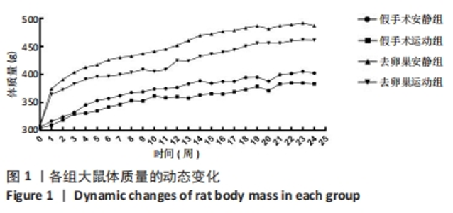
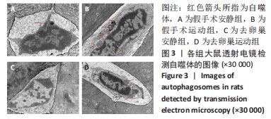
2.1 实验动物数量分析 共纳入48只SD大鼠,随机分为4组,全部进入结果分析,无脱失值。 2.2 大鼠体质量及骨量的变化 运动干预24周后,各组大鼠体质量均增加,且去卵巢安静组 > 去卵巢运动组 > 假手术安静组 > 假手术运动组。 在24周运动干预过程中,与假手术安静组相比,假手术运动组大鼠体质量降低,但无显著性差异(P > 0.05),去卵巢安静组大鼠体质量显著增加(P < 0.01),去卵巢运动组大鼠体质量在10,11周时显著增加(P < 0.05),在其余时间点极显著增加(P < 0.01)。与去卵巢安静组相比,去卵巢运动组大鼠在正式训练2,6,7,8,11,13,14,15,16,17周后体质量非常显著性降低(P < 0.01),在正式训练4,5,9,10,12,18,19,21,22,23周后体质量显著性降低(P < 0.05),各组大鼠体质量动态变化如图1所示。"
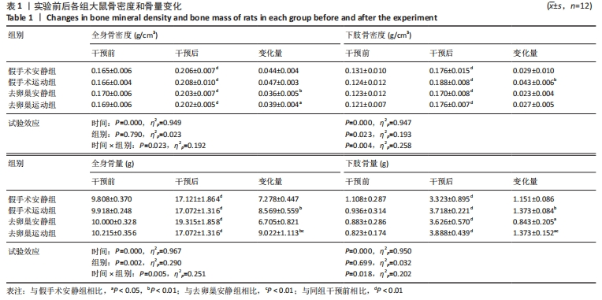
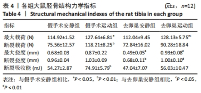
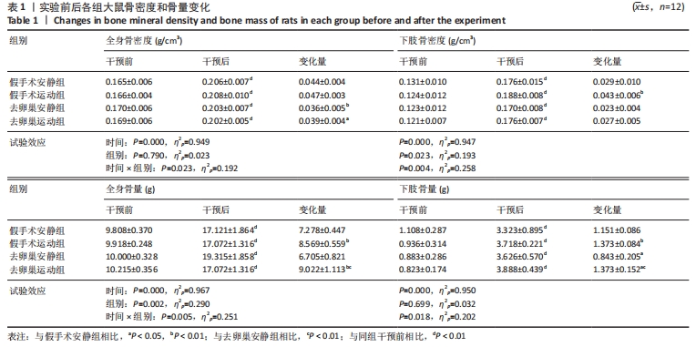
运动干预24周后,4组大鼠的全身及下肢骨密度、骨量均存在时间效应,且在时间×组别上均存在交互相应,各组大鼠全身及下肢骨密度、骨量均显著增加,见表1。骨密度变化量比较:去卵巢安静组、去卵巢运动组全身骨密度增加程度显著低于假手术安静组(P < 0.01,P < 0.05);假手术运动组下肢骨密度增加程度显著高于假手术安静组(P < 0.01)。骨量变化量比较:假手术运动组、去卵巢运动组全身骨量增加程度显著高于假手术安静组(P < 0.01,P < 0.01),去卵巢运动组全身骨量增加程度显著高于去卵巢安静组(P < 0.01);假手术运动组、去卵巢运动组下肢骨量增加程度显著高于假手术安静组(P < 0.01,P < 0.05),去卵巢安静组下肢骨量增加程度显著低于假手术安静组(P < 0.05),去卵巢运动组下肢骨量增加程度显著高于去卵巢安静组(P < 0.01)。表明去卵巢导致大鼠全身及下肢骨密度、骨量降低,中等强度跑台运动增加了全身及下肢的骨密度、骨量。"
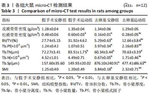
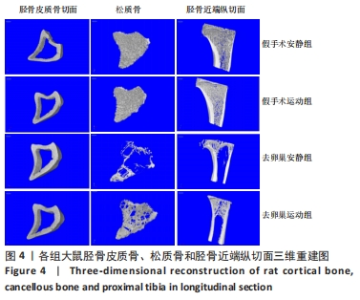
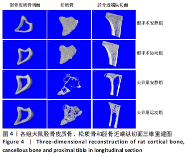
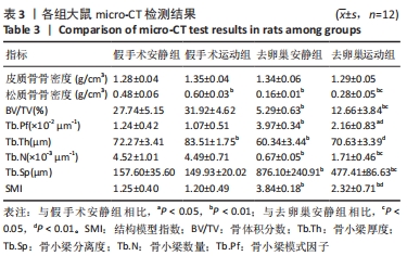
运动干预24周后,与假手术安静组相比,假手术运动组大鼠的胫骨松质骨骨密度、Tb.Th均显著增加(P < 0.01);去卵巢安静组大鼠松质骨骨密度、BV/TV、Tb.Th、Tb.N均显著降低(P < 0.01),Tb.Pf、Tb.Sp、SMI均显著增加(P < 0.01);去卵巢运动组大鼠松质骨骨密度、BV/TV、Tb.N显著降低(P < 0.01),Tb.Pf、Tb.Sp、SMI显著增加(P < 0.05或P < 0.01)。与去卵巢安静组相比,去卵巢运动组大鼠的松质骨骨密度、BV/TV、Tb.N、Tb.Th均显著增加(P < 0.05或P < 0.01),Tb.Pf、Tb.Sp、SMI均显著降低(P < 0.01),见表3。"
| [1] 吕遐,扶琼.原发性骨质疏松症的研究进展与最新指南解读[J].临床内科杂志,2020,37(5):319-322. [2] 陈祥和,陆鹏程,刘波,等.不同力学刺激方式对T2DM小鼠骨中Notch途径及骨形成代谢的影响[J].北京体育大学学报,2021,44(2):80-89. [3] 廖阳,屈萌艰,孙光华,等.运动对老年大鼠骨质疏松及骨髓间充质干细胞自噬的影响[J].中国康复医学杂志,2023,38(1):16-21+34. [4] 李荀.运动经雌激素-受体途径对骨量的影响效应及其机制[D].北京:首都体育学院,2012. [5] 崔国宁,刘喜平,董俊刚,等.微环境对骨髓间充质干细胞自噬水平的影响研究进展[J].转化医学杂志,2018,7(6):338-340+349. [6] 陆鹏程,陈祥和,杨康,等.自噬介导运动改善2型糖尿病骨代谢紊乱的作用机制[J].中国组织工程研究,2020,24(20):3256-3262. [7] 张林.骨质疏松大鼠运动模型的建立与训练效果评定[J].体育科学, 1999,19(6):72-76. [8] 彭珊,欧阳厚淦,赵志冬,等.去卵巢大鼠骨质疏松模型的制备与评价[J].中国骨质疏松杂志,2017,23(10):1327-1329+1358. [9] BEDFORD TG, TIPTON CM, WILSON NC, et al. Maximum oxygen consumption of rats and its changes with various experimental procedures. J Appl Physiol Respir Environ Exerc Physiol. 1979;47(6):1278-1283. [10] BARENGOLTS EI, LATHON PV, CURRY DJ, et al. Effects of endurance exercise on bone histomorphometric parameters in intact and ovariectomized rats. Bone Miner. 1994;26(2):133-140. [11] 李荀.运动经AR介导调节骨骼肌ERK1/2和mTOR通路的机理研究[D].北京:北京体育大学,2015. [12] 冯钰,史仍飞.运动与骨骼肌内雌激素代谢的研究进展[J].生命科学, 2019,31(10):1047-1053. [13] SHI R, TIAN X, FENG Y, et al. Expression of aromatase and synthesis of sex steroid hormones in skeletal muscle following exercise training in ovariectomized rats. Steroids. 2019;143:91-96. [14] GONZÁLEZ-GARCÍA I, TENA-SEMPERE M, LÓPEZ M. Estradiol Regulation of Brown Adipose Tissue Thermogenesis. Adv Exp Med Biol. 2017;1043: 315-335. [15] 仇业鹏,张庭然.跑步运动配合银杏叶提取物摄入对去卵巢肥胖骨质疏松大鼠骨密度、血脂和抗氧化能力影响研究[J].中国骨质疏松杂志, 2019,25(3):343-350. [16] QIAO D, LI Y, LIU X, et al. Association of obesity with bone mineral density and osteoporosis in adults: a systematic review and meta-analysis. Public Health. 2020;180:22-28. [17] SIEVERS W, RATHNER JA, KETTLE C, et al. The capacity for oestrogen to influence obesity through brown adipose tissue thermogenesis in animal models: A systematic review and meta-analysis. Obes Sci Pract. 2019;5(6): 592-602. [18] 陈晓红,郑陆,于召栋.不同持续时间中等强度运动对去卵巢大鼠骨量及骨微结构的影响[J].中国运动医学杂志,2015,34(6):571-577. [19] 王孝强,郑陆,王培林,等.长期递增负荷运动对大鼠股骨OPG和RANKL蛋白表达的影响[J].山东体育学院学报,2017,33(2):73-79. [20] 王金玲,黄夏荣,屈萌艰,等.运动训练老年骨质疏松大鼠骨量及骨微结构的变化[J].中国组织工程研究,2023,27(5):676-682. [21] 李荀,陈晓红,郑陆,等.中等强度跑台运动经Wnt/β-catenin途径对骨量的影响效应[J].上海体育学院学报,2016,40(5):57-62+69. [22] 孔存青,黄晓婷,姚志豪,等.中国与东南亚女大学生体力活动水平与骨密度比较研究[J].中国骨质疏松杂志,2018,24(10):1305-1309+1323. [23] 廖秀风,陈叶.基于MicroRNA介导Wnt/β-catenin通路防治绝经后骨质疏松的中西医研究进展[J].现代中西医结合杂志,2019,28(29)3298-3303. [24] 许祥云,陈书昌,孔德九,等.自噬相关基因LC3和ATG5在胃癌中的表达及临床意义[J].实用医学杂志,2020,36(14):1939-1945. [25] LIRA VA, OKUTSU M, ZHANG M, et al. Autophagy is required for exercise training-induced skeletal muscle adaptation and improvement of physical performance. FASEB J. 2013;27(10):4184-4193. [26] ZENG Z, LIANG J, WU L, et al. Exercise-Induced Autophagy Suppresses Sarcopenia Through Akt/mTOR and Akt/FoxO3a Signal Pathways and AMPK-Mediated Mitochondrial Quality Control. Front Physiol. 2020;11:583478. [27] BRANDT N, DETHLEFSEN MM, BANGSBO J, et al. PGC-1α and exercise intensity dependent adaptations in mouse skeletal muscle. PLoS One. 2017;12(10):e0185993. [28] GAVALI S, GUPTA MK, DASWANI B, et al. Estrogen enhances human osteoblast survival and function via promotion of autophagy. Biochim Biophys Acta Mol Cell Res. 2019;1866(9):1498-1507. [29] 王友华,陈伟.运动调节内质网应激和线粒体自噬防治衰老相关疾病研究进展[J].生理科学进展,2019,50(3):236-241. [30] 杨康,陈祥和,赵仁清,等.运动调控骨自噬中长链非编码RNA的作用机制[J].中国组织工程研究,2019,23(31):5065-5071. |
| [1] | Yang Yufang, Yang Zhishan, Duan Mianmian, Liu Yiheng, Tang Zhenglong, Wang Yu. Application and prospects of erythropoietin in bone tissue engineering [J]. Chinese Journal of Tissue Engineering Research, 2024, 28(9): 1443-1449. |
| [2] | Chen Mengmeng, Bao Li, Chen Hao, Jia Pu, Feng Fei, Shi Guan, Tang Hai. Biomechanical characteristics of a novel interspinous distraction fusion device BacFuse for the repair of lumbar degenerative disease [J]. Chinese Journal of Tissue Engineering Research, 2024, 28(9): 1325-1329. |
| [3] | Min Meipeng, Wu Jin, URBA RAFI, Zhang Wenjie, Gao Jia, Wang Yunhua, He Bin, Fan Lei. Role and significance of artificial intelligence preoperative planning in total hip arthroplasty [J]. Chinese Journal of Tissue Engineering Research, 2024, 28(9): 1372-1377. |
| [4] | Wang Menghan, Qi Han, Zhang Yuan, Chen Yanzhi. Three kinds of 3D printed models assisted in treatment of Robinson type II B2 clavicle fracture [J]. Chinese Journal of Tissue Engineering Research, 2024, 28(9): 1403-1408. |
| [5] | Feng Tianxiao, Bu Hanmei, Wang Xu, Zhu Liguo, Wei Xu. Interpretation of key points of International Framework for Examination of the Cervical Region for potential of vascular pathologies of the neck prior to Orthopaedic Manual Therapy (OMT) Intervention: International IFOMPT Cervical Framework [J]. Chinese Journal of Tissue Engineering Research, 2024, 28(9): 1420-1425. |
| [6] | Yu Weijie, Liu Aifeng, Chen Jixin, Guo Tianci, Jia Yizhen, Feng Huichuan, Yang Jialin. Advantages and application strategies of machine learning in diagnosis and treatment of lumbar disc herniation [J]. Chinese Journal of Tissue Engineering Research, 2024, 28(9): 1426-1435. |
| [7] | Weng Rui, Lin Dongxin, Guo Haiwei, Zhang Wensheng, Song Yuke, Lin Hongheng, Li Wenchao, Ye Linqiang. Abnormal types of intervertebral disc structure and related mechanical loading with biomechanical factors [J]. Chinese Journal of Tissue Engineering Research, 2024, 28(9): 1436-1442. |
| [8] | Zhang Xihui, Li Zhengrong, Li Shineng, Xing Zengyu, Wang Jiao. Effect of rehabilitation training guided by Pro-kin balance system on proprioception and balance function of the affected knee after anterior cruciate ligament reconstruction [J]. Chinese Journal of Tissue Engineering Research, 2024, 28(8): 1259-1264. |
| [9] | Sheng Siqi, Xie Lin, Zhao Xiangyu, Jiang Yideng, Wu Kai, Xiong Jiantuan, Yang Anning, Hao Yinju, Jiao Yun. Involvement of miR-144-3p in Cbs+/- mouse hepatocyte autophagy induced by high-methionine diet [J]. Chinese Journal of Tissue Engineering Research, 2024, 28(8): 1289-1294. |
| [10] | Qi Xue, Li Jiahui, Zhu Yuanfeng, Yu Lu, Wang Peng. Abnormal modification of alpha-synuclein and its mechanism in Parkinson’s disease [J]. Chinese Journal of Tissue Engineering Research, 2024, 28(8): 1301-1306. |
| [11] | Yue Yun, Wang Peipei, Yuan Zhaohe, He Shengcun, Jia Xusheng, Liu Qian, Li Zhantao, Fu Huiling, Song Fei, Jia Menghui. Effects of croton cream on JNK/p38 MAPK signaling pathway and neuronal apoptosis in cerebral ischemia-reperfusion injury rats [J]. Chinese Journal of Tissue Engineering Research, 2024, 28(8): 1186-1192. |
| [12] | Zhao Garida, Ren Yizhong, Han Changxu, Kong Lingyue, Jia Yanbo. Mechanism of Mongolian Medicine Erden-uril on osteoarthritis in rats [J]. Chinese Journal of Tissue Engineering Research, 2024, 28(8): 1193-1199. |
| [13] | Wang Ji, Zhang Min, Li Wenbo, Yang Zhongya, Zhang Long. Effect of aerobic exercise on glycolipid metabolism, skeletal muscle inflammation and autophagy in type 2 diabetic rats [J]. Chinese Journal of Tissue Engineering Research, 2024, 28(8): 1200-1205. |
| [14] | Liu Xin, Hu Man, Zhao Wenjie, Zhang Yu, Meng Bo, Yang Sheng, Peng Qing, Zhang Liang, Wang Jingcheng. Cadmium promotes senescence of annulus fibrosus cells via activation of PI3K/Akt signaling pathway [J]. Chinese Journal of Tissue Engineering Research, 2024, 28(8): 1217-1222. |
| [15] | Zhou Bangyu, Li Jie, Ruan Yushang, Geng Funeng, Li Shaobo. Effects of Periplaneta americana powder on motor function and autophagic protein Beclin-1 in rats undergoing spinal cord hemisection [J]. Chinese Journal of Tissue Engineering Research, 2024, 28(8): 1223-1228. |
| Viewed | ||||||
|
Full text |
|
|||||
|
Abstract |
|
|||||
