Chinese Journal of Tissue Engineering Research ›› 2024, Vol. 28 ›› Issue (23): 3686-3691.doi: 10.12307/2024.390
Previous Articles Next Articles
Mechanism by which lycium barbarum polysaccharides inhibit keratinocyte apoptosis in burn wounds via autophagy
Zhu Yongzhao, Fang Chao, Zhao Fang, Zhang Qing, Zhao Dan
- General Hospital of Ningxia Medical University, Yinchuan 750004, Ningxia Hui Autonomous Region, China
-
Received:2023-05-05Accepted:2023-06-28Online:2024-08-18Published:2023-09-13 -
Contact:Zhao Dan, Research assistant, General Hospital of Ningxia Medical University, Yinchuan 750004, Ningxia Hui Autonomous Region, China -
About author:Zhu Yongzhao, MD, Associate researcher, General Hospital of Ningxia Medical University, Yinchuan 750004, Ningxia Hui Autonomous Region, China Fang Chao, Attending physician, General Hospital of Ningxia Medical University, Yinchuan 750004, Ningxia Hui Autonomous Region, China -
Supported by:Natural Science Foundation of Ningxia Hui Autonomous Region, No. 2020AAC03412 (to ZD)
CLC Number:
Cite this article
Zhu Yongzhao, Fang Chao, Zhao Fang, Zhang Qing, Zhao Dan. Mechanism by which lycium barbarum polysaccharides inhibit keratinocyte apoptosis in burn wounds via autophagy[J]. Chinese Journal of Tissue Engineering Research, 2024, 28(23): 3686-3691.
share this article
Add to citation manager EndNote|Reference Manager|ProCite|BibTeX|RefWorks

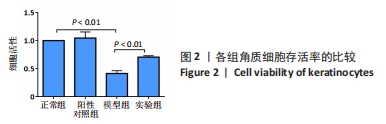
2.1 体外实验结果 2.1.1 各组角质细胞增殖情况 CCK-8检测结果显示,与正常组(100%)比较,阳性对照组细胞存活率无明显变化[(104.60±3.25)%,P > 0.05],模型组细胞存活率明显降低[(41.42±2.90)%,P < 0.01];与模型组比较,实验组细胞存活率升高[(70.61±4.12)%,P < 0.01],见图2,说明枸杞多糖能够保护角质细胞,减少肿瘤坏死因子α对角质细胞增殖的抑制作用。"
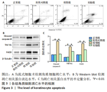
2.1.2 各组角质细胞凋亡情况 流式细胞术检测结果显示,正常组、阳性对照组、模型组与实验组的细胞凋亡率分别为(6.87±0.27)%,(6.25±0.32)%,(38.50±0.16)%,(23.89±0.23)%,见图3A。与正常组相比,肿瘤坏死因子α干预的角质细胞发生明显凋亡(P < 0.05);与模型组比较,实验组细胞凋亡率明显降低(P < 0.05)。 为了进一步确认枸杞多糖能够抑制肿瘤坏死因子α介导角质细胞的凋亡作用,利用Western blot检测细胞凋亡蛋白Cleaved caspase-8、TNF R1、FADD的蛋白水平,结果见图3B、C。与正常组比较,阳性对照组细胞内Cleaved caspase-8、TNF R1、FADD的蛋白表达均无明显变化(P > 0.05),模型组细胞内Cleaved caspase-8、TNF R1、FADD的蛋白表达均升高(P < 0.01);与模型组比较,实验组细胞内Cleaved caspase-8、TNF R1、FADD的蛋白表达均降低(P < 0.01)。"


为了进一步确认枸杞多糖通过抑制自噬保护炎性微环境下角质细胞凋亡,利用Western blot检测自噬蛋白LC3的表达水平,结果见图4。与正常组比较,阳性对照组细胞内LC3的蛋白表达均无明显变化(P > 0.05),模型组细胞内LC3的蛋白表达升高(P < 0.01);与模型组比较,实验组细胞内LC3的蛋白表达降低(P < 0.01)。"

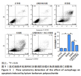
为了进一步确定自噬在角质细胞凋亡中的作用,使用自噬抑制剂进行干预,利用流式细胞术检测细胞凋亡情况。流式细胞术检测结果显示,正常组、自噬抑制剂组、模型组、实验组、自噬抑制剂+枸杞多糖组的细胞凋亡率分别为(7.83±0.29)%,(7.33±0.28)%,(43.00±0.24)%,(24.96±0.12)%,(15.90±0.19)%,见图5。与正常组比较,自噬抑制剂组细胞凋亡率无明显变化(P > 0.05),模型组细胞凋亡率升高(P < 0.01);与模型组比较,实验组细胞凋亡率降低(P < 0.01);与实验组比较,自噬抑制剂+枸杞多糖组细胞凋亡率降低(P < 0.01)。"

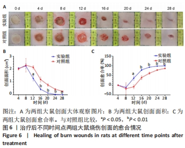
综上所述,枸杞多糖能够抑制肿瘤坏死因子α介导的角质细胞自噬。 2.2 体内实验结果 2.2.1 实验动物数量分析 对照组3只与实验组3只大鼠全部进入结果分析。 2.2.2 两组大鼠创面愈合率比较 随着治疗时间的延长,两组大鼠创面面积缩小,创面愈合率升高;与对照组相比,实验组治疗后12,16,20,24,28 d时的创面面积缩小明显(P < 0.05,P < 0.01),创面愈合率升高明显(P < 0.05,P < 0.01),见图6。"


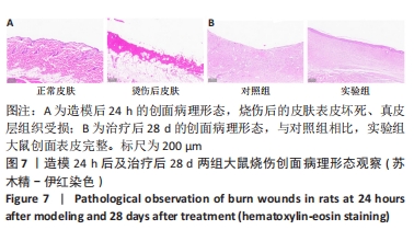
2.2.3 两组大鼠创面病理形态观察 造模后24 h的苏木精-伊红染色显示,正常皮肤组织完整、形态正常;烧伤后的皮肤表皮坏死、真皮层组织受损,提示造模成功(图7A)。治疗后28 d的苏木精-伊红染色显示,与对照组相比,实验组大鼠创面表皮完整、清晰(图7B)。"
| [1] 陈孝平,汪建平.外科学[M].北京:人民卫生出版社,2013. [2] ORYAN A, ALEMZADEH E, MOSHIRI A. Burn wound healing: present concepts, treatment strategies and future directions. J Wound Care. 2017;26(1):5-19. [3] SARKOZYOVA N, DRAGUNOVA J, BUKOVCAN P, et al. Preparation and processing of human allogenic dermal matrix for utilization in reconstructive surgical procedures. Bratisl Lek Listy. 2020;121(6):386-394. [4] SHAKHAKARMI K, SEO JE, LAMICHHANE S, et al. EGF, a veteran of wound healing: highlights on its mode of action, clinical applications with focus on wound treatment, and recent drug delivery strategies. Arch Pharm Res. 2023;46(4):299-322. [5] JING S, LI H, XU H. Mesenchymal Stem Cell Derived Exosomes Therapy in Diabetic Wound Repair. Int J Nanomedicine. 2023;18:2707-2720. [6] ZHAO D, XIAO J, QIANG L, et al. Walnut ointment promotes full-thickness burning wound healing: role of linoleic acid. Acta Cir Bras. 2022;37(9):e370902. [7] LIU J. A new way of repairing special wounds. Heliyon. 2023;9(5): e15755. [8] MARKS R. Seeing through the stratum corneum. Keio J Med. 2000; 49(2):80-83. [9] NORLÉN L. Skin barrier structure and function: the single gel phase model. J Invest Dermatol. 2001;117(4):830-836. [10] VOEGELI R, RAWLINGS AV, BRETERNITZ M, et al. Increased stratum corneum serine protease activity in acute eczematous atopic skin. Br J Dermatol. 2009;161(1):70-77. [11] FOLDVARI M, KUMAR P, KING M, et al. Gene delivery into human skin in vitro using biphasic lipid vesicles. Curr Drug Deliv. 2006;3(1):89-93. [12] HORVÁTH D, SIPOS A, MAJOR E, et al. Myosin phosphatase accelerates cutaneous wound healing by regulating migration and differentiation of epidermal keratinocytes via Akt signaling pathway in human and murine skin. Biochim Biophys Acta Mol Basis Dis. 2018;1864(10):3268-3280. [13] MORIYAMA M, MORIYAMA H, UDA J, et al. Beneficial Effects of the Genus Aloe on Wound Healing, Cell Proliferation, and Differentiation of Epidermal Keratinocytes. PLoS One. 2016;11(10):e0164799. [14] GRIEB G. Basic and Clinical Research in Wound Healing. Biomedicines. 2023;11(5):1380. [15] YU MS, LEUNG SK, LAI SW, et al. Neuroprotective effects of anti-aging oriental medicine Lycium barbarum against beta-amyloid peptide neurotoxicity. Exp Gerontol. 2005;40(8-9):716-727. [16] CHEN L, LI W, QI D, et al. Lycium barbarum polysaccharide protects against LPS-induced ARDS by inhibiting apoptosis, oxidative stress, and inflammation in pulmonary endothelial cells. Free Radic Res. 2018;52(4):480-490. [17] ZHANG M, TANG X, WANG F, et al. Characterization of Lycium barbarum polysaccharide and its effect on human hepatoma cells. Int J Biol Macromol. 2013;61:270-275. [18] NIU Y, ZHANG G, SUN X, et al. Distinct Role of Lycium barbarum L. Polysaccharides in Oxidative Stress-Related Ocular Diseases. Pharmaceuticals (Basel). 2023;16(2):215. [19] RAJKOWSKA K, OTLEWSKA A, BRONCEL N, et al. Microbial Diversity and Bioactive Compounds in Dried Lycium barbarum Fruits (Goji): A Comparative Study. Molecules. 2023;28(10):4058. [20] LI H, LI Z, PENG L, et al. Lycium barbarum polysaccharide protects human keratinocytes against UVB-induced photo-damage. Free Radic Res. 2017;51(2):200-210. [21] XIAO J, LIONG EC, CHING YP, et al. Lycium barbarum polysaccharides protect rat liver from non-alcoholic steatohepatitis-induced injury. Nutr Diabetes. 2013;3(7):e81. [22] LAM CS, TIPOE GL, SO KF, et al. Neuroprotective mechanism of Lycium barbarum polysaccharides against hippocampal-dependent spatial memory deficits in a rat model of obstructive sleep apnea. PLoS One. 2015;10(2):e0117990. [23] LENG H, PU L, XU L, et al. Effects of aloe polysaccharide, a polysaccharide extracted from Aloe vera, on TNF-α-induced HaCaT cell proliferation and the underlying mechanism in psoriasis. Mol Med Rep. 2018;18(3):3537-3543. [24] XIAO M, LI L, LI C, et al. Role of autophagy and apoptosis in wound tissue of deep second-degree burn in rats. Acad Emerg Med. 2014; 21(4):383-391. [25] SINSUEBPOL C, BURAPAPADH K, CHOWJAROEN V, et al. The radical scavenging activity of vanillin and its impact on the healing properties of wounds. J Adv Pharm Technol Res. 2023;14(2):99-104. [26] GALLEGOS-ALCALÁ P, JIMÉNEZ M, CERVANTES-GARCÍA D, et al. Glycomacropeptide Protects against Inflammation and Oxidative Stress, and Promotes Wound Healing in an Atopic Dermatitis Model of Human Keratinocytes. Foods. 2023;12(10):1932. [27] WOO YK, PARK J, RYU JH, et al. The anti-inflammatory and anti-apoptotic effects of advanced anti-inflammation composition (AAIC) in heat shock-induced human HaCaT keratinocytes. J Cosmet Dermatol. 2020;19(8):2114-2124. [28] DONG Y, CHEN H, GAO J, et al. Molecular machinery and interplay of apoptosis and autophagy in coronary heart disease. J Mol Cell Cardiol. 2019;136:27-41. [29] CAO Y, LI Q, LIU L, et al. Modafinil protects hippocampal neurons by suppressing excessive autophagy and apoptosis in mice with sleep deprivation. Br J Pharmacol. 2019;176(9):1282-1297. [30] YOUNG TM, REYES C, PASNIKOWSKI E, et al. Autophagy protects tumors from T cell-mediated cytotoxicity via inhibition of TNFα-induced apoptosis. Sci Immunol. 2020;5(54):eabb9561. [31] ZHU B, JIANG Q, QUE G, et al. Role of autophagy and apoptosis in atrophic epithelium in oral submucous fibrosis. J Oral Sci. 2020;62(2): 184-188. [32] ZHANG P, WANG H, CHEN Y, et al. DR5 related autophagy can promote apoptosis in gliomas after irradiation. Biochem Biophys Res Commun. 2020;522(4):910-916. [33] ZHU S, LI X, DANG B, et al. Lycium Barbarum polysaccharide protects HaCaT cells from PM2.5-induced apoptosis via inhibiting oxidative stress, ER stress and autophagy. Redox Rep. 2022;27(1):32-44. [34] WAJANT H. The Fas signaling pathway: more than a paradigm. Science. 2002;296(5573):1635-1636. [35] KISCHKEL FC, HELLBARDT S, BEHRMANN I, et al. Cytotoxicity-dependent APO-1 (Fas/CD95)-associated proteins form a death-inducing signaling complex (DISC) with the receptor. EMBO J. 1995;14(22):5579-5588. [36] DJAVAHERI-MERGNY M, AMELOTTI M, MATHIEU J, et al. NF-kappaB activation represses tumor necrosis factor-alpha-induced autophagy. J Biol Chem. 2006;281(41):30373-30382. [37] DING R, LIU Z, TAN J, et al. Advanced oxidation protein products mediate human keratinocytes apoptosis by inducing cell autophagy through the mTOR-Beclin-1 pathway. Cell Biochem Funct. 2022;40(8): 880-887. [38] TANG SC, KO JL, LU CT, et al. Chloroquine alleviates the heat-induced to injure via autophagy and apoptosis mechanisms in skin cell and mouse models. PLoS One. 2022;17(8):e0272797. [39] FERNANDO PDSM, PIAO MJ, KANG KA, et al. Hesperidin Protects Human HaCaT Keratinocytes from Particulate Matter 2.5-Induced Apoptosis via the Inhibition of Oxidative Stress and Autophagy. Antioxidants (Basel). 2022;11(7):1363. [40] YOUNCE CW, KOLATTUKUDY PE. MCP-1 causes cardiomyoblast death via autophagy resulting from ER stress caused by oxidative stress generated by inducing a novel zinc-finger protein, MCPIP. Biochem J. 2010;426(1):43-53. |
| [1] | Yang Yifeng, Ye Nan, Wang Lin, Guo Shuaicheng, Huang Jian. Signaling pathway of dexmedetomidine against ischemia-reperfusion injury [J]. Chinese Journal of Tissue Engineering Research, 2024, 28(9): 1464-1469. |
| [2] | Yue Yun, Wang Peipei, Yuan Zhaohe, He Shengcun, Jia Xusheng, Liu Qian, Li Zhantao, Fu Huiling, Song Fei, Jia Menghui. Effects of croton cream on JNK/p38 MAPK signaling pathway and neuronal apoptosis in cerebral ischemia-reperfusion injury rats [J]. Chinese Journal of Tissue Engineering Research, 2024, 28(8): 1186-1192. |
| [3] | Wang Ji, Zhang Min, Li Wenbo, Yang Zhongya, Zhang Long. Effect of aerobic exercise on glycolipid metabolism, skeletal muscle inflammation and autophagy in type 2 diabetic rats [J]. Chinese Journal of Tissue Engineering Research, 2024, 28(8): 1200-1205. |
| [4] | Zhou Bangyu, Li Jie, Ruan Yushang, Geng Funeng, Li Shaobo. Effects of Periplaneta americana powder on motor function and autophagic protein Beclin-1 in rats undergoing spinal cord hemisection [J]. Chinese Journal of Tissue Engineering Research, 2024, 28(8): 1223-1228. |
| [5] | Sheng Siqi, Xie Lin, Zhao Xiangyu, Jiang Yideng, Wu Kai, Xiong Jiantuan, Yang Anning, Hao Yinju, Jiao Yun. Involvement of miR-144-3p in Cbs+/- mouse hepatocyte autophagy induced by high-methionine diet [J]. Chinese Journal of Tissue Engineering Research, 2024, 28(8): 1289-1294. |
| [6] | Liu Qiwei, Zhang Junhui, Yang Yuan, Wang Jinjuan. Role and mechanism of umbilical cord mesenchymal stem cells on polycystic ovary syndrome [J]. Chinese Journal of Tissue Engineering Research, 2024, 28(7): 1015-1020. |
| [7] | Pan Xiaolong, Fan Feiyan, Ying Chunmiao, Liu Feixiang, Zhang Yunke. Effect and mechanism of traditional Chinese medicine on inhibiting the aging of mesenchymal stem cells [J]. Chinese Journal of Tissue Engineering Research, 2024, 28(7): 1091-1098. |
| [8] | Huang Yuxin, Liang Wenzi, Chen Xiuwen, Ni Na, Zhao Yinglin, Lin Changmin. Role of autophagy in hair regeneration [J]. Chinese Journal of Tissue Engineering Research, 2024, 28(7): 1112-1117. |
| [9] | Zhang Ya, Mu Qiuju, Wang Zilin, Liu Hongjie, Zhu Lili. Hydrogel loaded with platelet-rich plasma promotes wound healing in diabetic rats [J]. Chinese Journal of Tissue Engineering Research, 2024, 28(5): 690-696. |
| [10] | Shen Feiyan, Yao Jixiang, Su Shanshan, Zhao Zhongmin, Tang Weidong. Knockdown of circRNA WD repeat containing protein 1 inhibits proliferation and induces apoptosis of chondrocytes in knee osteoarthritis [J]. Chinese Journal of Tissue Engineering Research, 2024, 28(4): 499-504. |
| [11] | Chen Zepeng, Hou Yonghui, Chen Shudong, Hou Yu, Lin Dingkun. Tauroursodeoxycholic acid treats spinal cord injury by reducing apoptosis of spinal cord neurons under glucose and oxygen deprivation [J]. Chinese Journal of Tissue Engineering Research, 2024, 28(4): 528-534. |
| [12] | Xue Jingwen, Wang Fangfang, Zhang Xin, Pang Ruifeng, Wang Xiaoye, Ma Xiaoru. Effect of ganoderma spore on mitochondrial autophagy and apoptosis in testicular tissue of diabetic rats [J]. Chinese Journal of Tissue Engineering Research, 2024, 28(4): 562-568. |
| [13] | Yan Binghan, Li Zhichao, Su Hui, Xue Haipeng, Xu Zhanwang, Tan Guoqing. Mechanisms of traditional Chinese medicine monomers in the treatment of osteoarthritis by targeting autophagy [J]. Chinese Journal of Tissue Engineering Research, 2024, 28(4): 627-632. |
| [14] | Xie Ting, Liu Tingting, Zeng Xuehui, Li Yamin, Zhou Panghu, Yi Nianhua. Fucoxanthin alleviates glucocorticoid-induced osteoblast apoptosis by activating nuclear factor erythroid-2-related factor 2 [J]. Chinese Journal of Tissue Engineering Research, 2024, 28(23): 3609-3614. |
| [15] | Ma Wanli, Yang Hongsheng, Qu Bo, Zhang Zhengdong, Gong Kai, Lin Yanshui. Mechanisms by which baicalein protects against steriod-induced osteonecrosis of the femoral head in rats [J]. Chinese Journal of Tissue Engineering Research, 2024, 28(23): 3661-3668. |
| Viewed | ||||||
|
Full text |
|
|||||
|
Abstract |
|
|||||